Posts
A written blog for the public to gain the basic understanding of cancer.
Original author: Dr Hafsa Waseela Abbas
Original published date: 1st April 2014
Updated by: Dr Hafsa Waseela Abbas
Update date: April 2025
Review date: April 2027
Our bodies are made of millions of cells and are the smallest unit of living organisms. Cell division and growth is a regulatory process where cells of the same type become tissues (Cancer Research UK, n.d.; Cassidy et al., 2010). Different tissues form organs and many organs work together to produce organ systems. An ultimate living organism has different organ systems that carry out life processes. There are seven life processes: movement, respiration, sensitivity, nutrition, excretion, reproduction, and growth. Every cell has a lifespan and new cells replace old cells that become old and senescent. For instance, red blood cells have a lifespan of 120 days whereas, platelets have one to ten days.
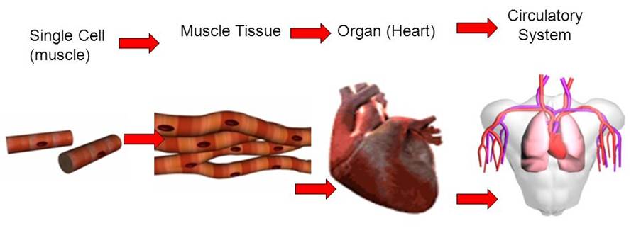
Figure 1: Levels of Organisations in humans
Cancer develops due to abnormal cellular growth that can evade the immune system, inhibitory signals, and apoptosis. Deoxyribonucleic acid (DNA) found in the nucleus to control the cell. A gene is a short section of the DNA where some genes called oncogenes promote cancer progression. On the other hand, some genes stop tumour growth and are known as tumour suppressor genes. Changes in the gene are referred to as mutations and can influence how normal cells grow and divide (Cancer Research UK, n.d.; Cassidy et al., 2010). For instance, a tumour suppressor gene that becomes mutated has oncogenic potential to increase cancer progression.
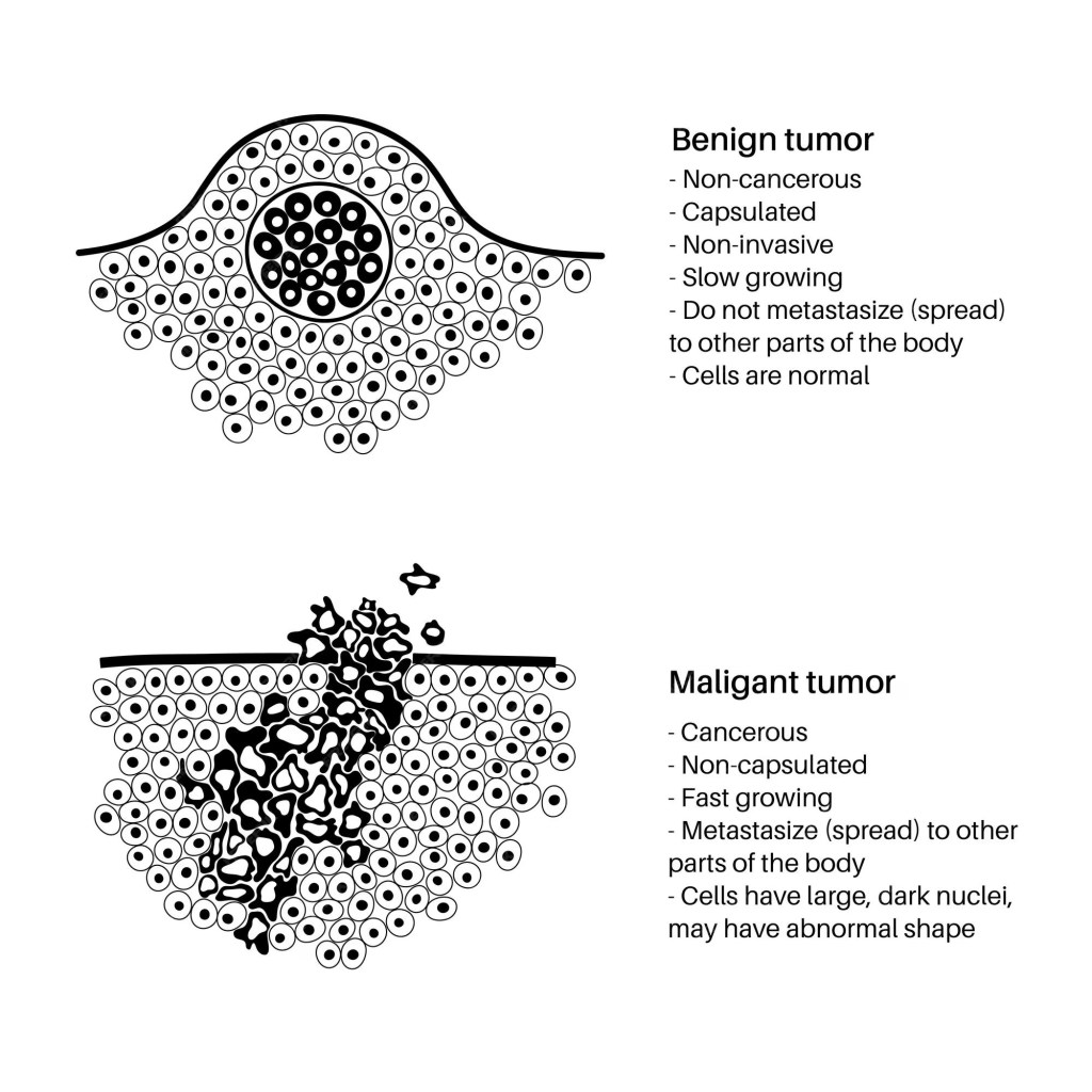
Benign tumours are cancers that remain in the primary site (organ where the cancer starts to grow). Many benign cancers are surgically removed to prevent cancer spread to other organs in the body. However, cancer cells that continue to grow uncontrollably because of the increased number of mutations can break from the primary site and travel to other organs via the blood, lymph, and cavities (pleural and peritoneal) to form secondary tumours (Cancer Research UK, n.d.; Cassidy et al., 2010). This process is called metastasis – please see Figure 2. The pleura covers the lungs via the thin, smooth serous membrane. There are two types of pleura: visceral pleura covers the lungs and, the parietal in the inner wall – See Figure 3A. The peritoneum is a thin layer of serous membrane and has two types: parietal is within the walls of the abdomen (belly). The visceral peritoneum covers the organs in the abdomen – see Figure 3B. Trans coelomic spread occurs in advanced cancers of the lungs, intestine, ovary, stomach, and womb (endometrium).
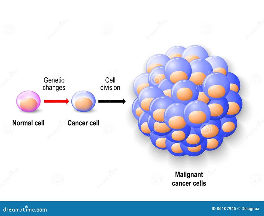
Figure 2: The transformation of a non-cancerous cell (benign) to a malignant cancer cell depends on the number of mutations expressed that alter how the cancer cell looks (phenotype).


Figure 3: Pleural cavity (A) Peritoneum (B).
The blood consists of red blood cells that transport oxygen, white blood cells and antibodies that fight off infection, hormones that regulate bodily functions, and platelets to clot blood and prevent blood loss. The lymph is a colourless liquid made up of white blood cells and it functions in draining the fluid and fighting infection.
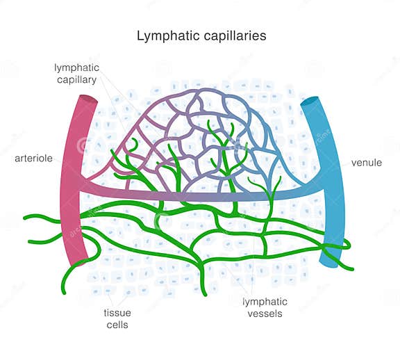
Figure 4: The blood and lymph
References
Cancer Research UK (n.d.) What is cancer? Available at: https://www.cancerresearchuk.org/about-cancer/what-is-cancer (Accessed: 15th April 2025)
Cassidy, J., Bissett, D., Spene, R. and Payne, M. (2010) Oxford Handbook of Oncology. Oxford: Oxford University Press.

Leave a comment