
Diagnosis of Ovarian Cancer
During the consultation with the doctor, there will be an initial discussion of the patient’s symptoms and how long they have experienced them. Questions will also be asked about their lifestyle and family history. The doctor will conduct a physical examination and the relevant further tests to detect the cause.
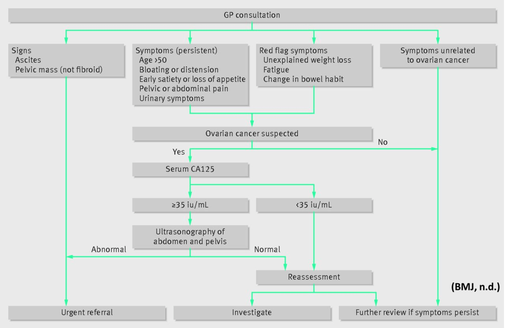
NICE Guidelines For Ovarian Cancer
After discussing the symptoms, the healthcare professional would perform a general check-up, including a temperature and blood pressure check.
Blood Test
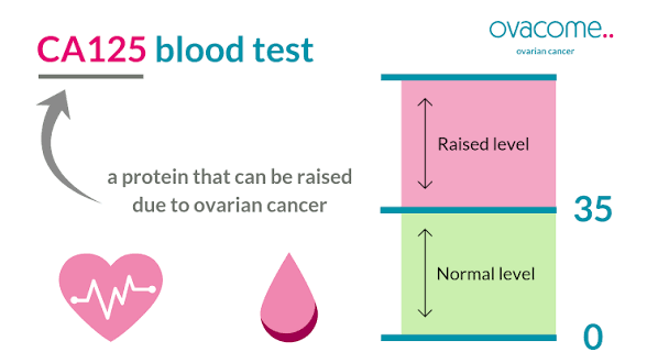
CA125
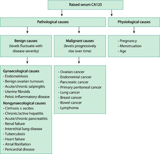
CA125 is a protein biomarker found in the blood. When there is inflammation, cancer, or another issue in the ovaries, the level of this biomarker increases.
The reference range for normal levels is less than 35 IU/ml.
So if it is above 35 IU/ml, further investigations need to be performed.
Other Blood Tests
Female patients below the age of 40, levels of alpha feta protein (AFP) and beta human chorionic gonadotrophin (HcG), and a CA125 to determine the type of ovarian cancer.
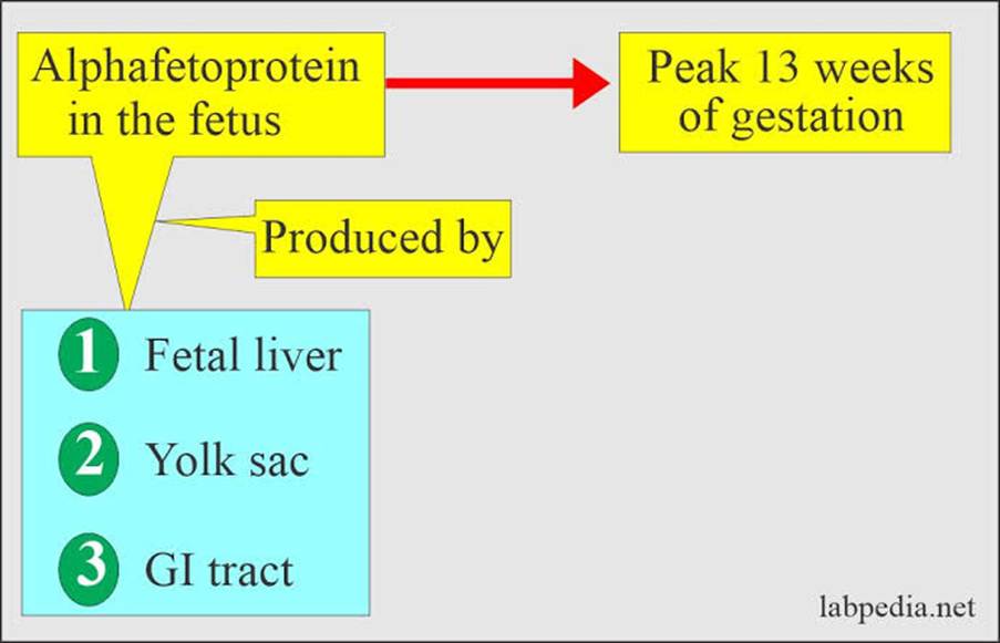
Alpha Feta-protein
AFP is a glycoprotein produced in the fetal liver, yolk sac, and digestive system. It helps to detect several cancers (testicular and ovarian) and other forms.
It also helps to detect liver conditions such as cirrhosis, liver cancer, and other hepatic-related and gastrointestinal issues.
Human chorionic gonadotropin (hCG)
This is a hormone produced by the placenta during pregnancy. A placenta is the food tube from the mother to the foetus.
It is a signal biomarker for determining how well the pregnancy is progressing. Serum hCG is useful for detecting early pregnancies.

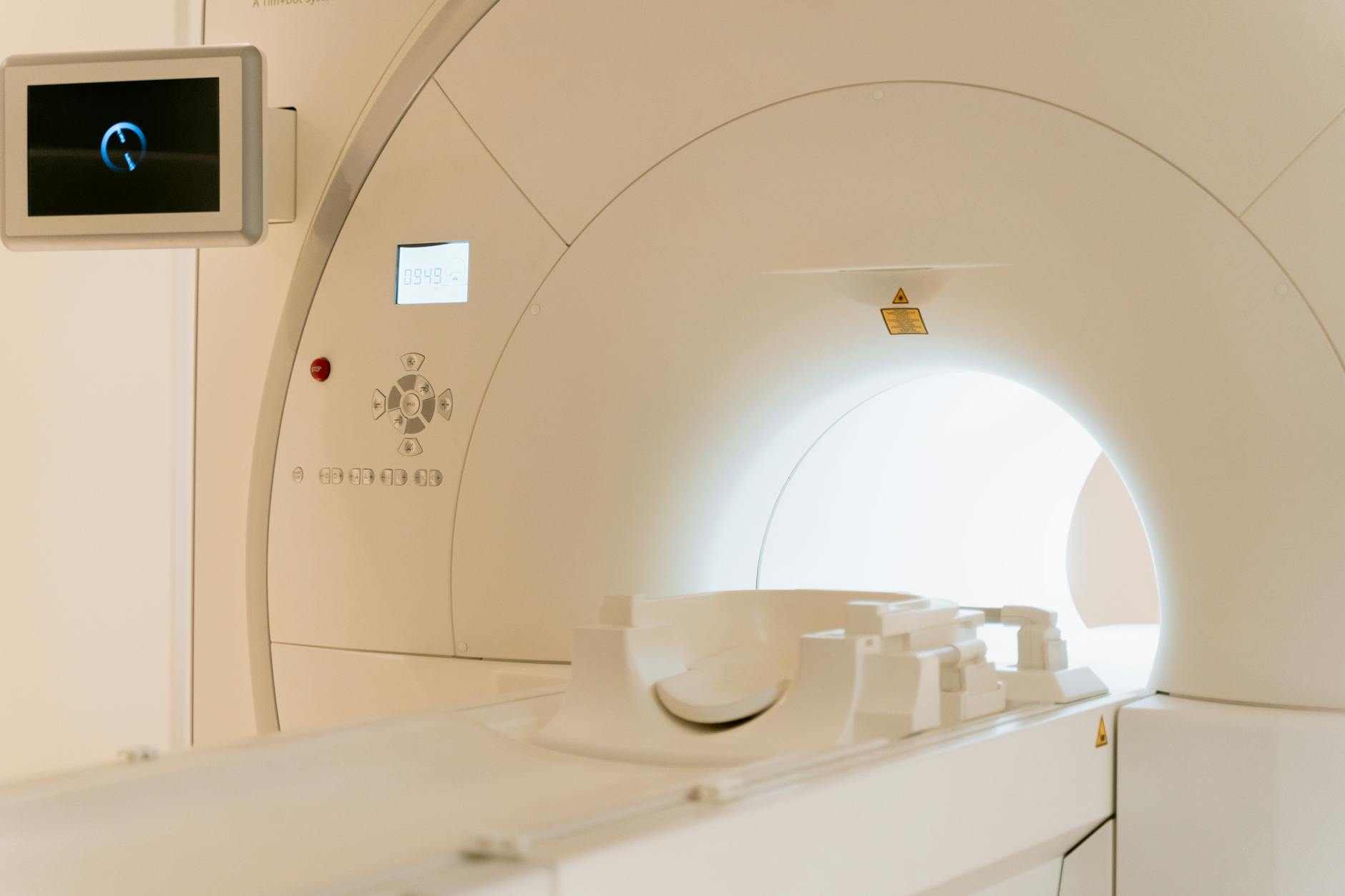
Imaging
The type of imaging is dependent on the area or the assessment.
This is commonly an ultrasound or computer tomography (CT scan) to determine whether there is cancer and how far it has spread.
Ultrasound
The first type of imaging technique for suspected ovarian cancer cases is ultrasound. Ultrasound uses sound waves to create images.
Ultrasound on the belly (abdomen) and pelvis is commonly performed.
After the imaging procedure, a risk of malignancy index (RMI I)
If cancer is found, the patient is referred to the suspected cancer pathway.
Dr Saeed Ahmad: Ultrasound of Ovarian Cancer
CT Scan
This uses X-rays to provide a deeper view of the area under assessment. CT is performed when it is confirmed that ovarian cancer is present via the level of CA125 and the ultrasound outcome.
If there are cases where there is a normal ultrasound but high or normal levels of CA125
- It is important to assess the cause of clinical symptoms
- If no other clinical cause is found, the patient is returned to the GP for frequent/ persistent symptoms.
Biopsy
Whilst performing imaging, ovarian cancer needs to be confirmed by extracting a tissue sample called a biopsy.
Biopsies are assessed by a doctor who specialises in examining tissues under the microscope, called a pathologist, to see whether they are cancerous.
If inappropriate, then the assessment of cells under the microscope is conducted by a cytologist.
There are various types of biopsies; the most common type is percutaneous image-guided biopsy, under the skin using a small incision/cut, and a specialised needle is added to extract the sample.
Dr Jay Mehta: Guided Core Biopsy

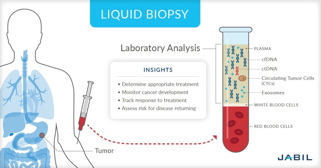
Other types include liquid biopsy.
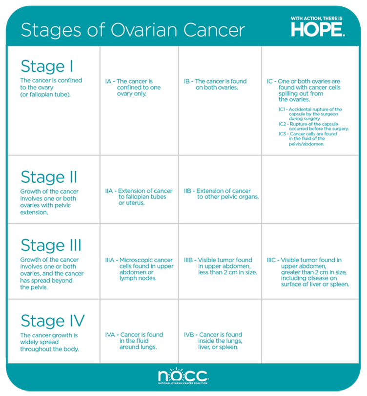
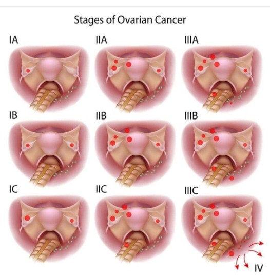
Staging Of Ovarian Cancer

Useful Resources And Further Reading
Updated April 2026 Next Review March 2027

Leave a comment