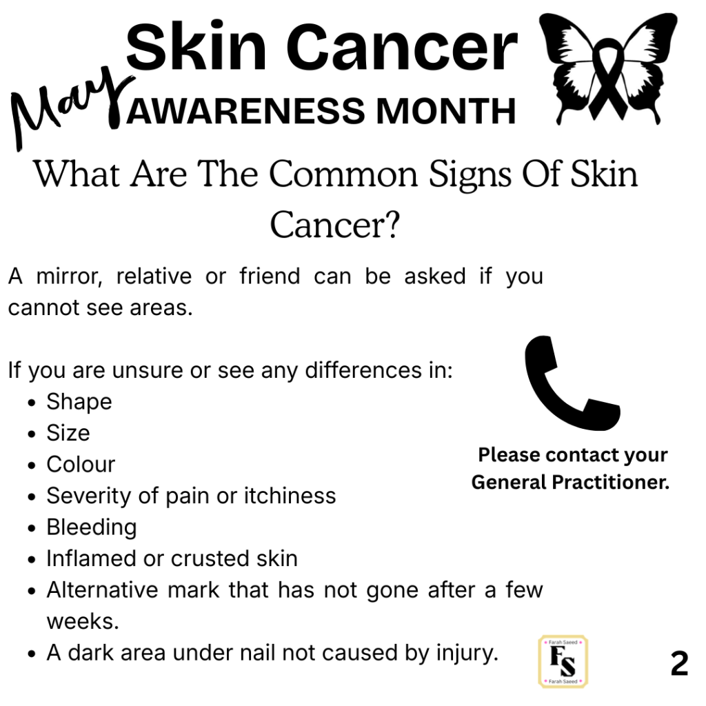
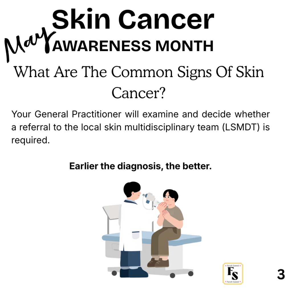
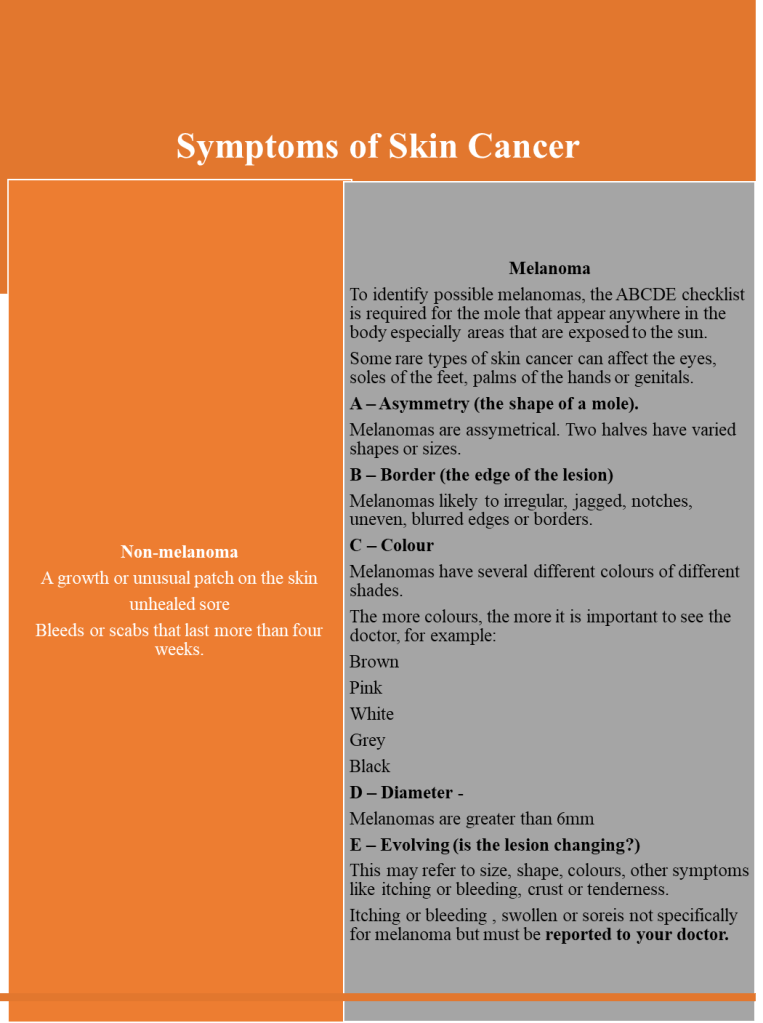
What Are The Signs And Symptoms Of Skin Cancer?
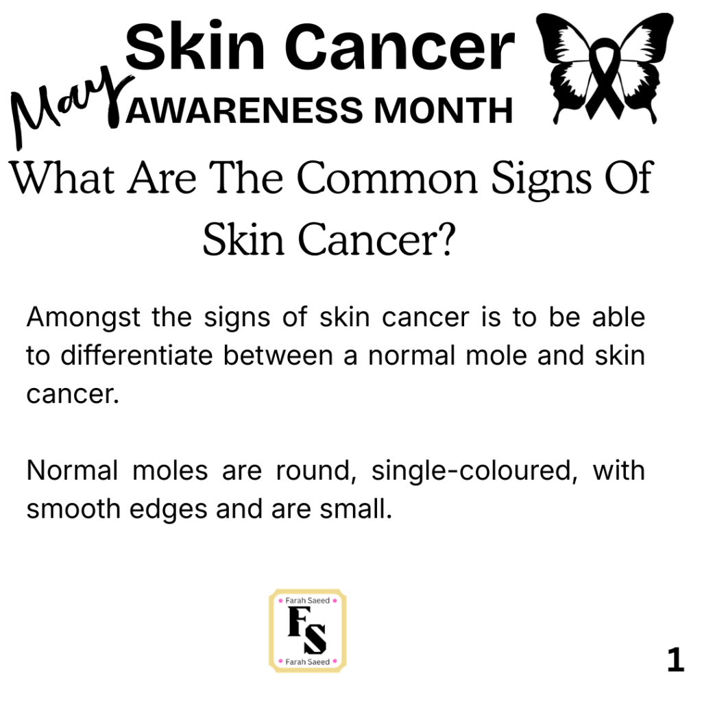
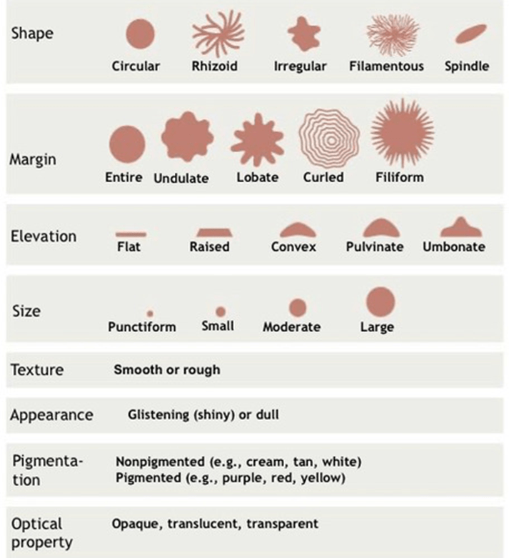
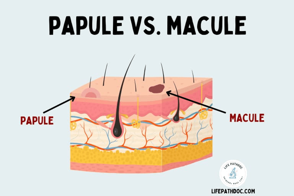
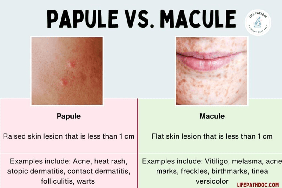
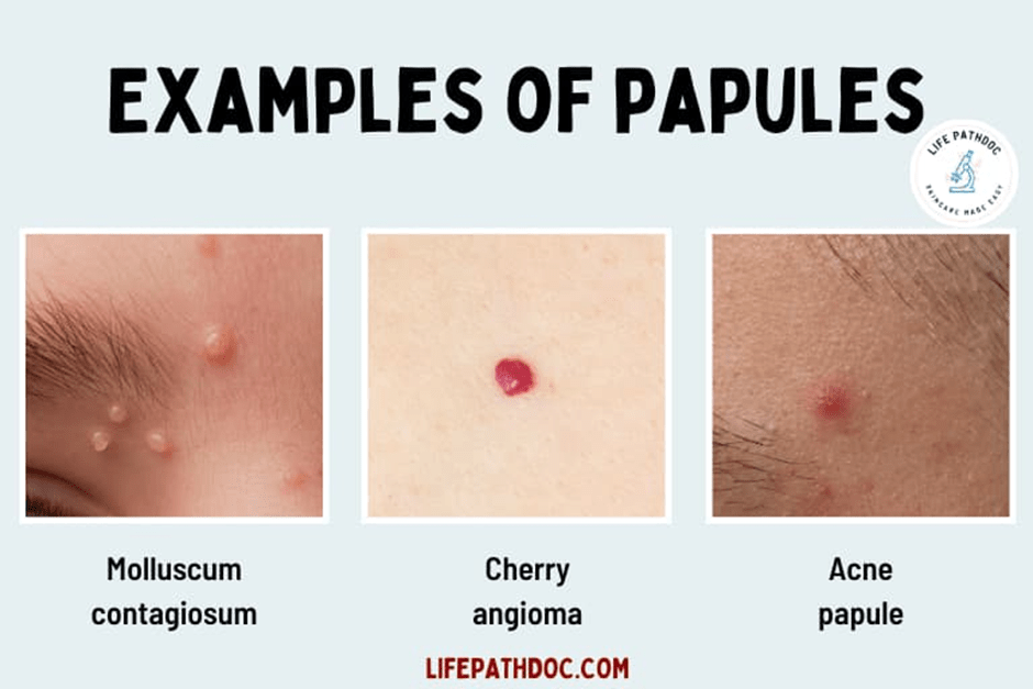
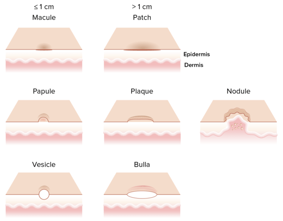
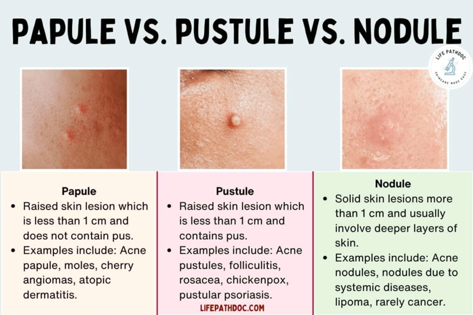
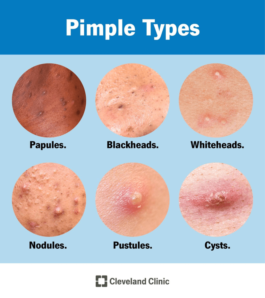
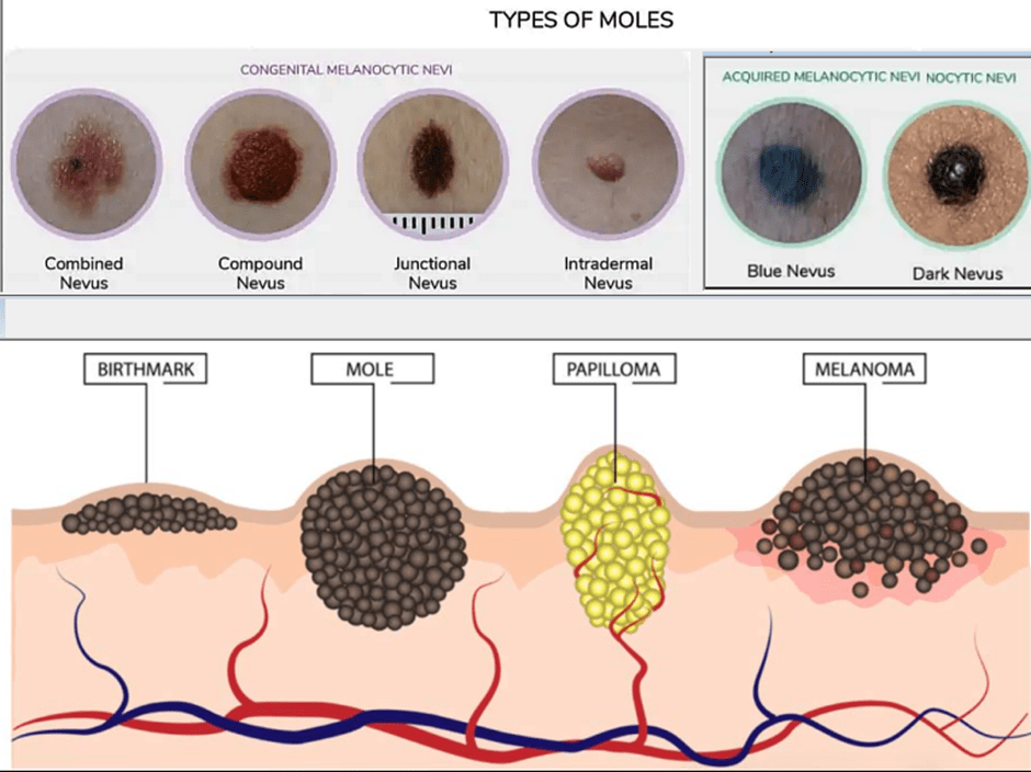
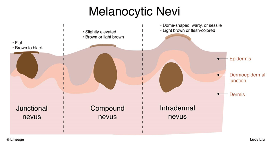
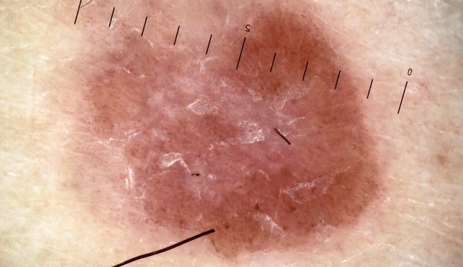
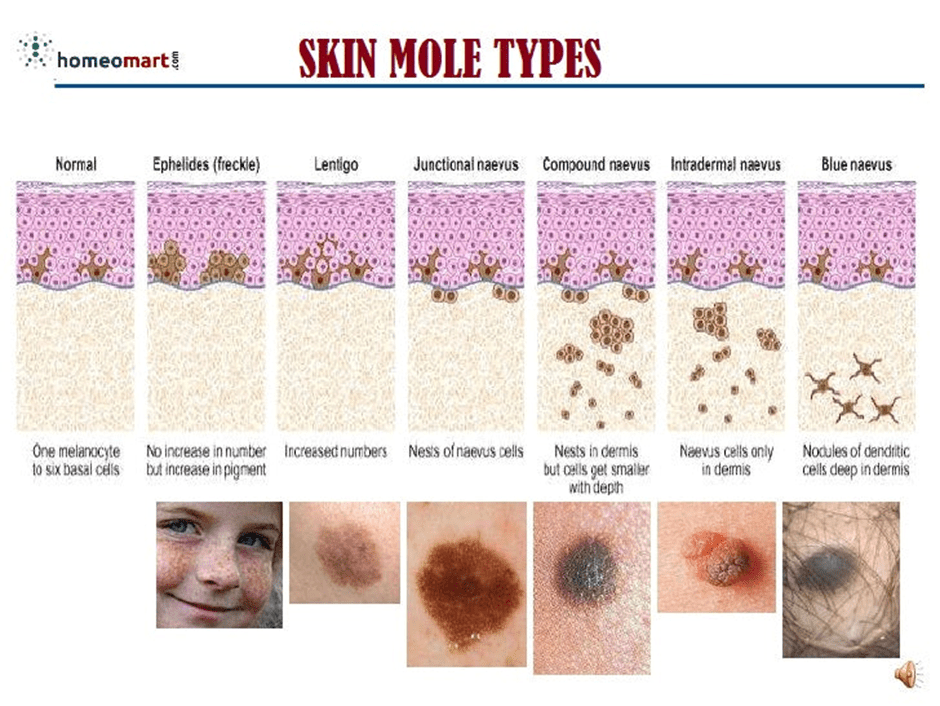
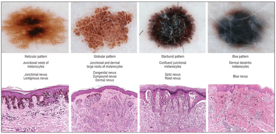
Describing features of the mole
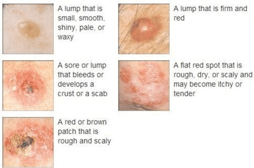
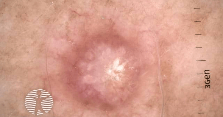
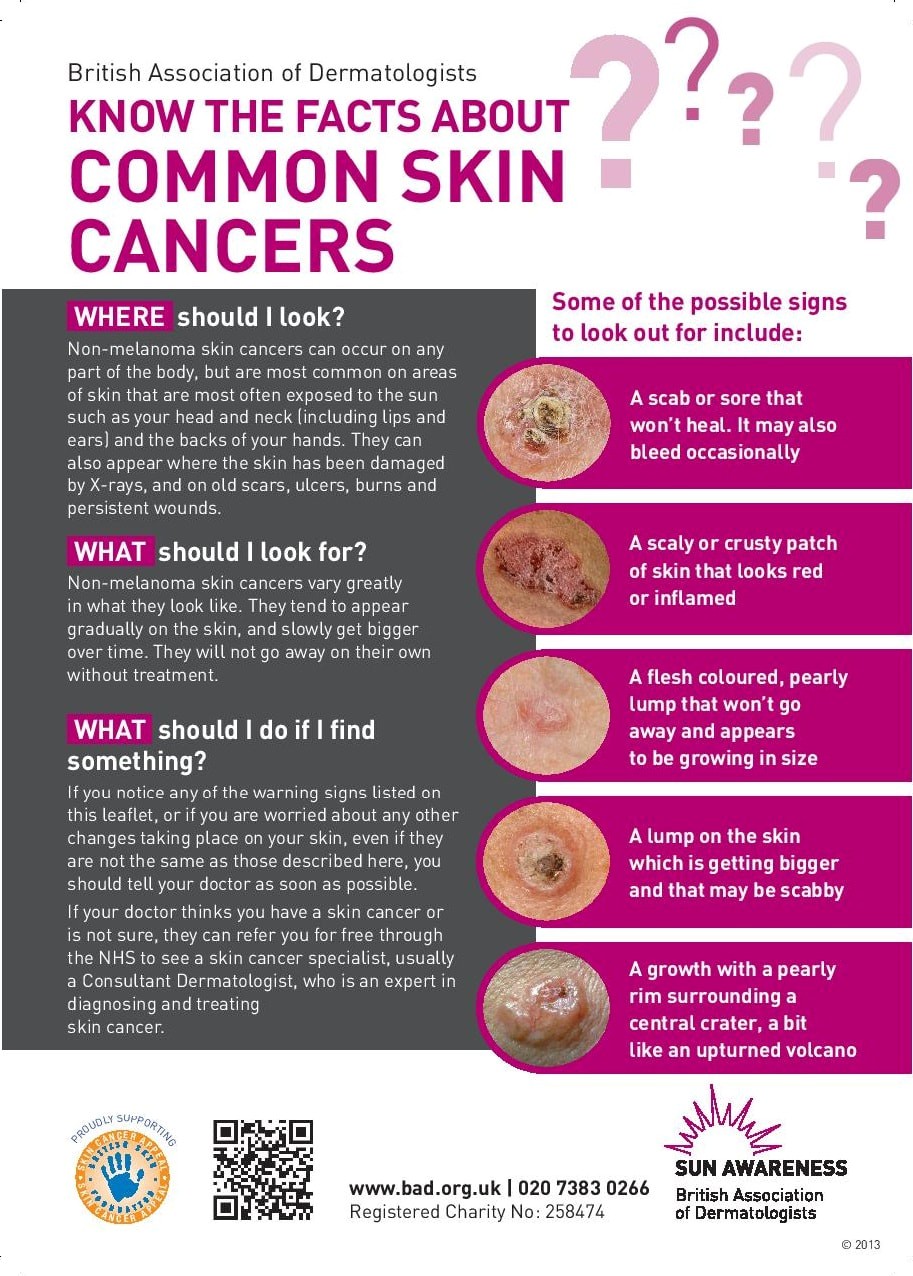
Examination Of Non-Melanoma
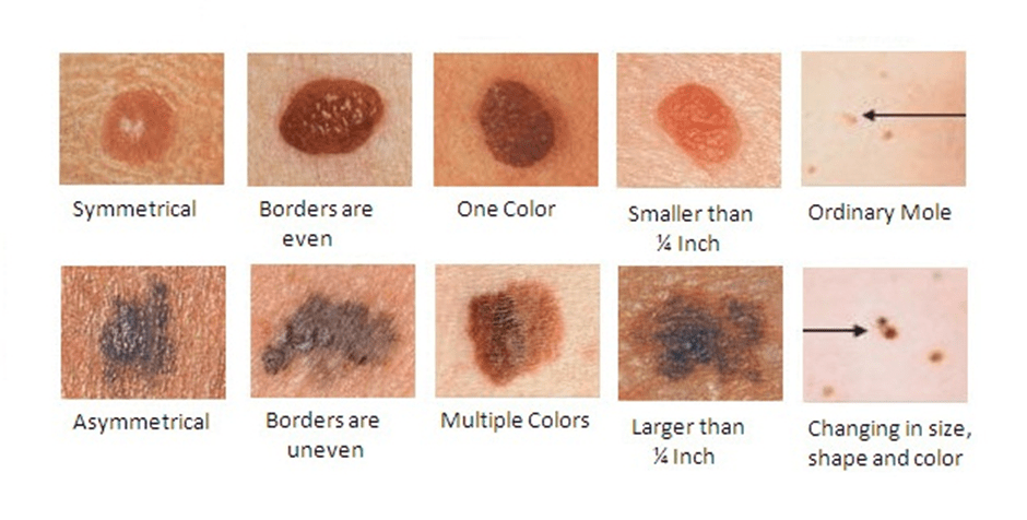
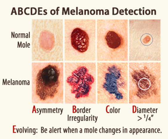
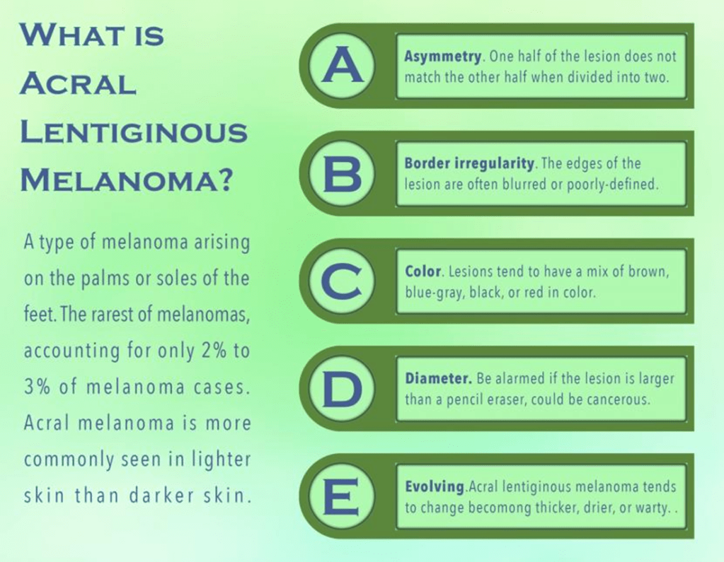
The ABCDE Checklist
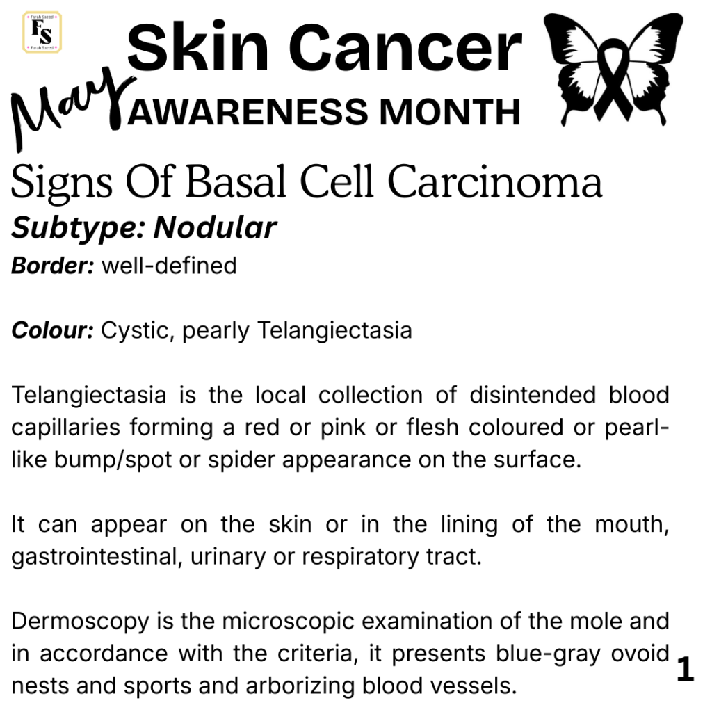
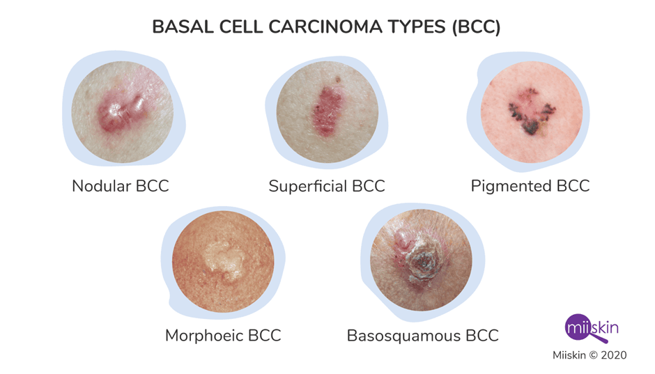
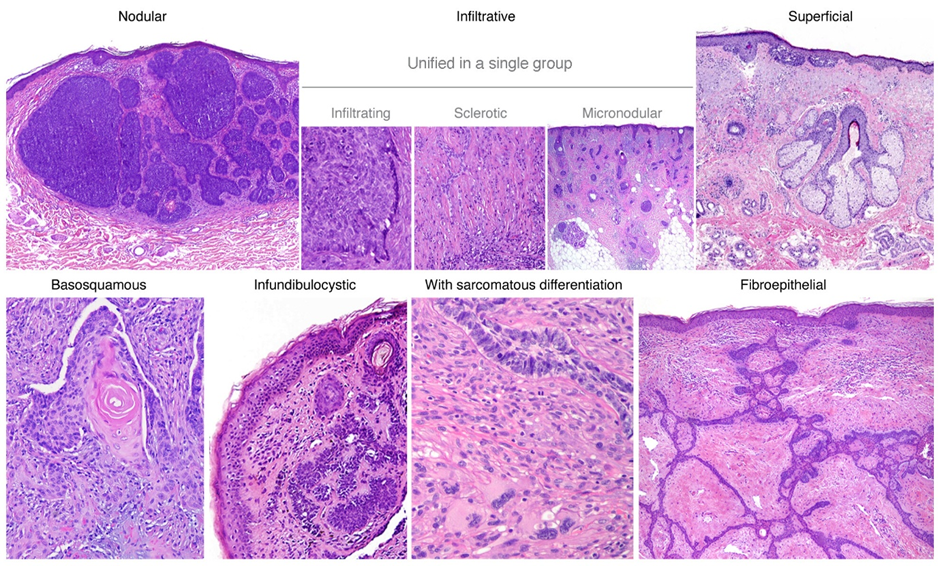
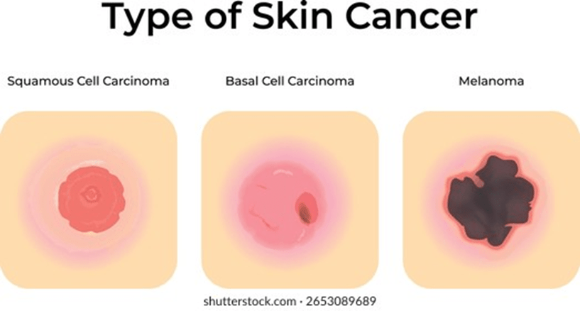
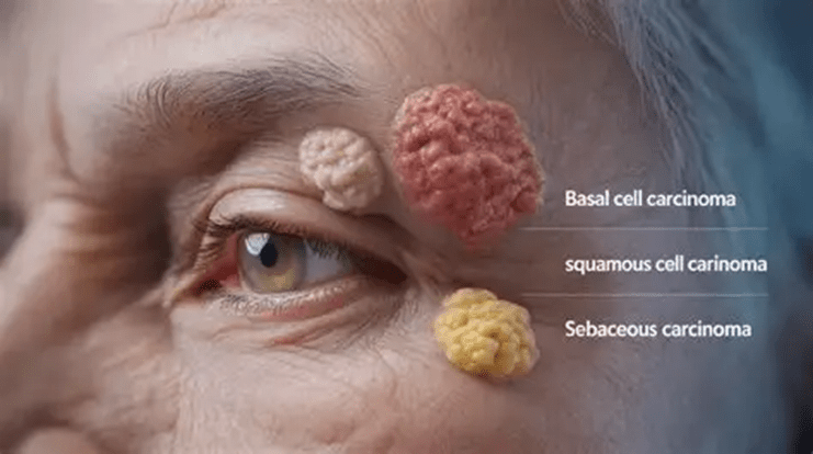
Signs Of Basal Cell Carcinoma

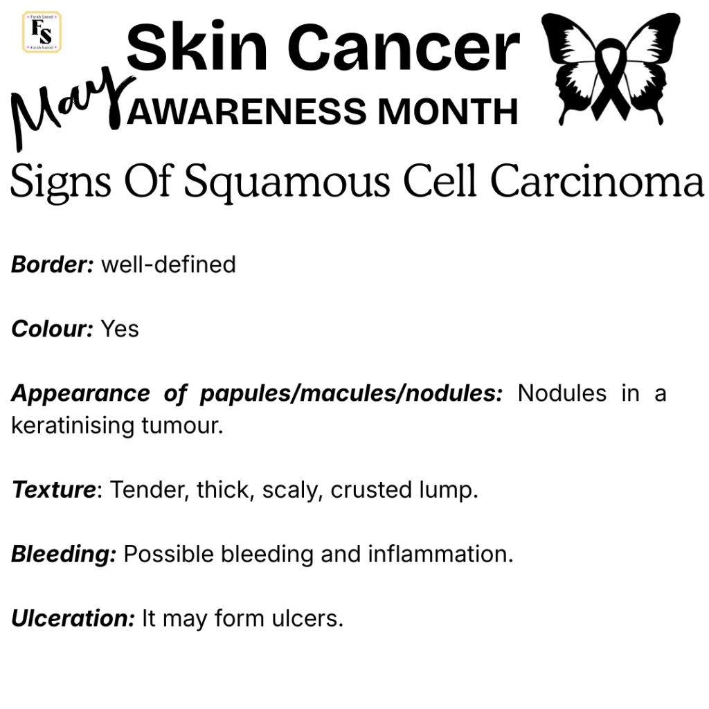
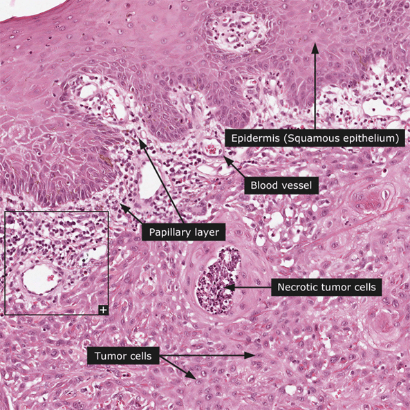
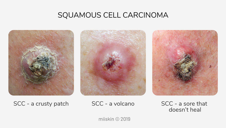
Signs Of Squamous Cell Carcinoma
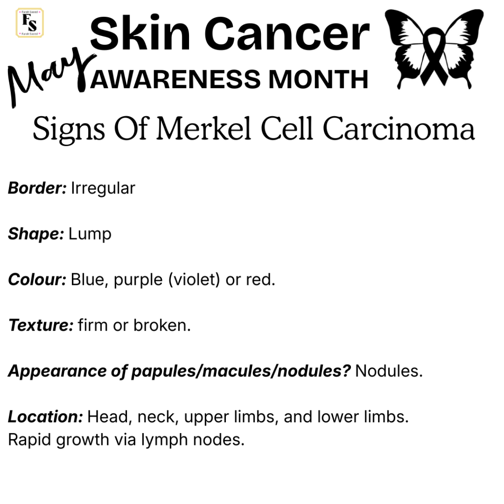
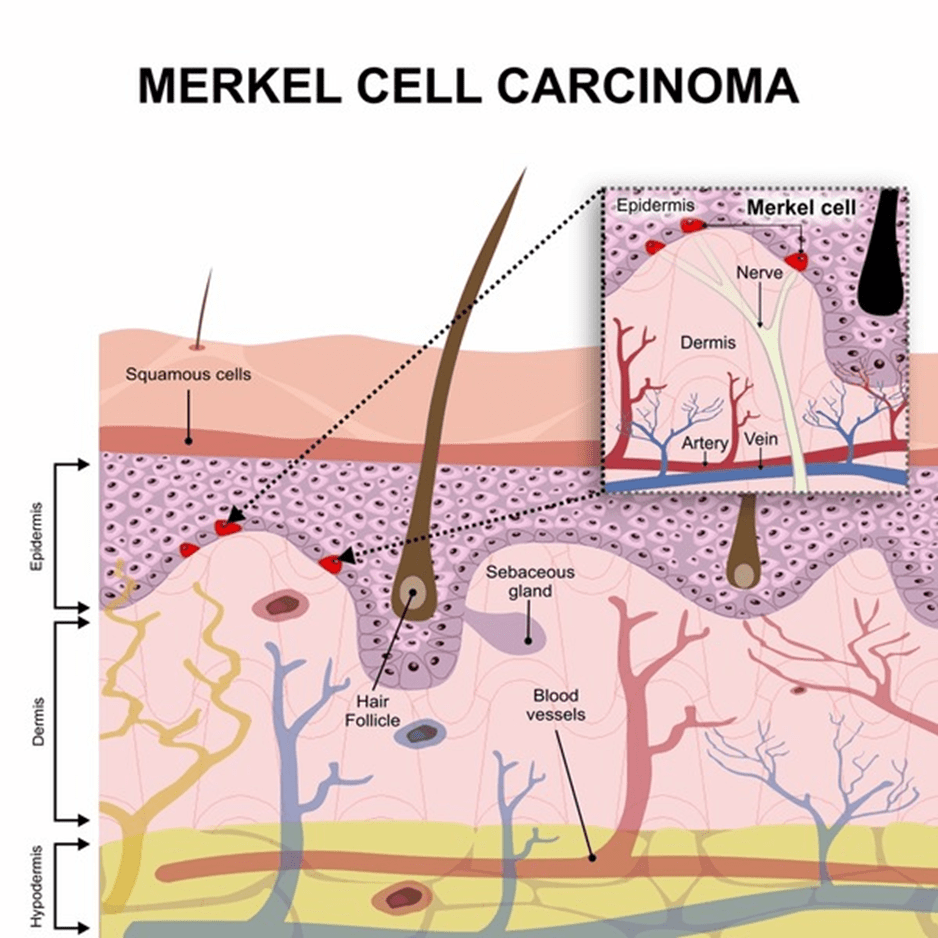
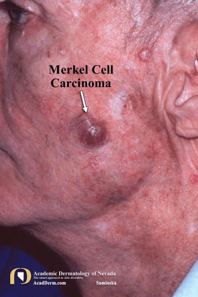
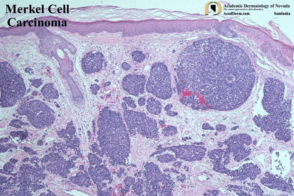
Merkel Cell Carcinoma
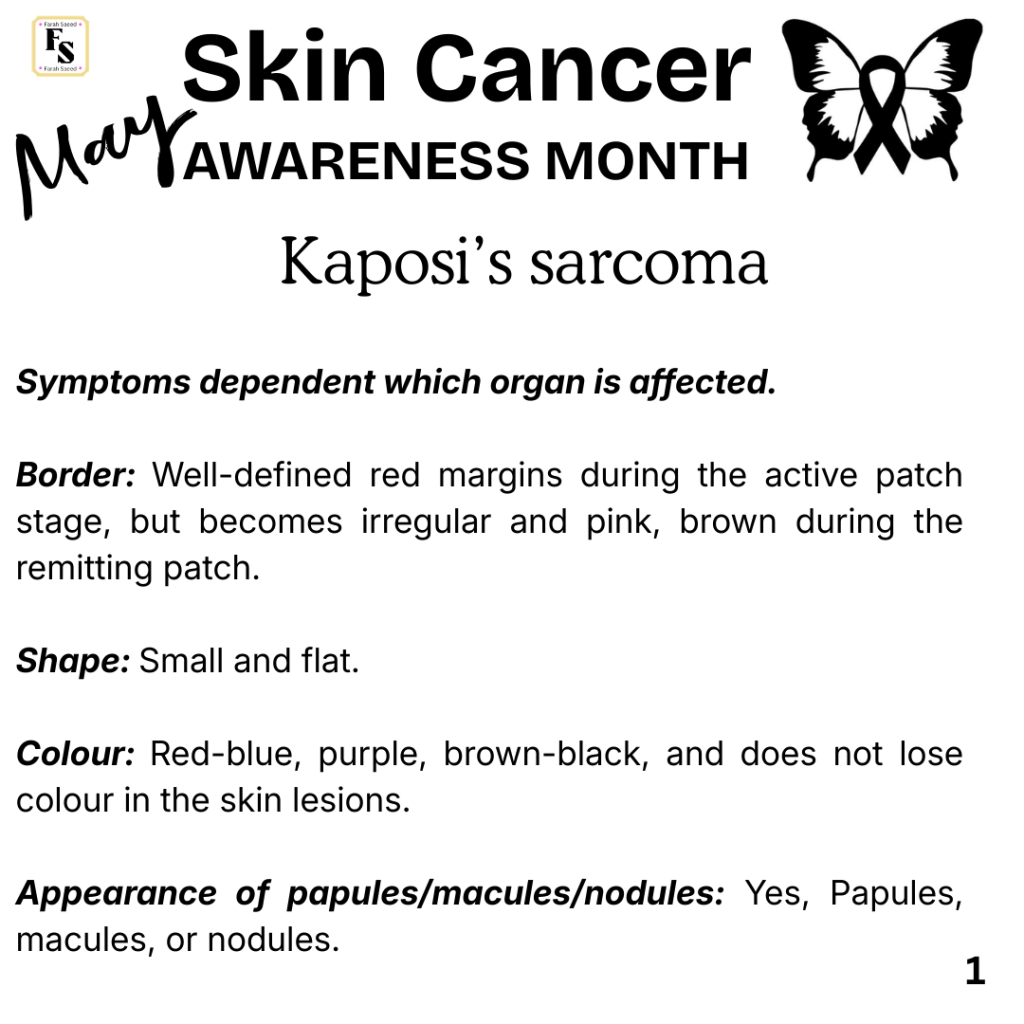
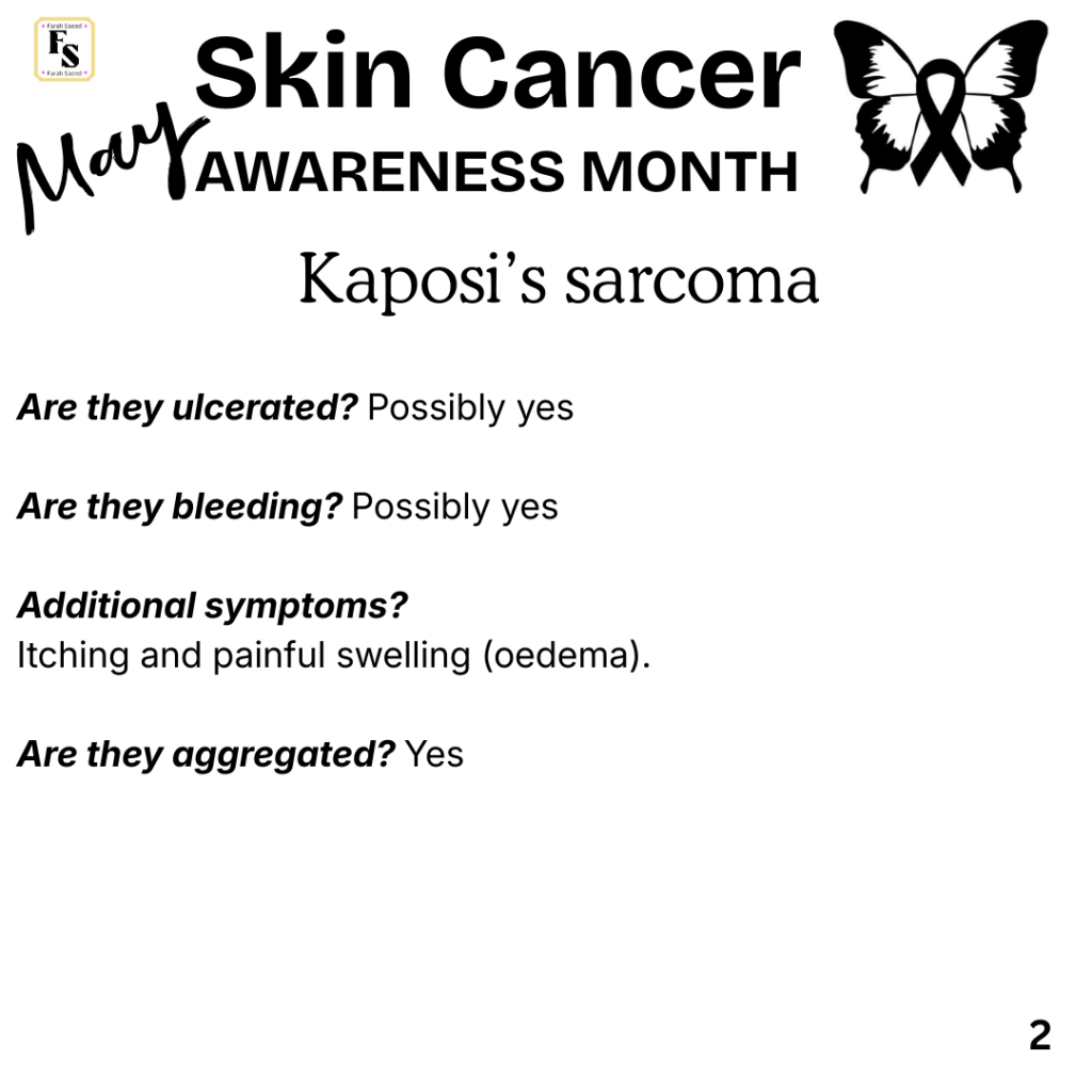
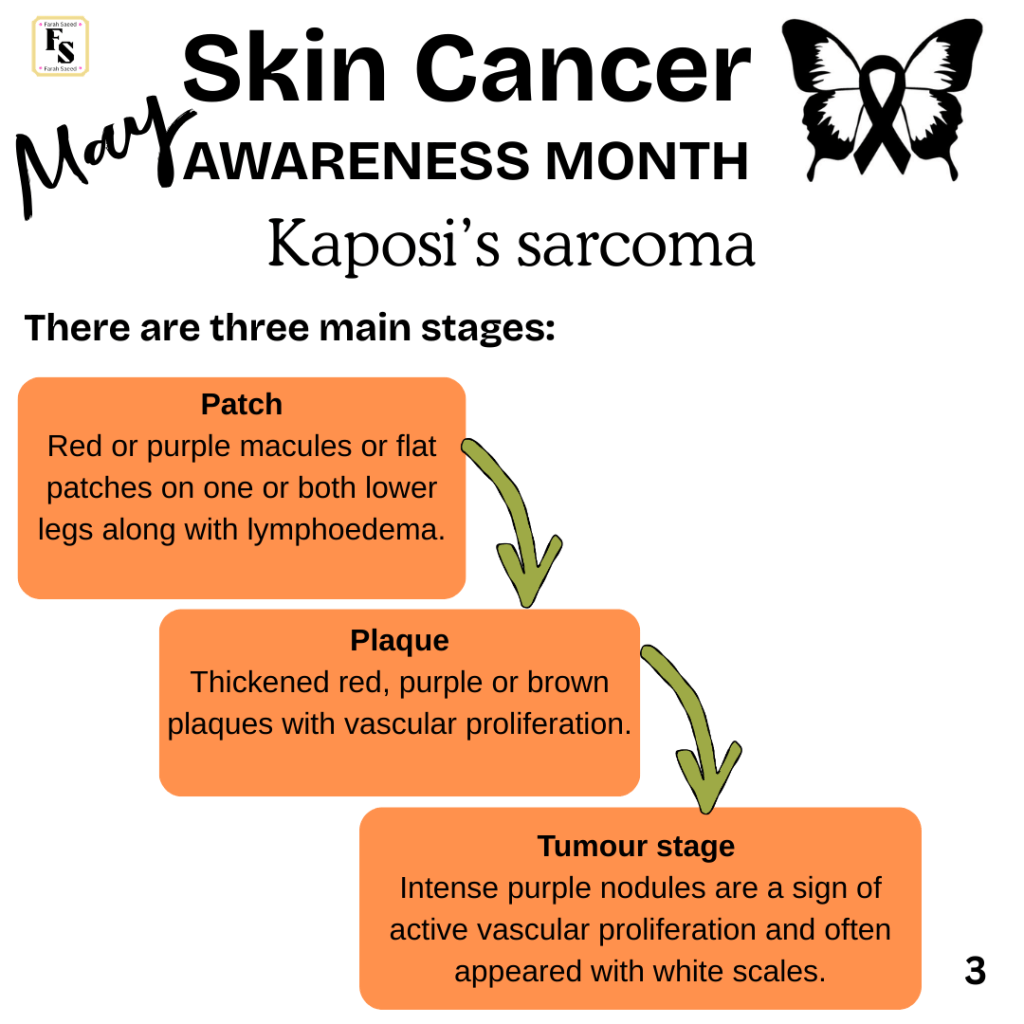
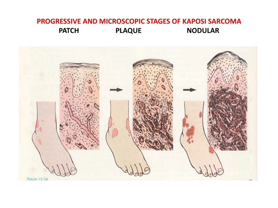
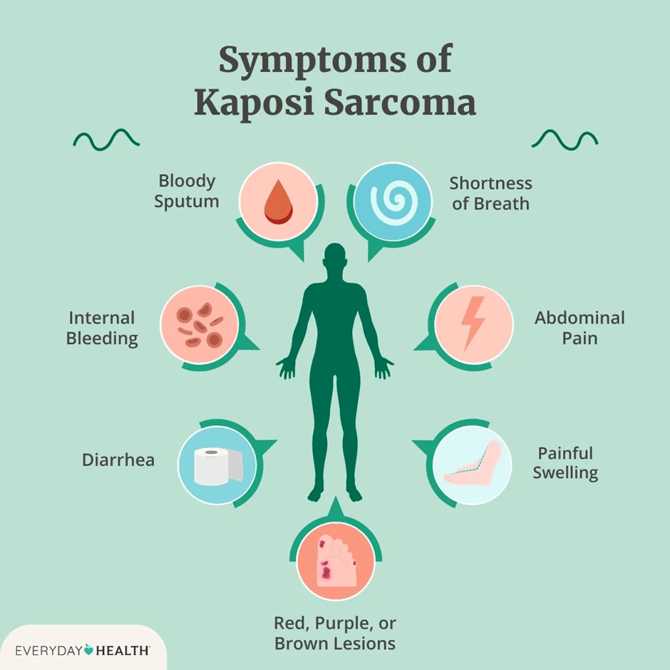
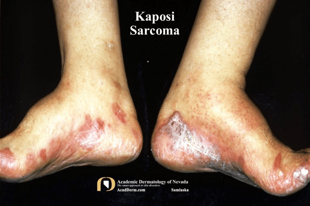
Kaposi’s Sarcoma
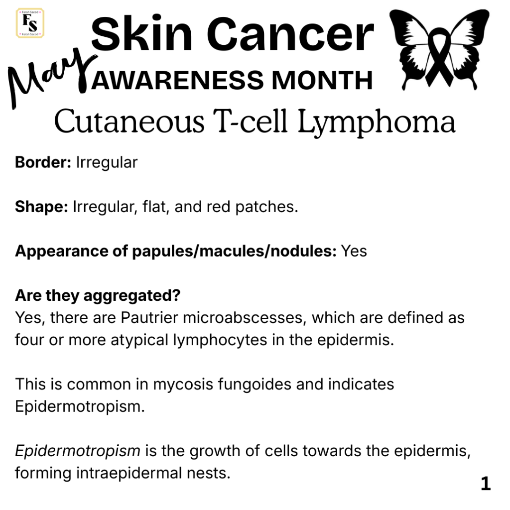
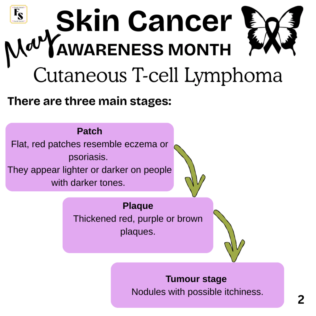
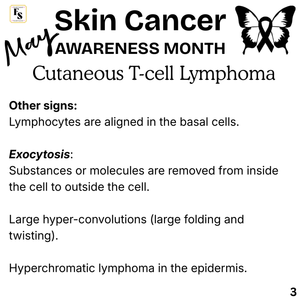
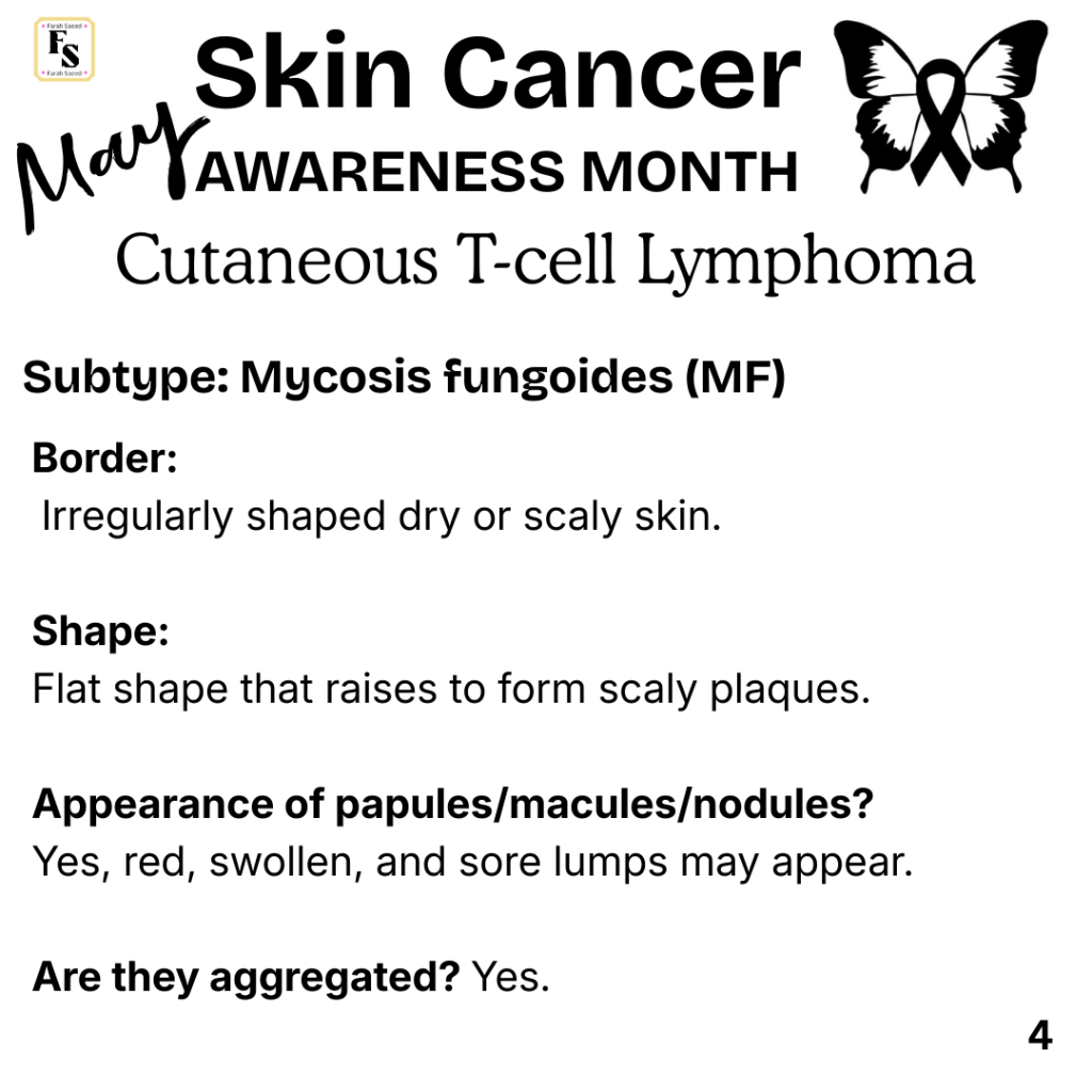
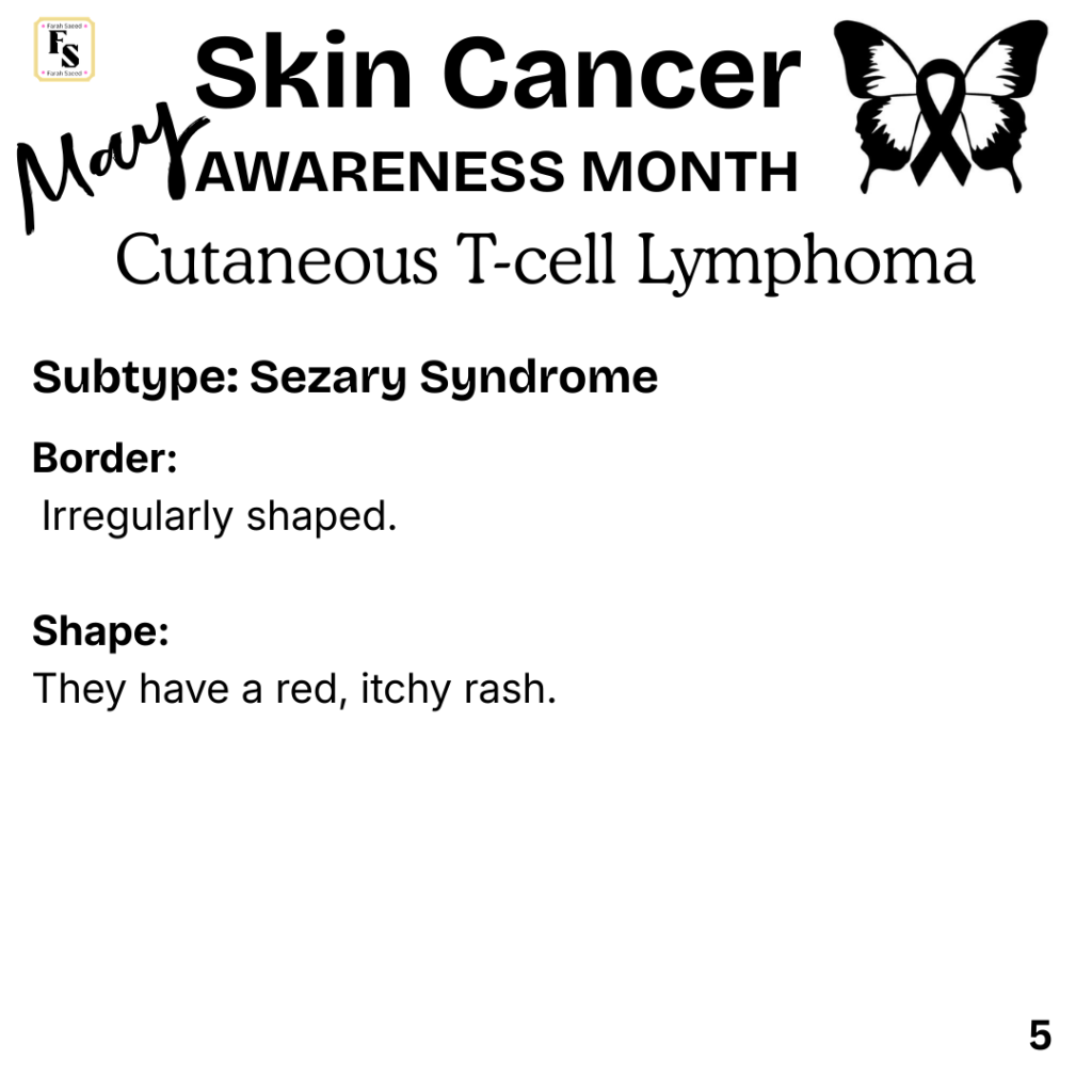
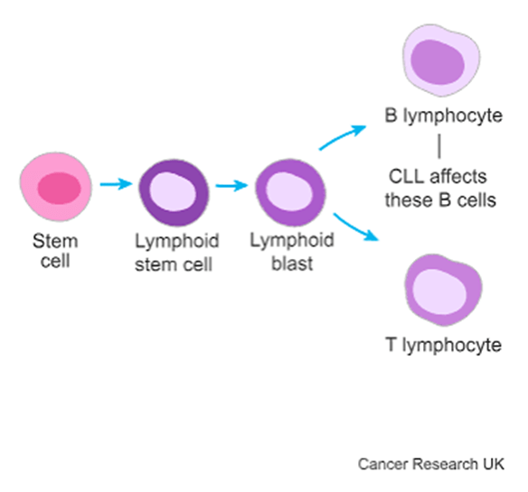
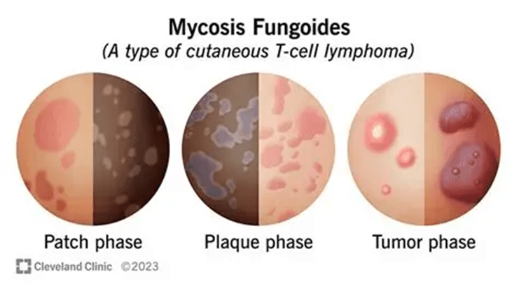
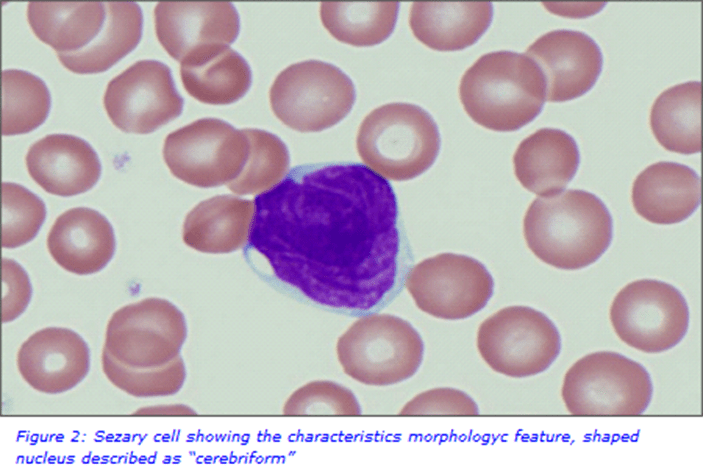
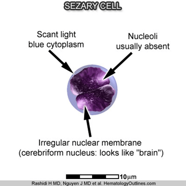
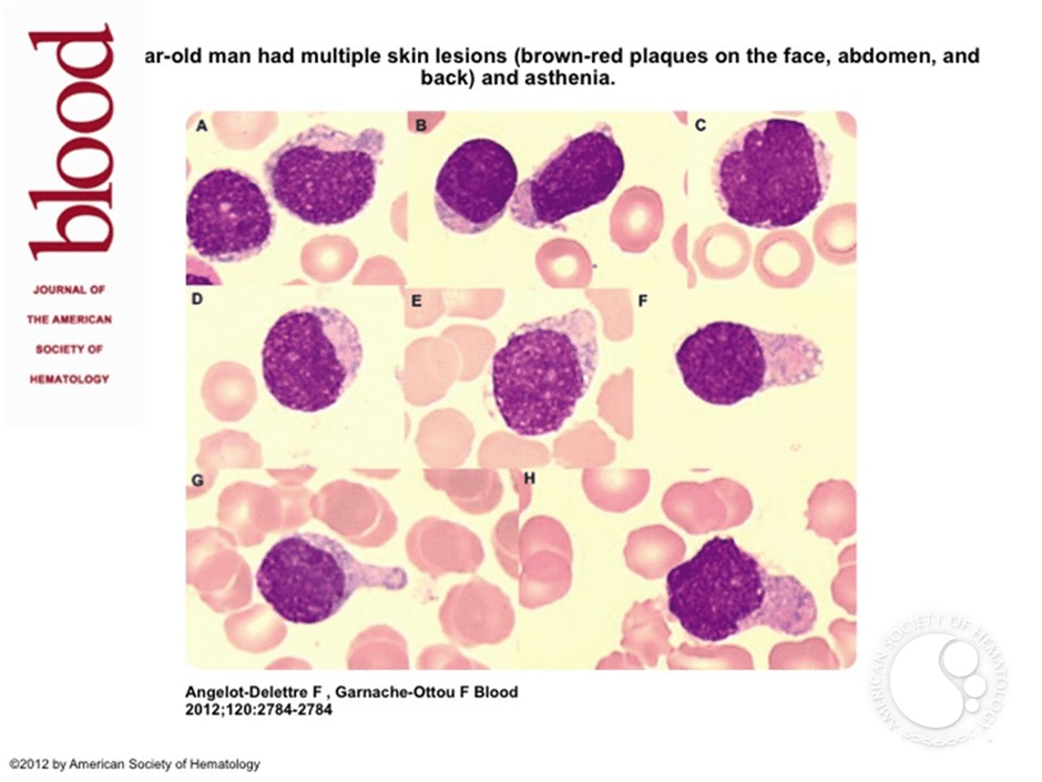
Cutaneous T-cell lymphoma
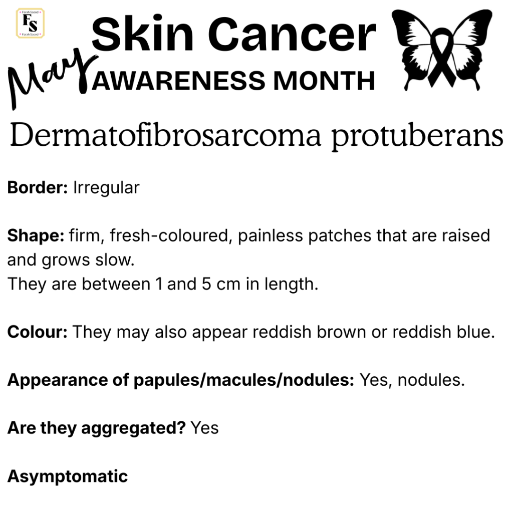
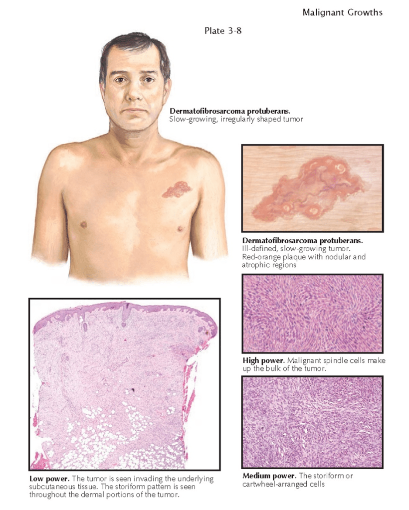
Dermatofibrosarcoma protuberans

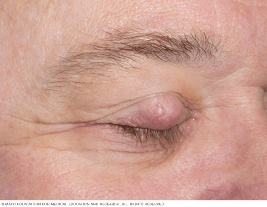
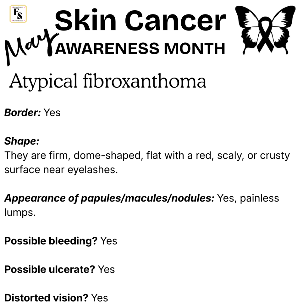
Atypical Fibroxanthoma

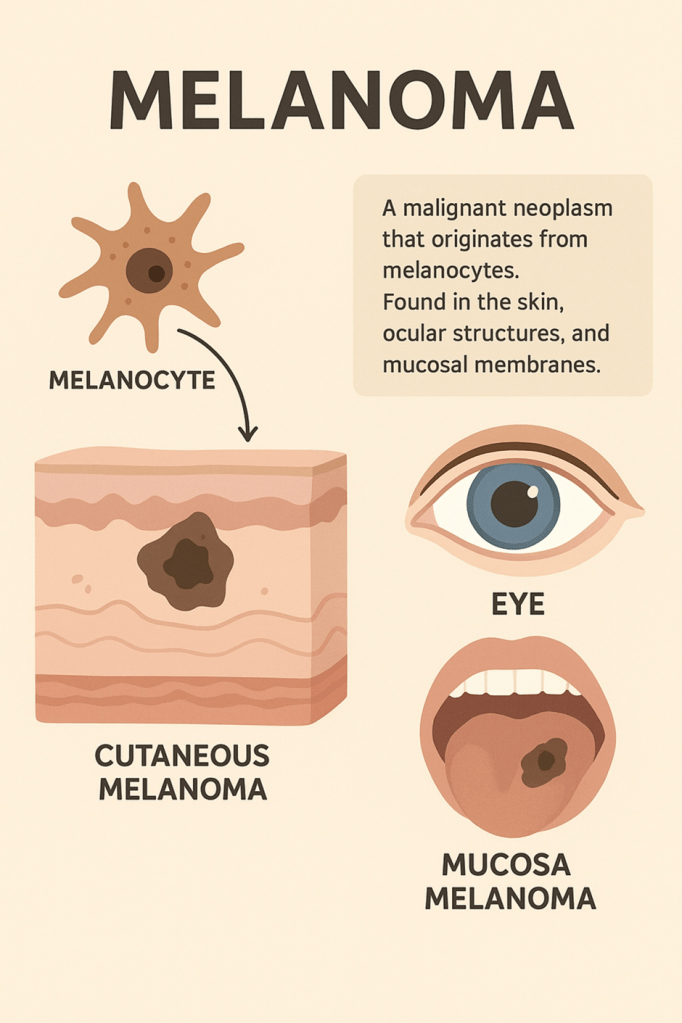
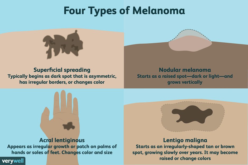
Examination Of Malignant Melanoma
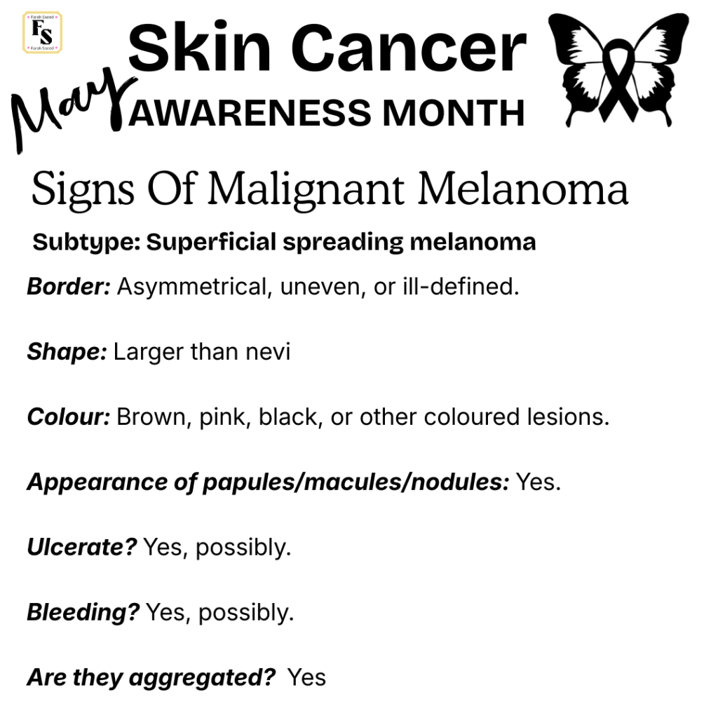
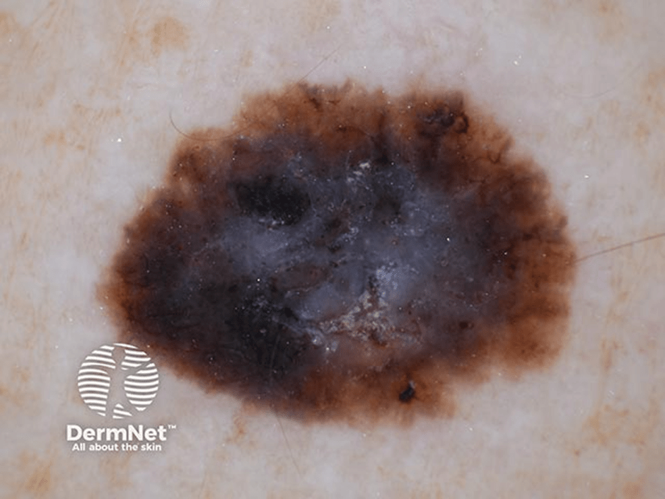
Superficial Spreading Melanoma
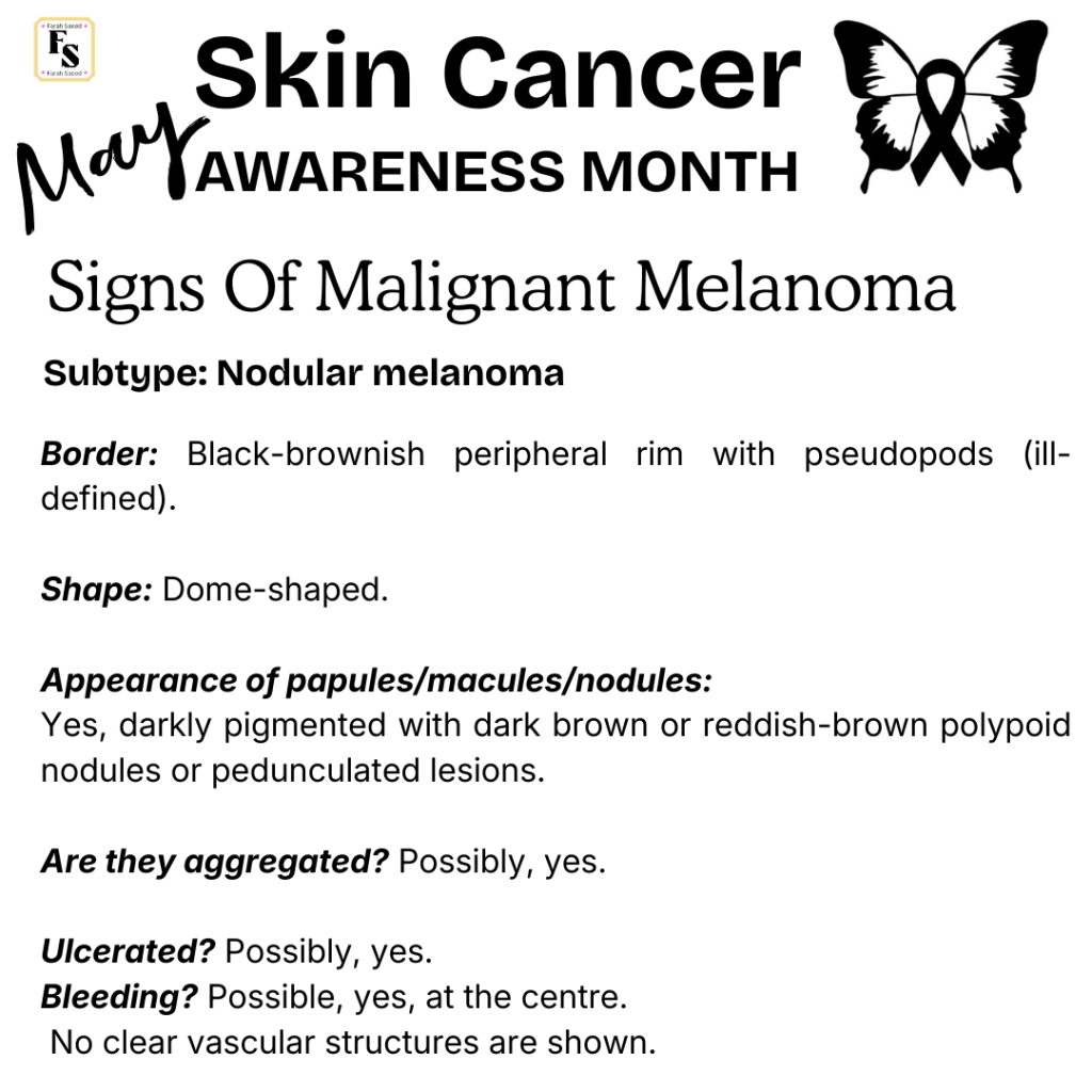
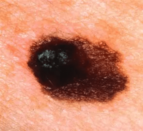
Nodular Melanoma
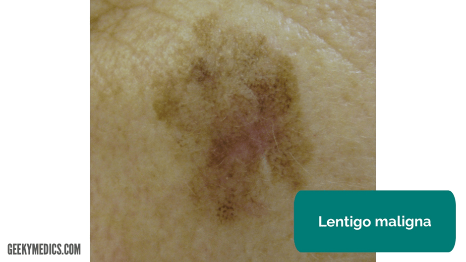
Lentigo Malignant Melanoma


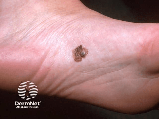
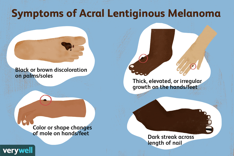
Acral Lentiginous Melanoma

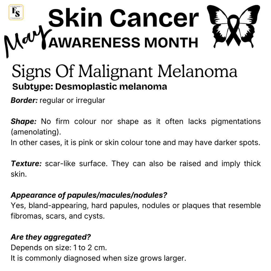
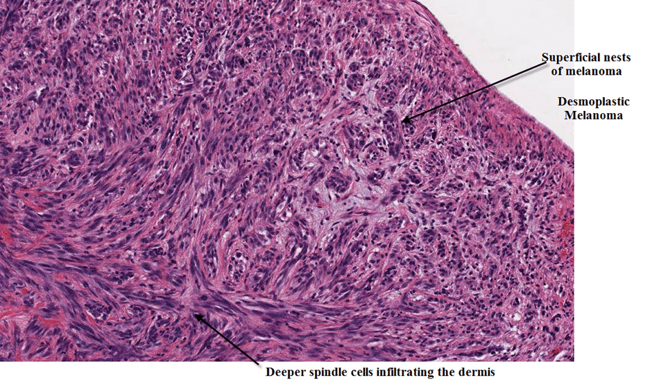
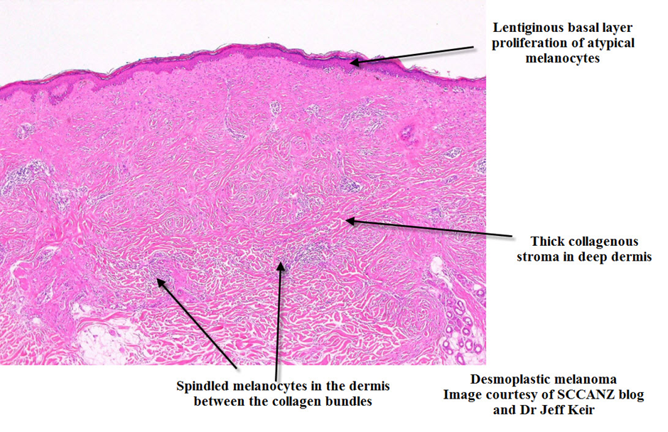
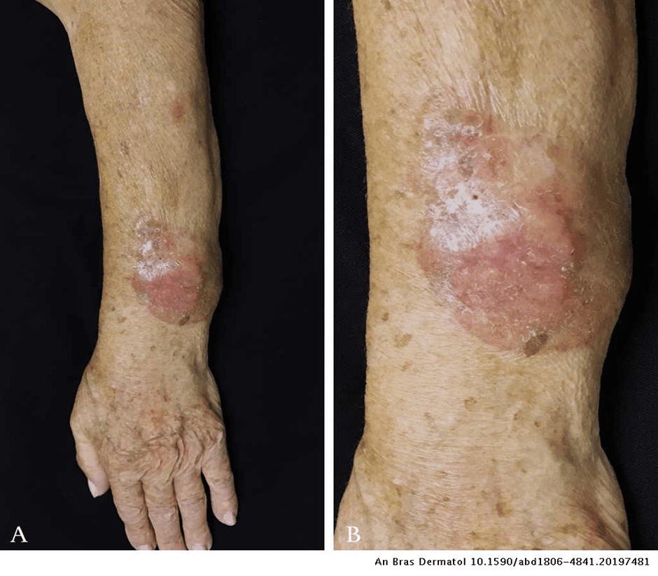
Desmoplastic melanoma
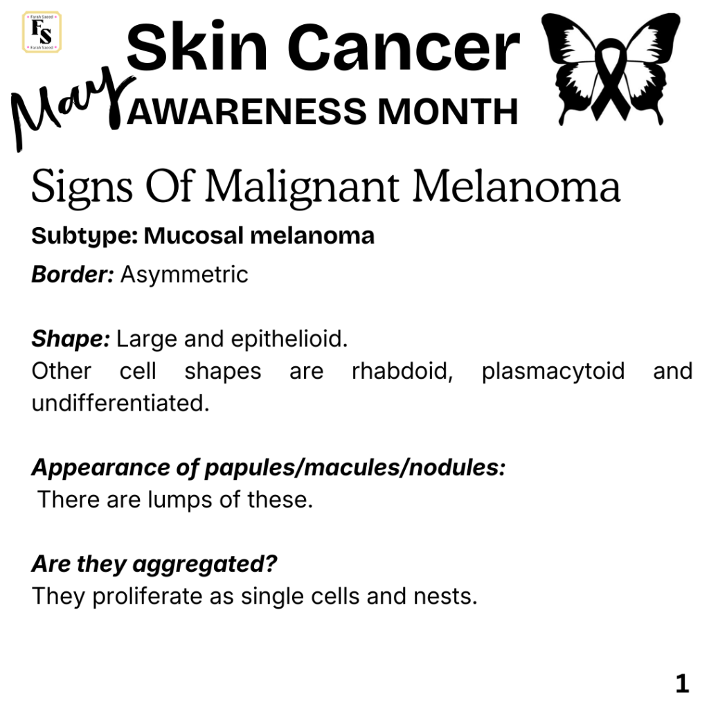
Mucosal Melanoma

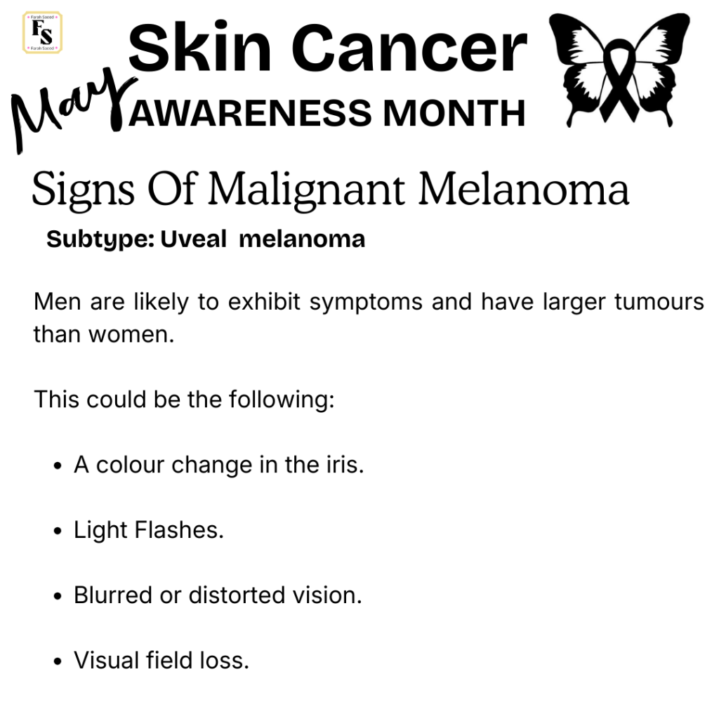
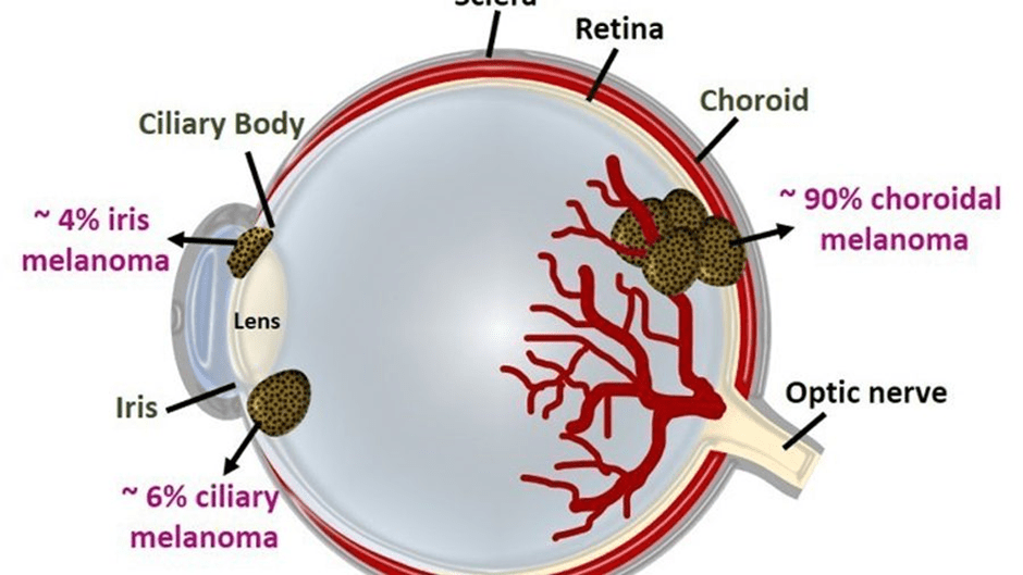
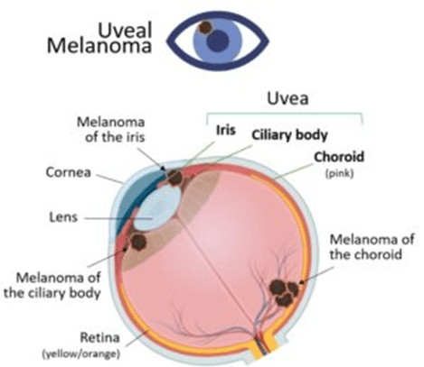
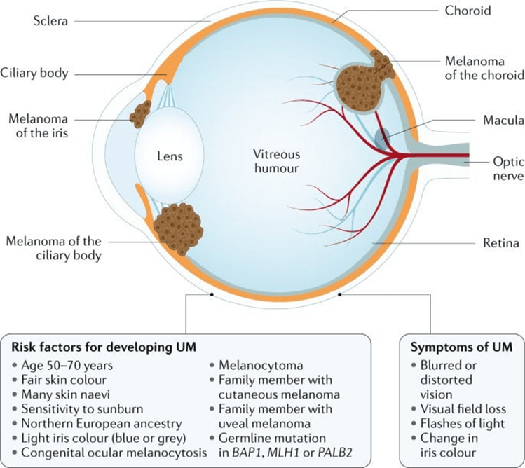
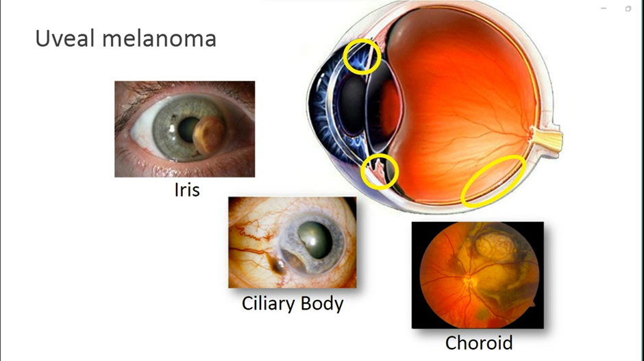
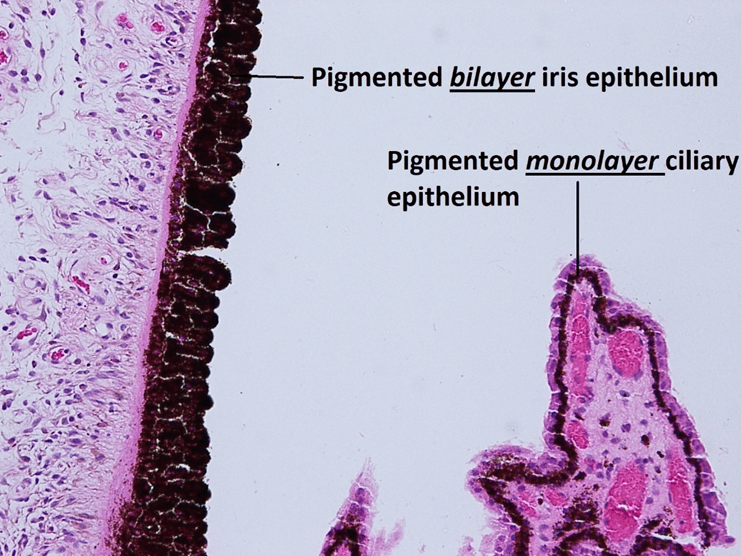
Uveal Melanoma
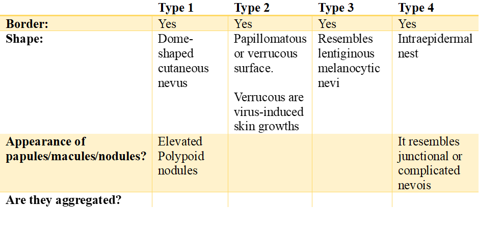
Naevoid melanoma
The table below presents the major types of Naevoid melanoma
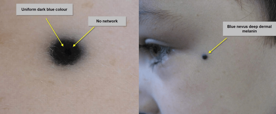
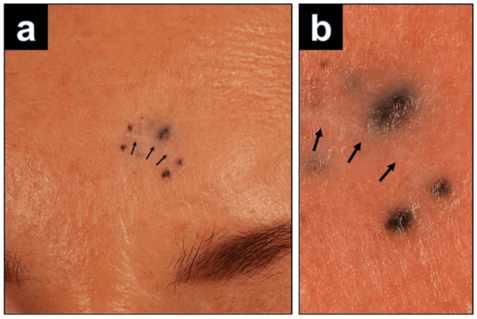
Blue Nevi Melanoma


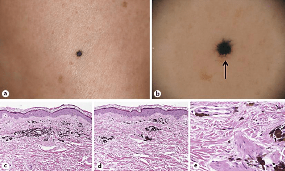
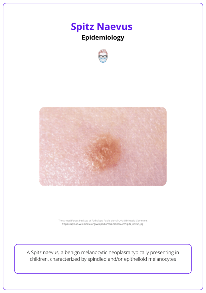
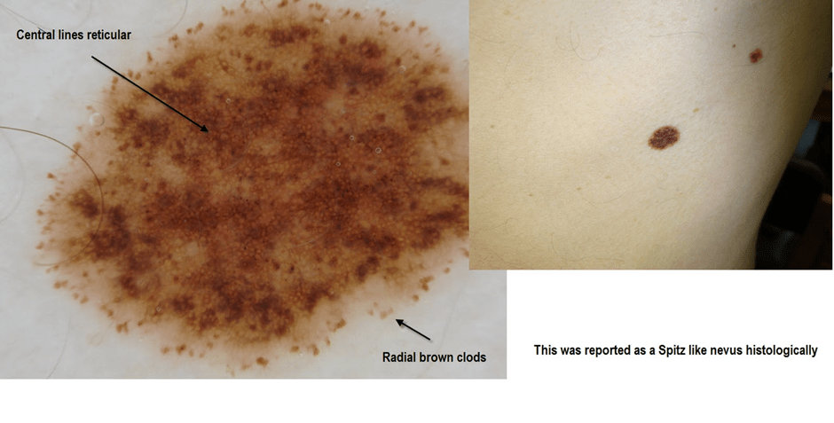
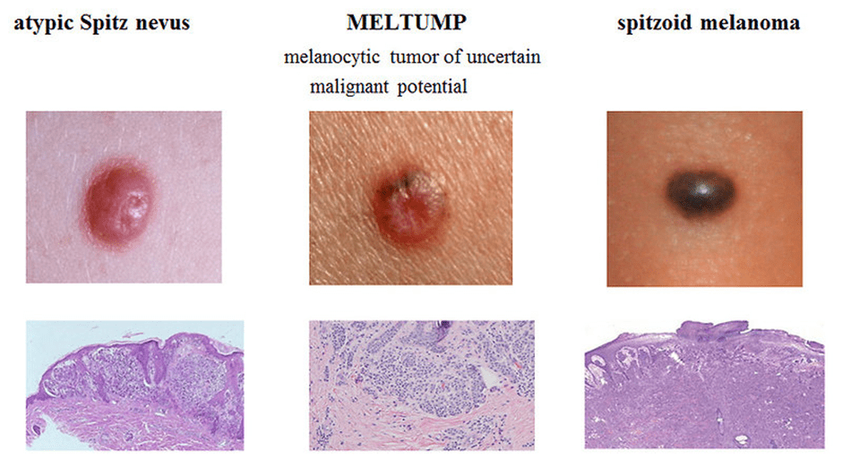
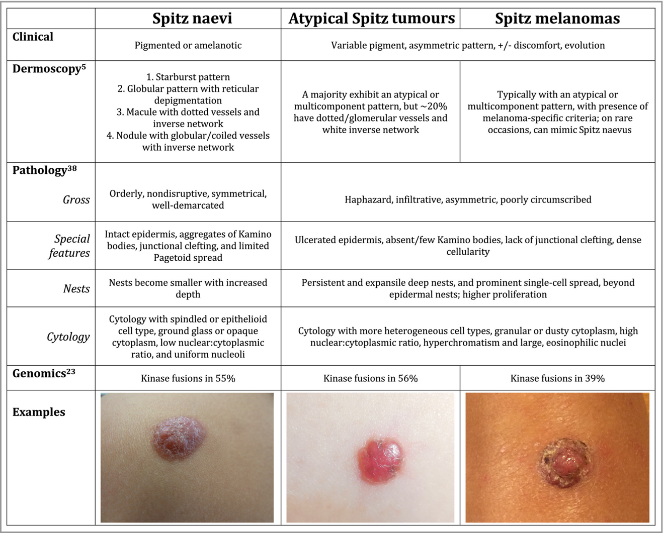
Spitzoid Melanoma
Recommended Sources
Spotting Cancer: Skin Cancer Guide
References
Abdulameer, S. and Mantilla, J. (2023) Soft Tissue Vascular Intermediate (locally aggressive / rarely metastasizing) Kaposi sarcoma. Available at: https://www.pathologyoutlines.com/topic/softtissuekaposi.html (Accessed: 9th February 2026)
Alani, A.M., Desar, S., Torres-Cabala, C. (2023) Skin melanocytic tumor, blue nevi, dermal melanocytoses and associated neoplasms, blue nevus / cellular blue nevus. Available at: https://www.pathologyoutlines.com/topic/skintumormelanocyticbluenevus.html (Accessed: 10th February 2026)
Barnhill, R. and Kim, J. (2026) Spitz nevus, atypical Spitz tumor (Spitz melanocytoma), and Spitz melanoma. Available at: https://www.uptodate.com/contents/spitz-nevus-atypical-spitz-tumor-spitz-melanocytoma-and-spitz-melanoma (Accessed: 10th February 2026)
Bartenstein, D.W., Fisher, J.M., Stamoulis, C., Weldon, C., Huang, J.T., Gellis, S.E., Liang, M.G., Schmidt, B., and Hawryluk, E.B. (2018). Clinical features and outcomes of spitzoid proliferations in children and adolescents. British Journal of Dermatology, 181(2), pp.366–372. doi: https://doi.org/10.1111/bjd.17450.
Biology Insights (2025a) Nevoid Melanoma: What It Is, Appearance, and Treatment. Available at: https://biologyinsights.com/nevoid-melanoma-what-it-is-appearance-and-treatment/ (Accessed: 10th February 2026)
Biology Insights (2025b) Melanophages: Their Role in Pigmentation and Skin Health. Available at: https://biologyinsights.com/melanophages-their-role-in-pigmentation-and-skin-health/ (Accessed: 10th February 2026)
Cambridge University Hospitals (2026) Skin cancer. Available at: https://www.cuh.nhs.uk/our-services/cancer-services/cancer-and-types-of-treatment/cancer-types-a-z/skin-cancer/ (Accessed: 5th February 2026)
Cleveland Clinic (2022a) Sebaceous Carcinoma Available at: https://my.clevelandclinic.org/health/diseases/24087-sebaceous-carcinoman (Accessed: 10th February 2026)
Cleveland Clinic (2022b) Spitzoid Melanoma. Available at: https://my.clevelandclinic.org/health/diseases/24153-spitzoid-melanoma (Accessed: 10th February 2026)
Cleveland Clinic (2023) Mucosal Melanoma. Available at: https://my.clevelandclinic.org/health/diseases/24643-mucosal-melanoma (Accessed: 10th February 2026)
Durden, F. (2023).Desmoplastic melanoma. Available at: https://www.cancercenter.com/cancer-types/melanoma/types/desmoplastic-melanoma (Accessed: 10th February 2026)
Fahrner, L. (2022). Melanoma arising from Blue Nevus. Available at: https://www.dovemed.com/diseases-conditions/melanoma-arising-blue-nevus (Accessed: 10th February 2026)
Kalmukykova, A. and McKee, P. (2021) Skin nonmelanocytic tumor Carcinoma (nonadnexal) Basal cell carcinoma. Available at: https://www.pathologyoutlines.com/topic/skintumornonmelanocyticbcc.html (Accessed: 10th February 2026)
Kneitz, H., Rose, C., Glutsch, V., and Goebeler, M. (2022). Recurrence of a Cellular Blue Nevus with Satellitosis: A Diagnostic Pitfall with Clinical Consequences. Dermatopathology (Basel, Switzerland), [online] 9(4), pp.361–367. doi: https://doi.org/10.3390/dermatopathology9040042.
Madhumita, M. and Bhat, R. (2020). Pautrier’s microabscess: An eponym by mistake. Indian Journal of Dermatology Venereology and Leprology, 86(6), pp.747–747. doi: https://doi.org/10.4103/ijdvl.ijdvl_1100_19.
Mahon, C. (2023). Acral lentiginous melanoma. Available at: https://dermnetnz.org/topics/acral-lentiginous-melanoma (Accessed: 10th February 2026)
Morgan, J. (202.. Kaposi sarcoma. Available at: https://dermnetnz.org/topics/kaposi-sarcoma (Accessed: 10th February 2026)
National Health Service (2023). Symptoms -Melanoma skin cancer. Available at: https://www.nhs.uk/conditions/melanoma-skin-cancer/symptoms/ (Accessed: 9th February 2026)
Roky, A.H., Islam, M.M., Fuad, M., Mostaq, M.S., Mahmud, M.Z. Amin, M.N., and Mahmud, M.A. (2024). Overview of skin cancer types and prevalence rates across continents. Cancer Pathogenesis and Therapy, 3(2). doi: https://doi.org/10.1016/j.cpt.2024.08.002.
Swali, R., Limmer, A., and Tyring, S.K. (2020). Kaposi Sarcoma of the Medial Foot in an MSM, HIV-Negative Man: A Fifth Clinical Variant. Journal of Clinical and Aesthetic Dermatology, 13(10), pp.42–44.
Tidy, C. (202.. Basal cell carcinoma. Available at: https://patient.info/doctor/dermatology/basal-cell-carcinoma (Accessed: 10th February 2026)
Ting, S. (2025). Blue naevus. Available at: https://dermnetnz.org/topics/blue-naevus (Accessed: 10th February 2026)
Tourlaki, A., Nazzaro, G., Wei, Y., Stefano Buffon, Mattioli, M.A., Marzano, A.V. e Brambilla, L. (2022). Clinical, Dermoscopic, Ultrasonographic, and Histopathologic Correlations in Kaposi’s Sarcoma Lesions and Their Differential Diagnoses: A Single-Center Prospective Study. Journal of Clinical Medicine, 12(1), pp.278–278. doi: https://doi.org/10.3390/jcm12010278.
Updated February 2026 Next Review May 2028
