
Lecture Ten: TGF-Beta
The lecture focuses on two signaling pathways of the proinflammatory cytokine and receptor, Transforming growth factor β (TGF-β) dependent and independent of SMAD proteins. SMAD proteins transduce the signal post-receptor activation to regulate growth and differentiation. In the SMAD-independent pathway, the enzyme Rho GTPase associates with guanosine triphosphate (GTP) and hydrolyzes to guanosine diphosphate (GDP) to regulate cell migration, invasion, and metastasis. The structure and function of key components were reviewed. The structure, function, and classification of key components were reviewed. For instance, the TGF has three main family groups: Mammalian TGF-β inhibin/activin, TGF-β, BMP (bone morphogenic protein)/GDF (growth and differentiation factor) family. Further discussion is made on the crosstalk between TGF and other pathways, for instance, the GPCR signaling, particularly the GPCR ligands. A review of the causes of dysregulation of the TGF signaling pathway is mentioned with examples of cancer to elucidate the role of TGF in promoting and preventing cancer progression. For instance, TGF can prevent tumour proliferation in the early stages, whereas, in the advanced stage, it increases invasion, metastasis, and angiogenesis and induces chemotherapy resistance.
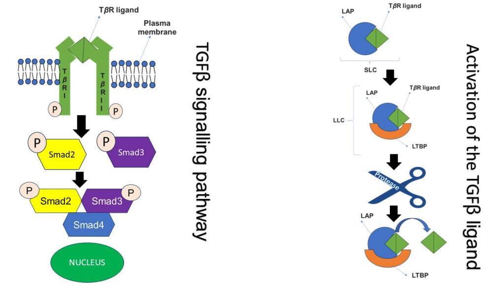
The TGF signalling pathway
This image presents an overview of the activation of the TGF ligand (green) and TGF signaling pathway. In the first image, there are three main types found in mammals with distinctive functions: TGF-β1, -β2, and -β3 but can activate the same cell surface receptor. The TGF contains nine cysteine amino acid residues. A dimer is formed between two TGF-β proteins using disulfide bonds. Different types of SMADs are involved in the TGF signaling pathway. SMAD1, 2, 3 and 5 regulate the receptors. SMAD4 helps with interaction with other SMADs to transduce signals. SMAD6 and 7 are inhibitors of TGF-β regulation. TGFR receptors bind together in the plasma membrane. The activated receptor phosphorylates SMAD proteins SMAD2 (yellow) and SMAD3 (purple). The phosphorylated SMAD 2 AND 3 bind to the SMAD4 (blue) protein to form a complex and dissociate from the receptor to translocate into the nucleus (dark green) and induce transcription of target genes particularly those involved in apoptosis (programmed cell death). For instance, it can inhibit the expression of both antiapoptotic genes and inhibit the expression of genes that encode for caspase 8 and 3 involved in promoting apoptosis.
The second image presents the activation of the TGF ligand (green). A complex is formed between the C-terminal prodomain latency-related peptide (blue; LAP) with the TGF ligand to form a small latency complex (SLC). The SLC binds with the TGF-β binding protein 1 (orange; LTBP1) to produce a large latent complex (LLC). LLC interacts with protease enzymes. The LLC releases the TGF-β ligand to become active. The TGF-β can bind with the receptor by stimulating LAP or LTBP. To activate the receptor. A larger size of the image can be found in the resource list.
Resource List For Lecture Ten
Youtube video
Glossary
Quiz
PDF formats of the images

Leave a comment