Lecture Eleven: BCR-ABL
This lecture explores the structure and function of ABL and BCR and how the BCR-ABL fusion can transduce a signal and elicit cellular response in regulated and dysregulated pathways. The main signal transduction methods discussed are Src kinases (Hck and Lyn) in myeloid cell lines, signal transducers and activators of transcription (JAK/STAT), and RAS proteins. Causes and examples of cancers are present in the lecture and a specific focus on mutations and overexpression of ABL and SH domains that affect protein-to-protein interaction in different ABL fusions and blood cancers. The blood cancers discussed are chronic myeloid leukaemia, B-cell acute lymphoblastic leukaemia (B-ALL), and acute lymphoblastic leukaemia (A-LL) with varying risk factors, age of incidence, and symptom presentation. The link between PI3K kinase and BCR-ABL is examined and is possibly associated with the proto-oncogene casitas B-lineage lymphoma protein (CBL) phosphorylated via tyrosine residues. The BCR-ABL tyrosine inhibitor, imatinib (Gleevec) partially inhibits BCR-ABL-expressing leukaemic cells and patients can develop drug resistance.
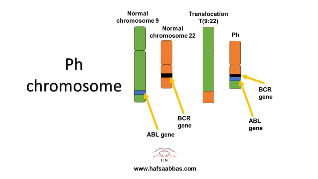
Structure of Ph chromosome
The image presents the structure of the ABL1 gene (blue) on chromosome 9 (green) and the breakpoint cluster region (BCR) gene (black) on chromosome 22 (orange). The BCR gene encodes for two proteins: 160 kd and 190 kd. The ABL gene encodes for a 145 kd non-receptor tyrosine phosphokinase in the cytoplasm. The tumour-specific fusion chimeric protein called BCR-ABL tyrosine kinase occurs. The human Philadelphia (Ph) chromosome arises from a translocation between chromosomes 9 and 22 t(9;22) and facilitates the hallmarks of cancer and survival. A larger size of this image is found in the resource list.
Resource List For Lecture Eleven
Youtube video
Glossary
Quiz
PDF format of the images

Leave a comment