Lecture Two: Hallmarks of Cancer
This lecture aims to review the hallmarks of cancer developed by Hanahan and Weinberg. They are: sustained proliferative signaling, evading growth suppressors, resisting apoptosis, enabling replicative immortality, invasion and metastasis, angiogenesis, reprogramming energy metabolism, and avoiding immune destruction. There are two contributing factors: genomic instability and tumor-promoting inflammation that underlies most of the hallmarks of cancer. These features and characteristics occur in normal cellular processes and have undergone dysregulation to become malignant.
In addition, a review of the cell cycle is discussed and is monitored by proteins called cyclins and their corresponding enzymes called cyclin-dependent kinases. An insight into how cancer cells continue to proliferate is presented. Techniques for memorizing these hallmarks are presented as images, flowcharts, and tables.
Hallmarks of Cancer
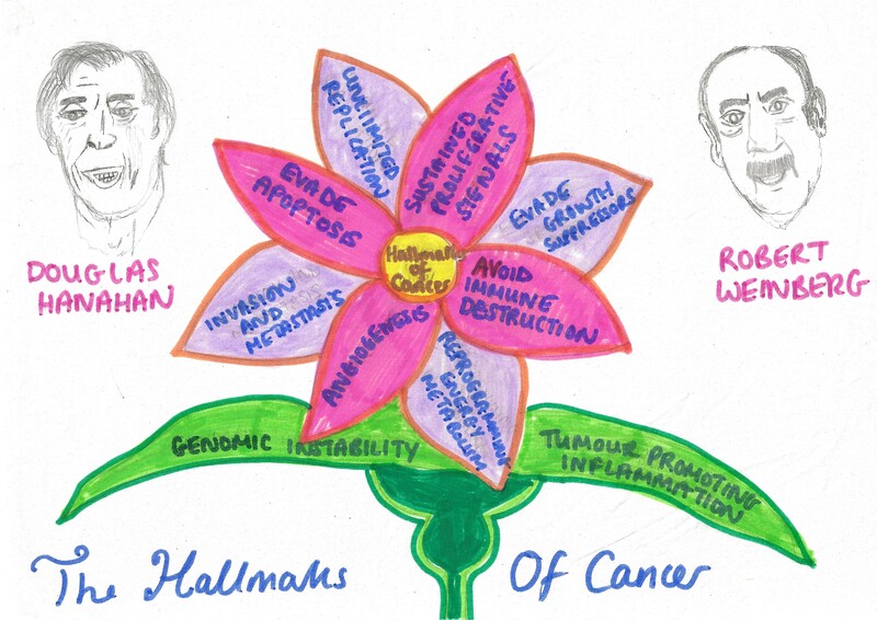
This image presents the hallmarks of cancer developed by Hanahan and Weinberg. The concept presented as a form of a flower was initiated and designed by the author to illustrate how the hallmarks of cancer is propagated (pink and purple petals) and supported by genomic instability and tumour inflammation (green). A large size of this image is found in the resource list.
Comparison of Normal and Cancer cells.
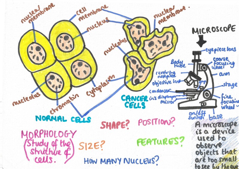
This image presents how the morphology of normal and cancer cells (dark yellow) vary based on shape, size, position, structure, and features. Cells are observed using a microscope and cannot be seen by a naked eye. There are various types of microscopes: a simple light microscope (black) is labelled. A large size of this image is found in the resource list.
Mitosis And The Cell Cycle
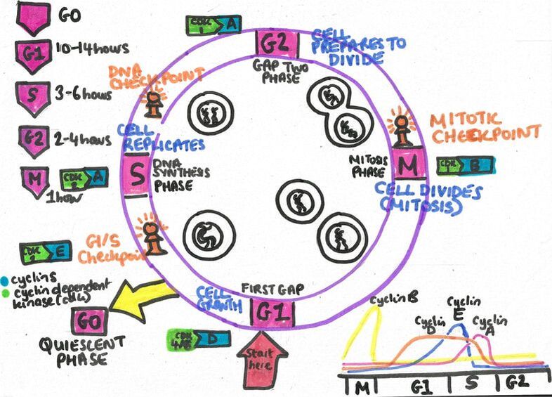
Mitosis is one of the main types of cell division and functions in growth and repair. The cell cycle (pink cycle) presents the phases in how normal cells divide and grow. It is regulated by proteins called cyclins (dark yellow circles) and its specific enzymes: cyclin-dependent kinases (light pink). The enzyme becomes activated when it undergoes phosphorylation [adding a phosphate group (red)]. The regulatory process is prevented by inhibitors (light yellow). A large size of this image is found in the resource list.

The Cell Cycle
This image presents a closer look at the cell cycle. The cell cycle contains four phases: Gap 1 (G1), S phase, Gap 2 (G2) and Mitosis (M). G1 is involved in growth. The S phase is where DNA undergoes replication (copy). The G2 phase is where further growth occurs. Mitosis is where the cells undergo cell division. A larger size of this image is found in the resource list.
The Structure Of The Eukaryotic DNA
Eukaryotes are namely animal, plant and fungi. The DNA (blue) found in eukaryotes are stabilised by histone proteins (pink) that form part of the chromatin found in the nucleus (lime green) of the cell. A large size of this image is found in the resource list.


Replication And Telomeres
Telomeres are found at the end of chromosomes (orange). They undergo a cut via the enzyme telomerase for every round of replication. The shelterin complex controls the telomere length
and protect the chromosomal ends. A large size of this image is found in the resource list.
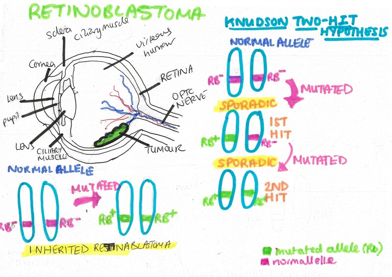
Retinoblastoma
Retinoblastoma is the cancer of the eye and can be inherited by parents or sporadically (without family history). Both parents need to have the mutated Rb tumour suppressor gene (light green) to get retinoblastoma. However, if it occurs via sporadic mutation, the Knudson Two-Hit Hypothesis will take place where at during the first mutation, one allele is affected (green) and at the second hit both alleles are affected. Retinboblastoma is autosomal recessive at a genetic level but inherited as autosomal dominant phenotypically. A large size of this image is found in the resource list.
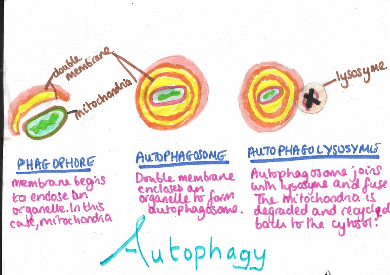
Autophagy
Autophagy is one of the process that is involved in degrading and recycling cellular components. A large size of this image is found in the resource list.
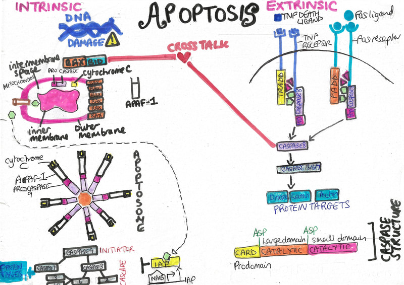
Apoptosis
Each cell has a lifespan and undergoes programmed cell death when reaching senescence and loss of function. There are two types of apoptosis: Intrinsic and extrinsic vary in how they initiate their respective pathways but depend on specific enzymes called caspases that cleave target proteins. Both pathways undergo crosstalk between the BAX-BID complex (orange-blue) of the intrinsic pathway and Caspase 8 (light purple) of the extrinsic pathway. Cancer cells can evade apoptotic signals and continue to grow. A larger size of this image can be found in the resource list.
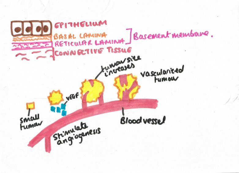
Basement Membrane And Angiogenesis
The basement membrane is a thin lining under the epithelium (brown). The epithelium is the inner lining found in many organs. Angiogenesis is the production of new blood vessels (red) to transport nutrients for growth. Cancer cells can stimulate angiogenesis as a nutrient supply for growth and tumour development forming a vascularized tumour. A larger size of this image is found in the resource list.
Resource List For Lecture Two
Youtube video
Glossary
Quiz
Summary table of the Apoptotic proteins
PDF formats of the images

Leave a comment