The most common gynaecological malignancies that are often treated with radiation therapy are cervical and endometrial cancers. In contrast, surgical wide local excision sparing the normal tissue is required in the treatment of vulval and vaginal cancers. Thereafter, post-operative external beam radiation therapy is required for the vulval and vaginal tumours that are recurrent and also those that have remained post-surgery (residual disease). Radiation oncology uses ionizing radiation, most commonly high-energy X-ray beams, to treat malignant disease. The clinical and biological effects of radiotherapy depend on the radiation dose and delivery timing. The purpose of radiotherapy is to deliver small doses in fractions of radiotherapy to help elucidate a safe and effective dose of radiation that can be safely given as a single fraction to maximise the eradication of cancer cells and improve the survival and quality of life of cancer patients. This is referred to as fractionation. The aim is to review the application of radiotherapy using the technique of Volumetric Modulated Arc Therapy (VMAT) as an advanced technique from intensity-modulated radiation therapy (IMRT) for the treatment of vaginal and vulval cancers.
What is the vagina and vulva?
The vaginal is a muscular tube estimated to be 7.5 inches long and connects the vulva and uterus. The vagina is in front of the rectum, where the poo/stools/faeces are stored. The vagina is in the back of the bladder, where urine/wee/pee is stored. In other words, the bladder is in front of the vagina, and behind the vagina lies the rectum, as presented in Figure 1. The vulva is the external genitalia of the female reproductive system, which contains the inner and outer lips known as labia minora and majora, respectively. Other parts of the vulva include the orifices (openings) of the vagina and urethra. It holds the vestibules where glands like Bartholin’s and Skene’s (preurethral) are found. Patients with vulvar cancer are a small population of patients with vulvar cancer in comparison to organs elsewhere: breast, lung, colon of the large intestine, and the prostate gland.
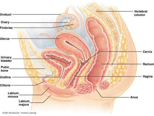
Figure 1: The mid-sagittal image of the female urogenital system.’
What type of radiotherapy is given for vulval and vaginal cancers?
Post-surgical radiotherapy (adjuvant therapy) aims to target the tumour and regional lymph nodes and pelvic disease when there are inadequate resection margins, with a dose of 45-50 Gy at 1.8-2.0 Gy per day, with an electron boost of 10-20 Gy. For electron treatments, the dose is 50-60 Gy, which includes the boost. This can be incorporated with internal radiotherapy, like intracavitary brachytherapy, to 80 to 85 Gy. Treatments are either intracavitary using an applicator called a cylinder, or interstitial using needles and catheters. As there are doses beyond 40 Gy, experts in the field commonly recommend treatment breaks due to the risk of moist desquamation. Moist desquamation is the thinning of the outermost layer of the skin, known as the epidermis where it loses its strength and architecture in exposed areas to the sun, highest skin dose, friction that disrupts the effects of healing. If there is no infection or exudates, it is treated topically using Plancentrex ointment, silver sulfadiazine, 1% gentian violet, or amniotic membrane.
What is VMAT, its function, and its risks?
VMAT and IMRT, illustrated in Figure 2, are the most common vulvar and vaginal radiation techniques to help shape the radiation beams and limit the dose to healthy tissues in the surrounding areas, namely the bowel, rectum, and bladder. For vulvar cancers, the planning technique aims to increase the dose to the vulvar area where the multi-leaf collimators do not close down on the skin surface. Multi-leaf collimators (MLC) are devices made up of individual “leaves” of a high atomic-number material, usually tungsten. Originally used as a surrogate for alloy blocks to shape a field, the leaves are placed in the path through which the beam exits. Each leaf of the multileaf collimators can move independently in and out of the path of the beam, selectively blocking portions of it and helping refine the dose delivered, i.e., providing conformal shaping of the beam.
VMAT is an advanced method that was first introduced in 2007 but has, in recent years, been added for the treatment of gynaecological cancers. VMAT aims to rotate the gantry around the patient so there is continuous radiation positions that delivers conformation radiation in a single continuous arc. Other parameters that boost its effectiveness include rotation speed, intensity, beam shape that moves the multi-leaf collimators (MLC), and radiation dose rate, which increase speed and accuracy. Arc therapy delivers radiation from a radiation source that is continually rotating, allowing a highly conformed dose distribution and treatment to be administered from a full 360° beam angle, improving patient comfort and lowering exposure to healthy tissue, though there is enhanced tumour dose coverage.
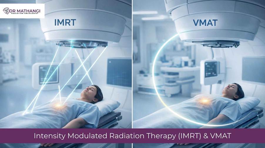
The aim is for VMAT to gain a satisfactory score compared to tomotherapy superimposed arcs. Tomotherapy is a method of delivering rotational IMRT offering various advantages, notably for complex and large targets such as the cranio-spinal axis. Its objective is to precisely shape radiation doses to conform to the tumour while sparing healthy tissues. The intensity level of each beam in a segment is controlled. Another advantage of IMRT is the ability to lower radiation dose.
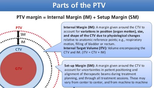
On the other hand, IMRT utilises computer-controlled linear accelerators to deliver precise radiation doses using a small ionization chamber dosimeter in a tank of water and requires a five-step procedure towards malignant tumours. At first, simulation and imaging is done by standardised imaging techniques likes the form of computed tomography, magnetic resonance imaging and positron emission tomography-computed tomography (PET-CT) to map the tumour and nearby organs. The target area is then defined, i.e., the volume of tissue to be irradiated. This includes the gross tumour volume (GTV) that can be visualised using a CT scan, together with potential tissues that may have microscopic invasion of tumour cells (clinical target volume). An additional margin is the planning target volume (PTV) that serves to lower potential risks that may include positioning of patients, organ movement that varies, the calibration of the machine, dosimetric uncertainties, transfer set-up errors from CT simulators to the treatment unit, and human errors, intra- and interfraction motion, setup error, progressive lymph edema, and swelling during treatment.
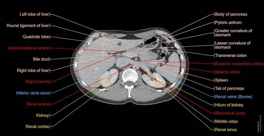
The target volumes near the skin surface, like the vulva, do not meet the International Commission on Radiation Units and Measurements safety guidelines. There is a risk that the treated dose differs from the planned dose due to expansions and setup uncertainties. The PTV served by the clinical doctor aims to ensure that the GTV and/or CTV will receive adequate dose coverage. Please see Figure 3 that presents the composition of PTV. Figure 4 presents the GTV, PTV, and CTV on an abdominal scan. Figure 5 labels the CT abdomen scan on a normal patient. Thus, there is a dire need to know the organs that are at risk or have a lower tolerance to radiation. Amongst the common examples are the kidneys and spinal cord.
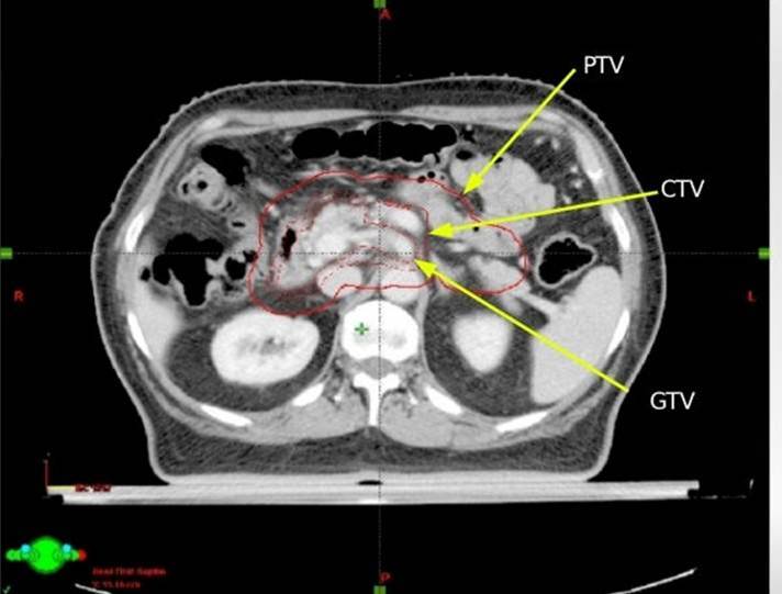
One of the techniques that help overcome this limitation is the use of advanced software and computerised data sets of measured beam, where calculations are made depending on the algorithms that allow the attenuation to vary and the scattering of the X-rays in tissues that have different densities. Thus, the need for treatment planning can help the optimisation of the beam angles and intensity. Quality assurance is then needed to ensure that the planned dose is uniformly irradiated, the critical organs do not exceed the tolerance doses, and the correct size, shape, direction, and number of the beams are verified by the use of radiographs on a radiotherapy simulator prior to treatment. The fifth step is for the radiation oncologist to prescribe and deliver the treatment radiation painlessly over multiple sessions implicated in a fractionation schedule. This is achieved by entering the dataset into the computer system on the linear accelerator for each treatment. Overall, this systematic approach ensures there is high accuracy and minimal collateral damage whilst the addition of maximum tumour targeting.
However, the difference between VMAT and IMRT focuses on several features: dose modulation, treatment time, precision, and organ sparing, and is summarised in Table 1.
Table 1: A comparison between IMRT and VMAT.
| Feature | Conventional RT | VMAT radiotherapy |
| Dose modulation | Limited | Highly dynamic |
| Treatment time | Longer | Shorter |
| Precision | Moderate | Very high |
| Organ sparing | Basic | Advanced |
Another method to help overcome these uncertainties is the use of integrated skin flash (ISF) with virtual bolus to step out the optimiser and improve the coverage of the skin surface that has an absence of PTV margins. Please see Figure 6.
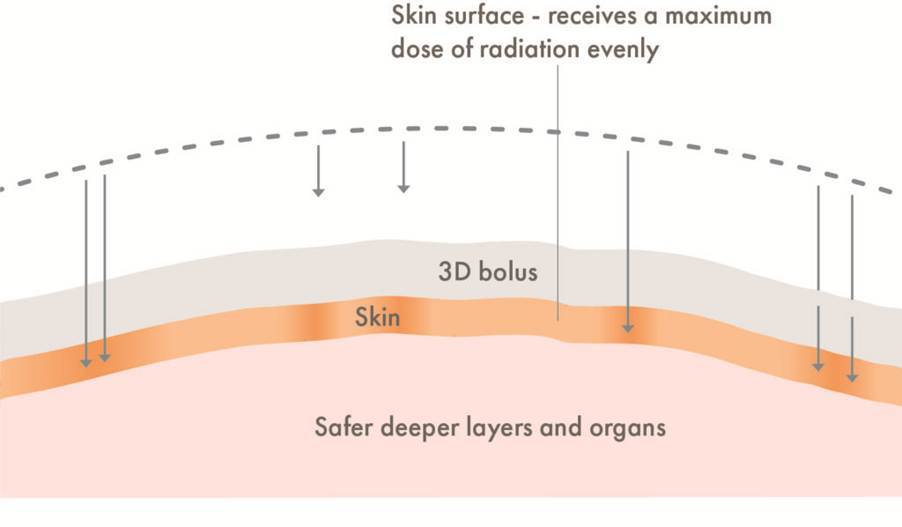
Inshetski et al. (2022) evaluated the use of VMAT in gynaecological cancers, where they developed a 1.5 to 2.0 virtual bolus that was used as a template where the planning structure was developed. The outer edge was retracted 5.0 mm from the outer edge. The virtual bolus and planned structure were then optimised to help control the higher dose gradient at the interface of the virtual bolus surface and air. This helps to prevent the multileaf collimators from being close to the vulvar skin surface and to maintain adequate dose during therapy.
On the contrary, those ISFs have overcome many limitations from systematic and gross errors in VMAT. It also has its disadvantages when the virtual bolus is removed and replaced with a clinical bolus of 3.0 to 5.0 mm, where there is an increased observation of hotspots along the skin surface of the perineum and vulva. These hotspots vary day to day and commonly occur when applying the ISF planning technique. There is also a risk of higher skin toxicity as the treatment planning system equates the absence of virtual bolus as missing tissue in the final calculation.
What’s New?
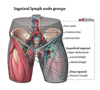
Inshetski et al. (2022) assessed five patients between the ages of 34 and 78 years with International Federation of Gynecology and Obstetrics (FIGO) staging IB and higher in their study. They delineated tumour volumes, confirmed the exact positions, and labelled them on the anatomic area. The bilateral inguinal and regional lymph nodes (para-aortic, common iliac, external and internal iliac nodes and even pre-sacral nodes in advanced cases), as well as the vagina and vulva, were treated using risk factors and relapse. Please see Figure 7. Fractionation regimens ranged from 25 to 34 fractions. Dose to the vulvar surface varied from 50 to 67.7 Gy depending on staging.
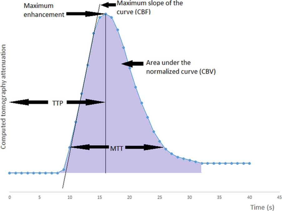
They developed a technique known as robust optimisation that aims to increase the dose and homogeneity at the skin surface without the need for ISF and virtual bolus. The treatment planning system (TPS) applies the defined shifts in X, Y, and Z directions and movements in the CT density curve to set up the uncertainties of the selected structure. An example of a CT density curve is presented in Figure 8.
In other words, the optimiser accounts for uncertainties whilst optimising the dose to the structure. The uncertainties are then displayed in multiple planning scenarios to correct the target volume. Robust optimisation helps to lower variation of dose across vulvar skin while increasing the treatment planning efficiency. In other words, the TPS was able to spread or perform dose distribution over an area of vulvar skin surface and deliver 95% of the prescription dose to 100% of the skin surface in the least desirable setup scenario, (2) deliver ≥ 100% of the prescription dose to 95% of the skin surface in the least desirable scenario, and (3) not deliver ≥ 110% of the prescription dose to 0.03 cubic centimeters (cc) at the skin surface.
Overall, robust optimisation has helped make VMAT a more effective and advanced radiotherapy technique when treating gynaecological cancers, especially the vulva and vagina. This is due to parameters such as dose delivery and reduced risk of toxicity. Treatment planning and quality assurance are crucial for patient safety and outcomes.
Useful Link To Skin Flashing Technique In Radiotherapy
References
Dr. Mathangi (n.d.) Intensity modulated radiation therapy (IMRT) & VMAT by Dr Mathangi. Available at: https://drmathangi.com/imrt-vmat-radiation-therapy-bangalore/ (Accessed: 1st November 2026)
Dyer, B.A., Jenshus, A. and Mayadev, J.S. (2019). Integrated skin flash planning technique for intensity-modulated radiation therapy for vulvar cancer prevents marginal misses and improves superficial dose coverage. Medical Dosimetry, 44(1), pp.7–10. doi:10.1016/j.meddos.2018.01.003.
Inshetski, R., Lenards, N. and Hunzeker, A. (2022). Robust optimization used to mitigate setup uncertainties in vulvar patients receiving VMAT. Medical Dosimetry, 47(2), pp.129–135. doi:10.1016/j.meddos.2021.11.003.
Kurz, K. D, Ringstad, G., Odland, A., Advani, R., E. Farbu and Kurz, M.W. (2015). Radiological imaging in acute ischaemic stroke. European Journal of Neurology, 23(S1), pp.8–17. doi:10.1111/ene.12849.
Micheau, A. and Hoa, D. (2008). CT axial male abdomen and pelvis. e-Anatomy. doi:10.37019/e-anatomy/181.
Pacelli, R., Caroprese, M., Palma, G., Oliviero, C., Clemente, S., Cella, L. and Conson, M. (2019). Technological evolution of radiation treatment: Implications for clinical applications. Seminars in Oncology, 46(3), pp.193–201. doi:10.1053/j.seminoncol.2019.07.004.
Sharma, S.C. (n.d) New Techniques in Radiotherapy Available at: https://www.slideshare.net/slideshow/new-techniques-in-radiotherapy/156323 (Accessed: 1st November 2026)
Stiles, J. (n.d.) 6.4 Female Pelvis. Available at: https://med.libretexts.org/Bookshelves/Allied_Health/Localization_and_Treatment_Procedures_in_Radiation_Therapy_(Stiles)/06:_Pelvis/6.04:_Female_Pelvis Accessed: 1st November 2026
Waghmare, C.M. (2013). Radiation burn—From mechanism to management. Burns, 39(2), pp.212–219. doi:10.1016/j.burns.2012.09.012.


Leave a Reply