It is common that after the operation, the patient will stay for several days due to:
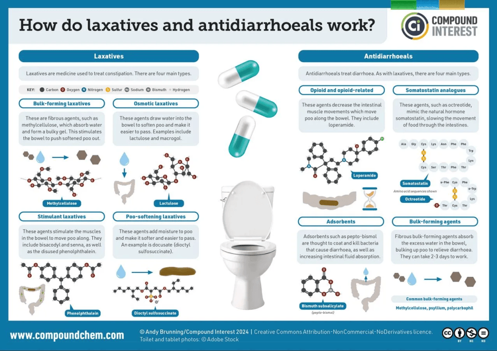
Pain management of the surgical incision and to get the bowels working – constipation is commonly characterised by dyschezia (difficulty passing poo/stool/faeces) or hard poo because of poor abdominal muscles and coordination in the pelvic floor. This can be overcome with laxatives to soften stool and eating wholemeal and fibre.



Making sure the kidneys are working…
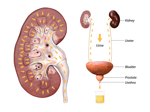
This pair of kidney-shaped organs is important for maintaining the levels of ions and water in the body and also produces urine. The patient must release a sufficient amount of urine (void volume) for the catheter to drain while recovering.
Reference ranges can be found in the link below:
https://www.bladderjournal.com/articles/normal-urine-volume-per-void


Complications Associated With Vaginectomy Needing Immediate Assistance
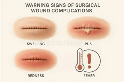
1. Surgical Site Infection
- Infection in the arm or leg (pain, redness, swelling, and warmth).
- There could also be discharge or fluid coming from the wound, incision, or vagina, with redness, chills, and fever (100.4 degrees). The type of infection and the site should be noted.
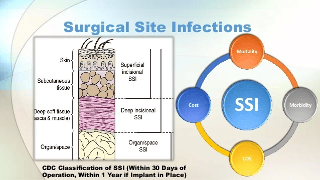
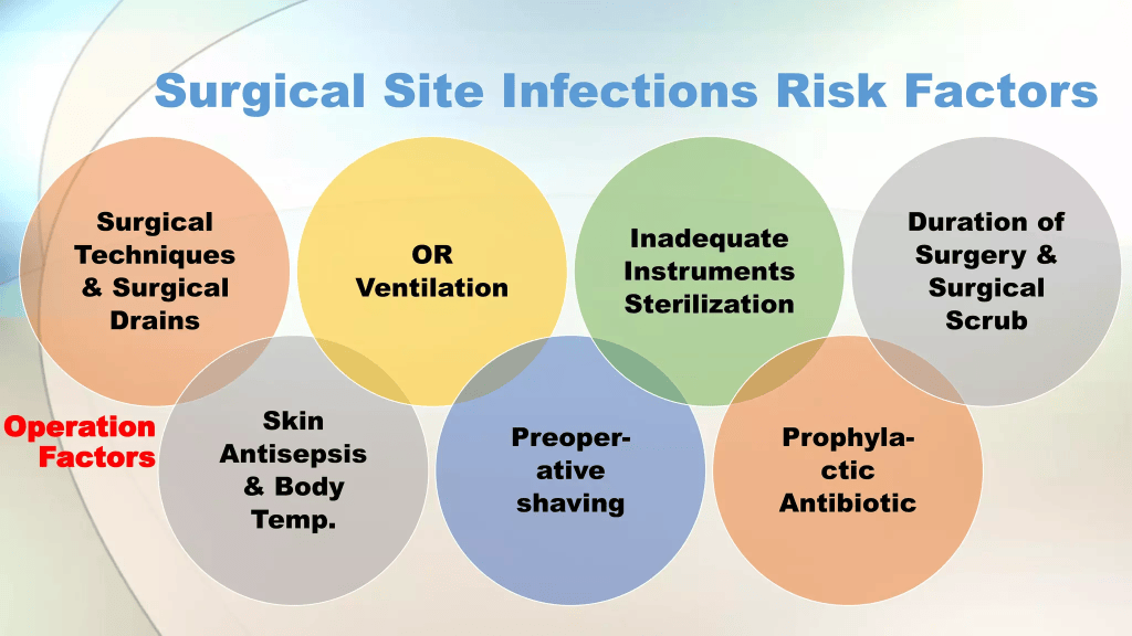
Recommended Lecture On Surgical Infections
https://www.slideshare.net/slideshow/surgical-site-infections-ssi/56316373#8
Diagnosis and Treatment of Surgical Site Infection By CitizenSurgeon
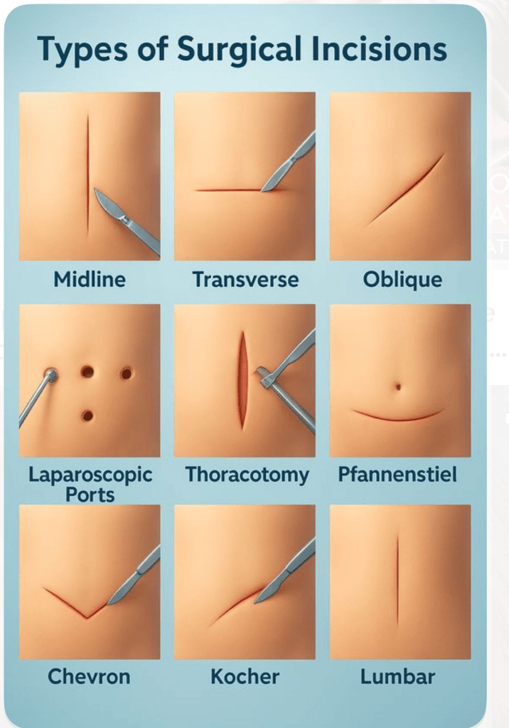
2. Incision Torn
3. Abnormal or Excessive Bleeding
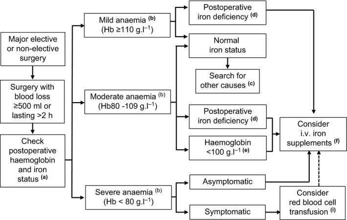
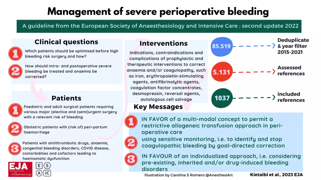
Peri-operative bleeding begins from before surgery (pre-operative state) to after surgery (post-operative state). There are several factors to consider, for instance, their blood status, how well their blood coagulates (changes from liquid to a jelly-like mass), and level of prothrombin (precursor protein) that switches on to form thrombin, which helps to convert a soluble (melts or dissolves easily) protein called fibrinogen to an insoluble protein called fibrin during the last moment of blood coagulation. The general health of the patient and other procedures. Management of bleeding is needed for trauma and non-trauma states.
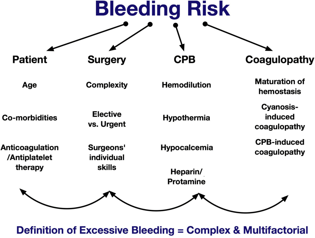
Abnormal or excessive bleeding puts patients at risk of anaemia. Anaemia is defined by the World Health Organisation (WHO) as a haemoglobin concentration < 130 g L-1 for men, < 120 g L-1 for non‐pregnant women and < 110 g L-1 for pregnant women. Haemoglobin is the protein found in red blood cells and joins with oxygen to form oxyhaemoglobin. Red blood cells travel around the body, giving cells and tissues oxygen for energy. Haemoglobin itself is made of iron and protein. This is why when there is a lack of iron, haemoglobin or folate are amongst the common causes of anaemia. These reference values may also be applied to patients after surgery. Post-operative anaemia is common in 80 to 90% of patients undergoing major surgery. The difference in the number is due to women having less circulating blood volume and less red blood cell mass compared to men. Even if both genders underwent the same surgery, women would need more blood transfusion to overcome the blood loss.
Patients experiencing anaemia BEFORE surgery or who had moderate to severe blood loss DURING surgery should be screened for anaemia AFTER surgery. This also includes patients who had blood loss > 500 ml or lasted for a minimum of two hours (> 2 h).
This flow diagram by Munoz et al. (2018) presents an overview of how to manage anaemia after surgery, whether it is mild, moderate, or severe anaemia. Several letters are found and correspond to the following. (a) Assess iron status within 24 hours after surgery if it has not been done before surgery. The haemoglobin levels should be monitored for 3 to four days after surgery. (b) This is according to the WHO classification. ( c ) Treatment should be considered. (d) Levels of ferritin, which reflects the iron stores in the liver, are detected as less than < 100 μg.l−1, ferritin < 300 μg.l−1 and transferrin saturation < 20% or reticulocyte haemoglobin content < 28 pg. Transferrin, otherwise known as siderophillin is a glycoprotein (sugar with protein) that carries iron in the blood. Reticulocytes are early-staged red blood cells and normally correspond to 1% of the total red cells. The level of reticulocytes corresponds to the level of red blood cells. The more red blood cells are produced, the higher the levels, and this is referred to as reticulocytosis. (e) This is due to pre-operative anaemia or heavy surgical bleeding irrespective of the iron status. (f) Total iron deficiency = (target haemoglobin − actual haemoglobin) × weight (kg) × 0.24. Add another 10 mg.kg−1 for replenishing/finishing iron stores, especially in patients with pre‐operative iron deficiency. Consider adding recombinant human erythropoietin (40,000 IU) for patients with severe anaemia or declining transfusion. Erythropoietin is the protein hormone that helps to make red blood cells. It is made in the renal tubules of the kidneys. Levels of this hormone increase if low levels of oxygen are detected to help increase the rate of red cell production. (i) Transfuse one red blood cell unit at a time, with post‐transfusion re‐assessment of further needs. Consider i.v. iron supplementation after transfusion, using post‐transfusion haemoglobin as actual haemoglobin for total iron deficiency calculation.
4. Damage To Internal Organs
5. Bloating – The patient feels full in their belly.
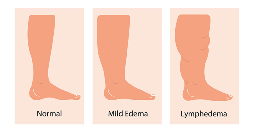
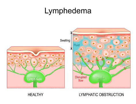
6. Lymphoedema
A build-up of fluid if lymph nodes have been removed. This is common in the legs and is commonly associated with mobility, itchiness, swelling, heaviness, or changes to the skin.
7. Alteration with bonding can cause emotional struggles.
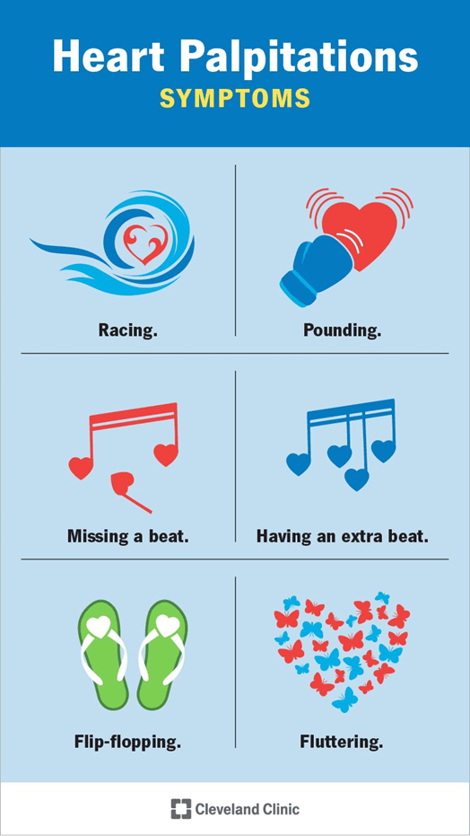
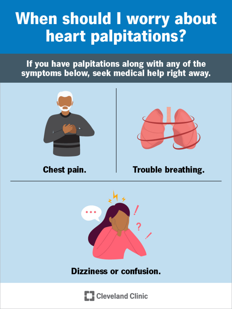
8. Chest Pain and heart palpitations
9. Feeling sick (nausea) or vomiting (emesis)
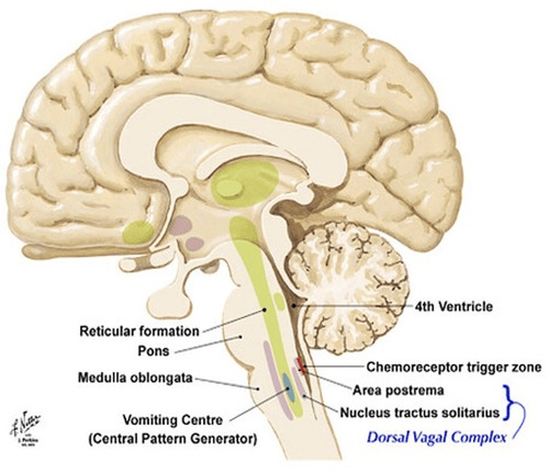
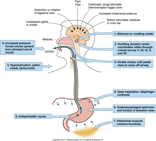
To find out the origin of the vomited material, does it have any blood (haematoemesis)? Then it is linked to the upper GI; it could also be linked to bile, where it could be green pigment (biliverdin) where there is absence or undigested food, or it could be yellow or orange (bilirubin) where there are small lumps. The vomiting centre is in the medulla and is activated by neurons.
10. Hernia
A hernia is defined as an abnormal external structure “that sticks out” of an organ or structure. To determine the type of hernia, it is important to consider the following factors:
- Lump
- Position
- Temperature
- Tenderness
- Shape
- Size
- Tension
- Contents/what it is made of (composition).
- Characteristics are noted over the overlying skin.
- The hernia should be palpated and felt when the patient makes a cough impulse.
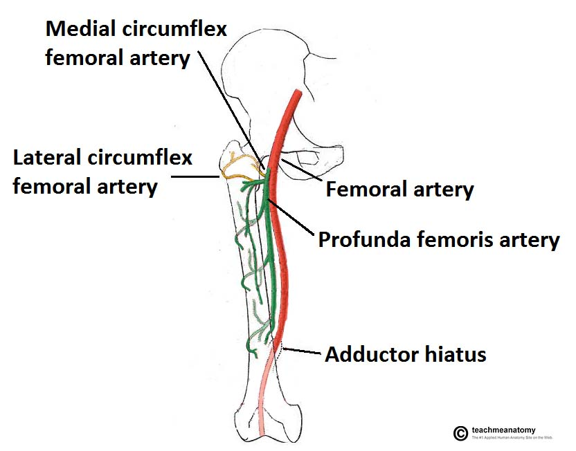
- Sounds of the bowel or bruits (harsh sounds) that may appear from the iliofemoral artery or the carotid artery due to obstruction.
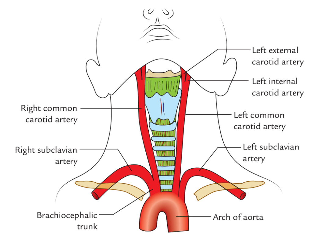
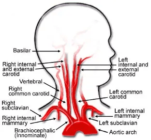
4. Examining the same site on the opposite side.
5. Attempt to make the hernia smaller. If it cannot be made smaller, it is referred to as strangulation, which is fixed and swollen, as there is a blockage of blood supply causing ischaemia (inadequate flow of blood supply) and necrosis (death) of the organ that is undergoing hernia.
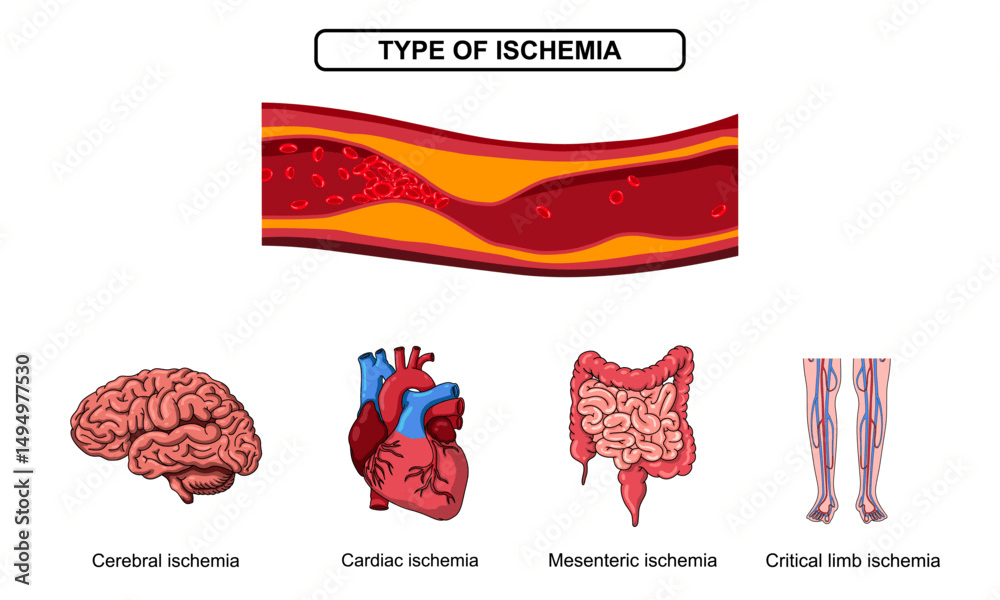
6. The hernias that appear in the abdomen are caused by bowel parts that protrude through a weakened area of the abdominal wall.
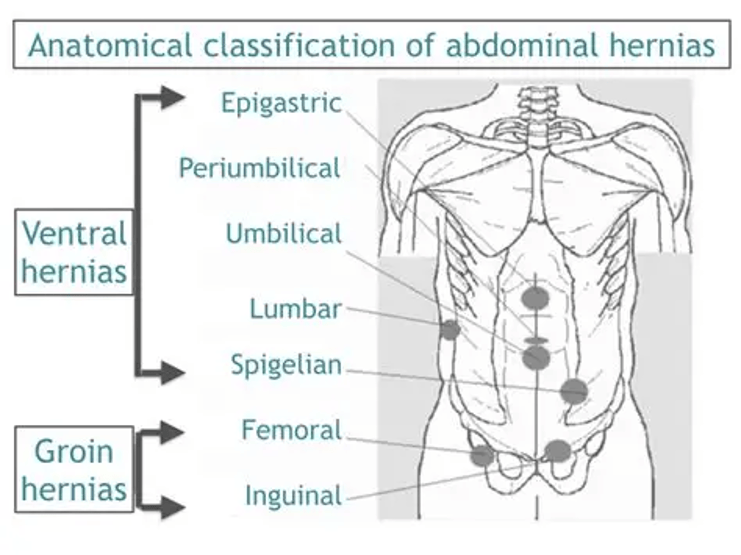
Types Of Hernia
Inguinal Hernia
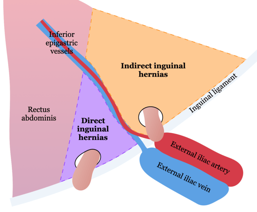
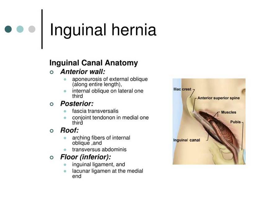
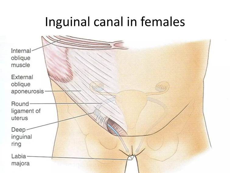
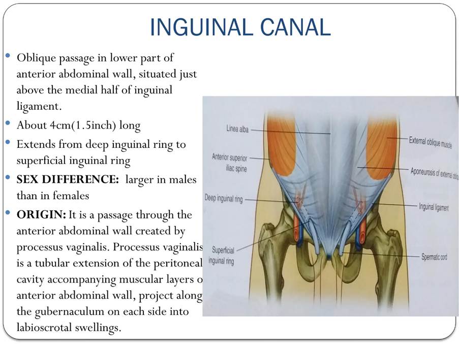
If it appears on the abdominal wall. It can be part of the Inguinal canal. This extends from the pubic bone to the anterior superior iliac spine. It also carries the round ligament of the uterus. Hernia can appear directly at the site of the external ring or indirectly, which is the most common form, in the internal ring, with bowel or omentum that travels down the inguinal canal and may even affect the external ring.
Femoral Canal Hernia
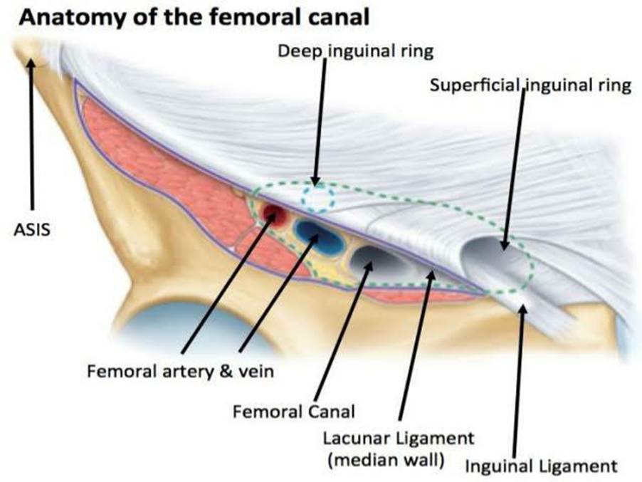
This is a small part of the femoral sheath medial to the femoral vessels and contains loose connective tissue and large lymph nodes. It is bordered anteriorly by the inguinal ligament. Lateral with the femoral vein and lacunar ligament medially and pectineal ligament posteriorly. The femoral hernia develops as protrusions of bowel or omentum through the space.
Umbilical/paraumbilical Hernia
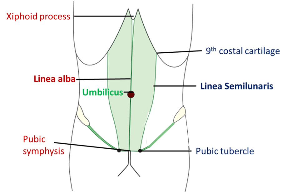
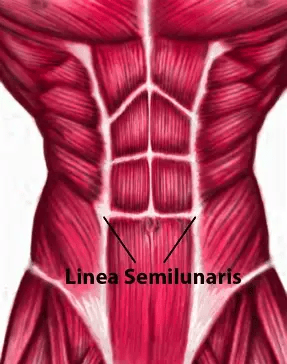
This is a hernia that appears through a defect near the umbilicus (navel). If there is a hernia above the umbilicus near the linea alba, it is referred to as epigastric. If it is below or to the side of the umbilicus near the linea semilunaris, it is referred to as Spigelian.
What Other Relevant Hernia Is Linked To Laparoscopic Or Intravaginal Vaginectomy?
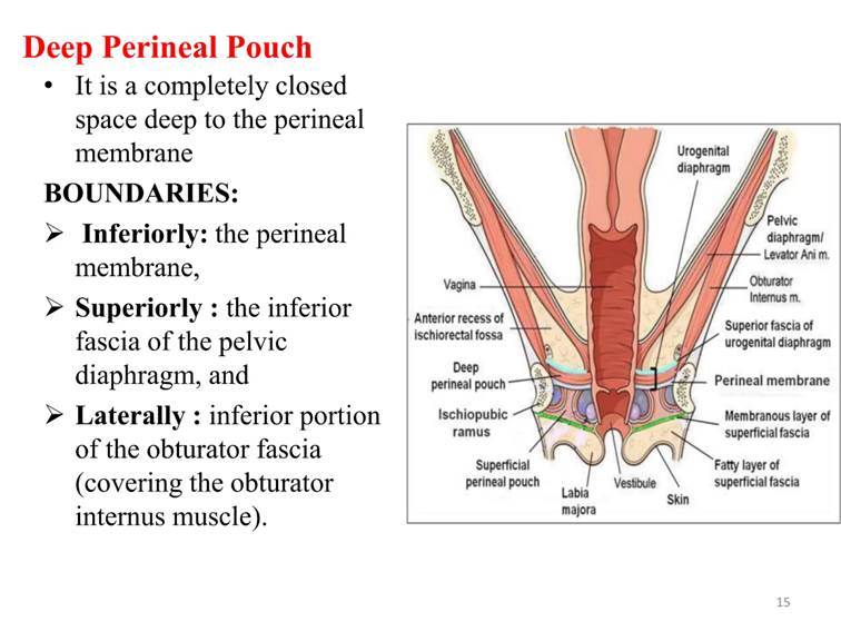
Perineal – Herniation through the pelvic floor, though rare.
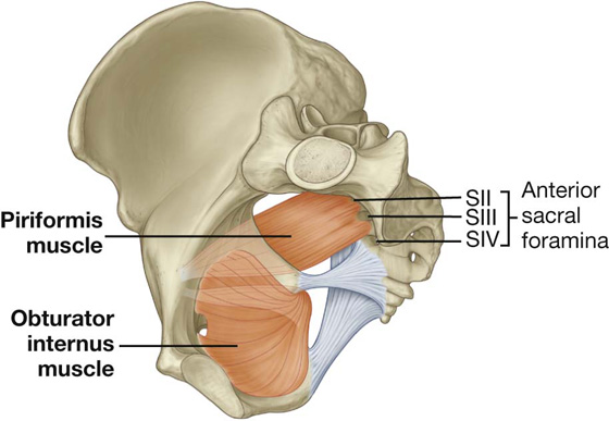
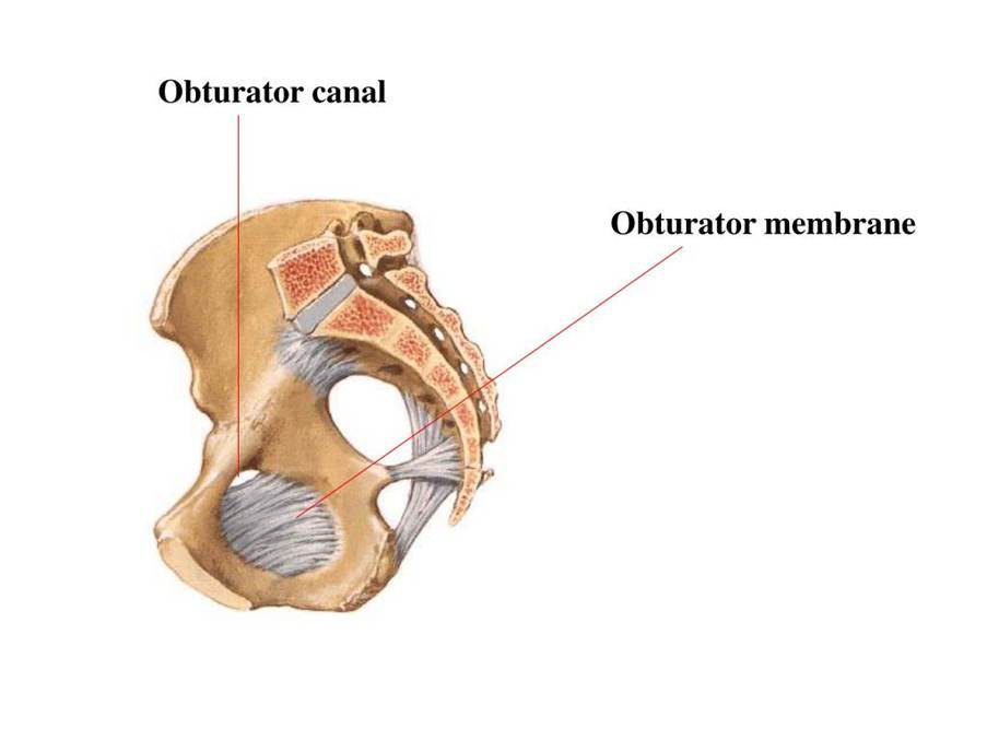
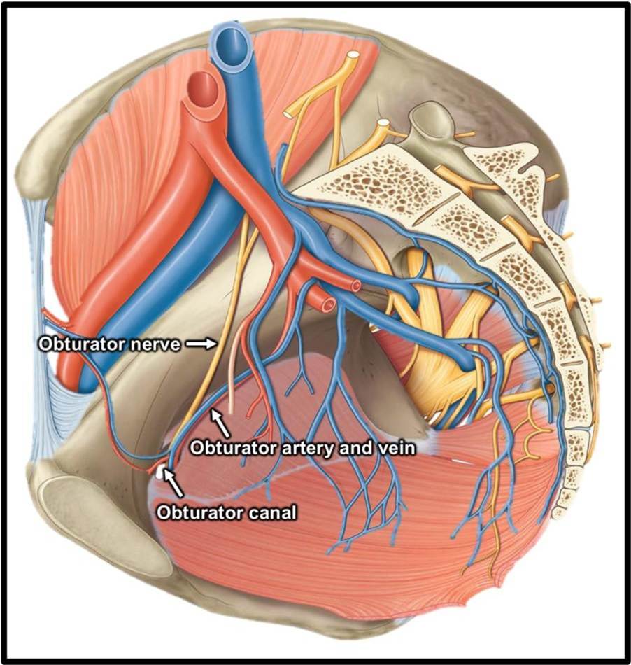
Obturator Hernia
This rises from obturator canal and it is common with those who have given lots of birth (multiparity) and increasing age.
Incisional Hernia
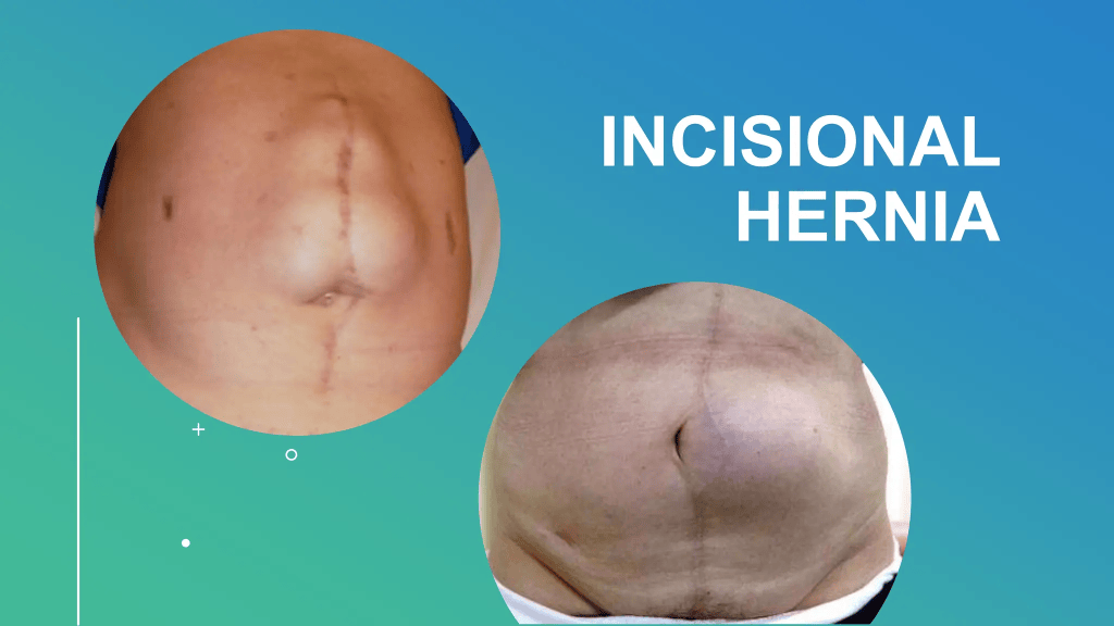
This is common in cases of recurrent surgery where a bulge may appear underlying a surgical scar. It may be caused by wound infection, inflammation in the fascia (covering), and is referred to as fasciitis. It can also be linked to death of the muscle (muscle necrosis).
Recommended lecture on Ventral and incisional hernias and management:
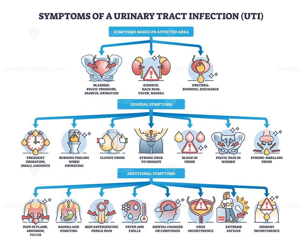
11. Urinary Symptoms
- Smell discharge from urethra
- Difficulty or pain to urinate (dysuria) – stinging pain felt from the urethra opening (meatus)
- Urine in blood (haematuria)
- Changes to the urine volume – lots of urine (polyuria). This is commonly linked to consuming lots of water, diabetes, and vasopression/ADH hormone that regulates the levels of water and ions. Dysfunction in the tubules and defects inthe the kidneys. All encourage more urine output.
- What is the difference between polyuria and urinary frequency?
It is important to differentiate polyuria from urinary frequency. Urinary frequency is the frequent passage of small amounts of urine. This varies with the number of times a day and whether the patient is producing more urine than normal or simply has an urge to urinate more than normal. - Urine at night – This may be linked to keeping the patient awake from sleep. How many times and what is the volume?
- Anuria – absence of urine formation, and this could be linked to renal dysfunction, shock, or obstruction.
- Little urine – oliguria < 300 ml in 24 hours – This is commonly linked to dehydration, shock, obstruction, or renal disease.
- Urgency to urinate (urinary incontinence) – There is loss of voluntary control of emptying the bladder.
- Dribbling, overflow, and stress (laughing, sneezing, coughing can add pressure).
12. Poo changes
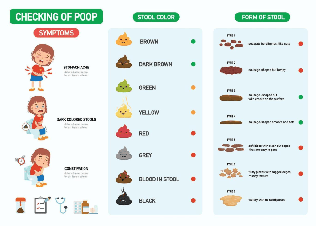
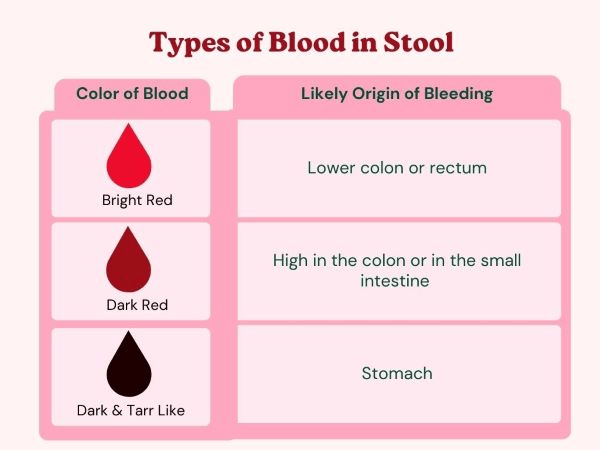
- Blood in stools varies with the type of colour (red, brown, or black):
Is it mixed with stools or on the stool?
Is it splatter or only on toilet tissue?
Is there any mucus that may appear clear?
Melaena – is it black with a pungent smell? This is linked to the upper GI tract. Or iron supplements that can cause blackened stools but not melaena smell or consistency.
It could also be haemorrhoids (piles), anal or GI issues – Blood coming from the back.
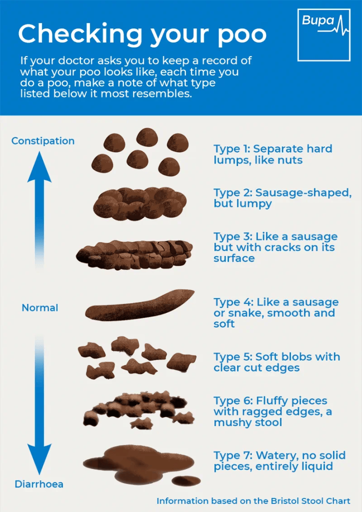
Constipation – this is the passage of stool that is hard or difficult to pass (dyschezia). The length of time, stool size, consistency, straining, other symptoms. Pain, whether there is blood. Fluid and fibre intake. Exercise level. What other diseases besides vaginal cancer does the patient have? Is it neurological, metabolic, or endocrine? Some medications can also cause constipation, for instance antidepressants, codeine, and aluminium and calcium antacids.
Diarrhoea – this is an increase in stool volume and frequency and a change of consistency to semi-formed or liquid, where there could be colour, consistency, smell, ease of flushing, the length og time, affect sleep, does it contain any blood, mucus, or pus, any other pain ot nausea, recent antibiotics, and other causes.
Surgical Complications With Vaginoplasty
Vaginoplasty recovery – a few weeks to a few months depending on the surgery.
- Light bleeding for 48 hours after surgery
- Infections – fever, yellow discharge
- No response to medications
- Vomiting
- Excessive bleeding -indicated by the blood-soaked dressing.
- Signs of blood clots – abnormal swelling in groin or lower legs.
- Swelling or bruising takes a few weeks.
- Itchiness or numbness – pain receptors.
- Scarring – lightens or changes colour.
- Painful intercourse (dyspareunia)
Some of the complications of Vaginoplasty can be overcome using the Vaginal Dilator.
A vaginal dilator is a tube-shaped plastic or silicone dilator. This is commonly applied after 6 days following vaginoplasty. Normally, after surgery, the vagina is kept open with gauze packs for six days. It is then removed and replaced with the vaginal dilator – this stretches the vagina to maintain the depth and width of the vaginal canal and activity of the muscles, as amongst the common complication is the loss of vaginal depth because the dilation is deducted or temporarily stopped especially during the first year of recovery. This is also used when there is pain during bonding (vaginismus). It is difficult to create additional depth after surgery. In some patients, dilation becomes difficult, and this is overcome by increasing the number of dilations, adding fragrance-free lubrication or oil-based lubricant, and taking pain medication an hour before dilating. There is some risk of increased pain, soreness,s or bleeding during the procedure, and it can be stopped; seek medical attention.

Please click here for further information, including a step-by-step approach:
The Leeds Teaching Hospitals NHS Trust (n.d.) Vaginal Dilators. Available at: https://www.leedsth.nhs.uk/patients/resources/vaginal-dilators/ (Accessed: 14th November 2026)
The Permanente Medical Group (2026) Vaginal Dilation After Vaginoplasty. Available at: https://mydoctor.kaiserpermanente.org/ncal/article/vaginal-dilation-after-vaginoplasty-1290049 (Accessed: 14th November 2026)
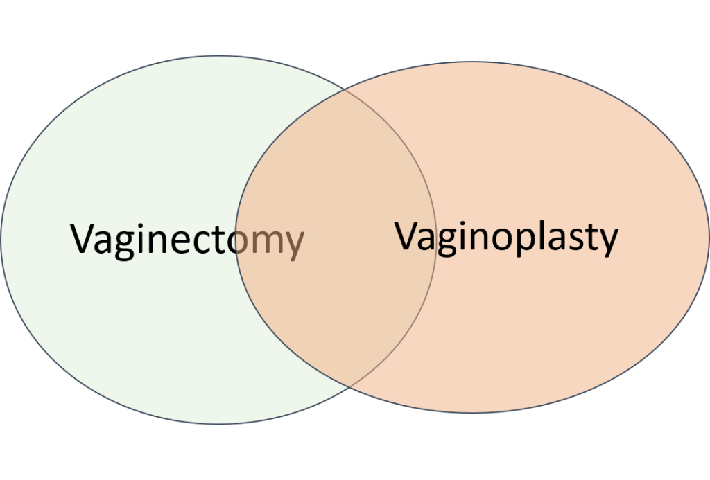
Overlapping Complications With Vaginectomy And Vaginoplasty
Most common symptoms are: Bleeding, Infections, feeling sick or vomiting, Blood clots, Swelling, scarring, and Painful intercourse (dyspareunia)
References
Adams, T.S., Maluleke, J.C. and Cuello, M.A. (2025). Cancer of the vagina: 2025 Update. International Journal of Gynecology & Obstetrics, 171(S1), pp.48–59. doi:10.1002/ijgo.70325.
Ball, T.L. and Douglas, R.G. (1949). Cryptomenorrhea. The American Journal of Surgery, 78(1), pp.40–49. doi:10.1016/0002-9610(49)90183-x.
Cancer Research UK (2027) Vaginal intraepithelial neoplasia (VAIN) Available at: https://www.cancerresearchuk.org/about-cancer/vaginal-cancer/vain (Accessed: 11th October 2026)
European Society of Anaesthesiology and Intensive Care (2022) Severe Perioperative Bleeding. Available at: https://esaic.org/guideline/severe-perioperative-bleeding-management-guideline-2nd-update/ (Accessed: 13th November 2026)
HafeezeAzee (n.d.) Basic surgical procedures. Available at: https://www.slideshare.net/slideshow/basic-surgical-procedures/198359135#2 (Accessed: 7th November 2026)
National Cancer Institute (2026) Vaginal Cancer Treatment (PDQ®)–Health Professional Version. Available at: https://www.cancer.gov/types/vaginal/hp/vaginal-treatment-pdq (Accessed: 17th August 2026)
Nout, R.A., Calaminus, G., Planchamp, F., Chargari, C., Lax, S., Martelli, H., McCluggage, W.G., Morice, P., Pakiz, M., Schmid, M.P., Stunt, J., Timmermann, B., Vokuhl, C., Orbach, D. and Fotopoulou, C. (2023). ESTRO/ESGO/SIOPe Guidelines for the management of patients with vaginal cancer. International Journal of Gynecologic Cancer, [online] 33(8). doi:10.1136/ijgc-2023-004695.
Oliveira, R., Bócoli, M.C., Saldanha, J.C., Murta, E.F.C. and Nomelini, R.S. (2013). Primary small cell carcinoma of the vagina. Case Reports in Obstetrics and Gynecology, 2013, pp.1–4. doi:10.1155/2013/827037.
The Eve Appeal (2026) Vaginal Cancer. Available at: https://eveappeal.org.uk/information-and-advice/gynaecological-cancers/vaginal-cancer/ (Accessed: 7th August 2026)
The Leeds Teaching Hospitals NHS Trust (n.d.) Vaginal Dilators. Available at: https://www.leedsth.nhs.uk/patients/resources/vaginal-dilators/ (Accessed: 14th November 2026)
The Permanente Medical Group (2026) Vaginal Dilation After Vaginoplasty. Available at: https://mydoctor.kaiserpermanente.org/ncal/article/vaginal-dilation-after-vaginoplasty-1290049 (Accessed: 14th November 2026)
Updated: November 2026. Next Review: September 2028


Leave a Reply