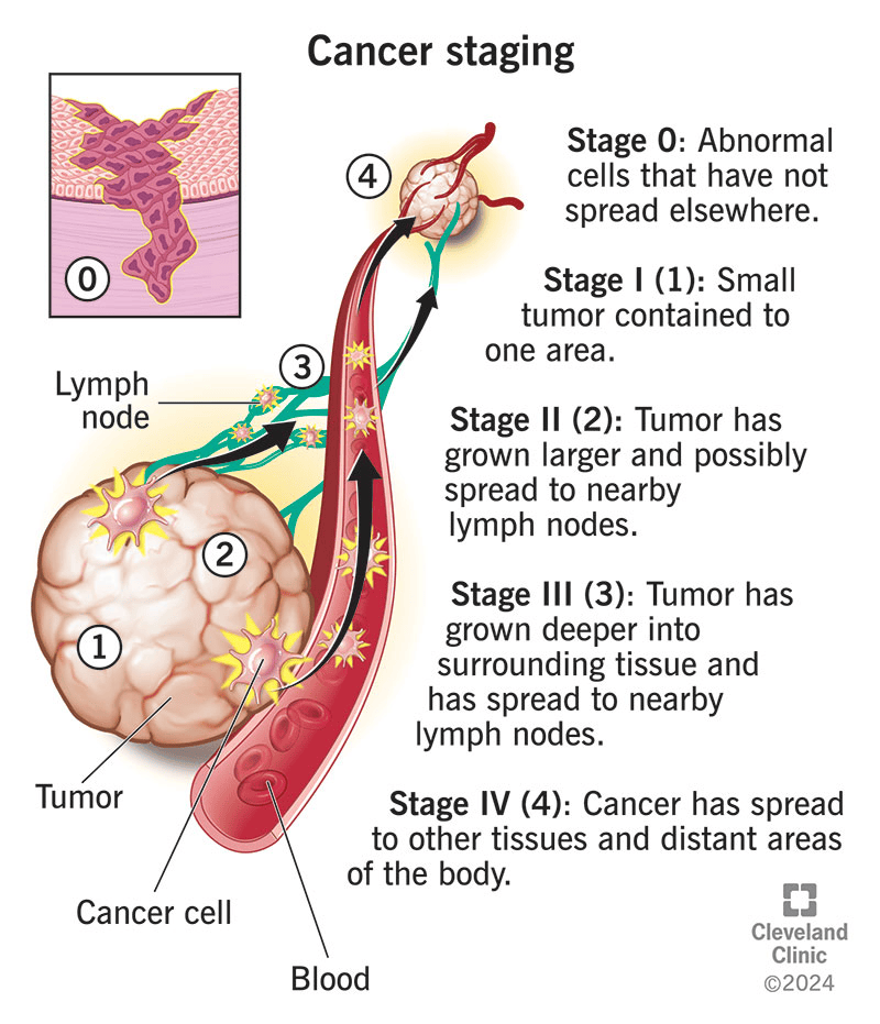

An overview of Staging and Grading
One of the primary ways to detect cancer is through staging and grading. Staging refers to the extent of the growth of the cancer: has it grown from its primary site (T), has it affected the lymph nodes that boost the immune system (N), and has it affected other organs – this is referred to as metastasis (M). This is commonly performed using a biopsy sample that is examined by a pathologist under the microscope. The other technique is through scans.
On the other hand, grading refers to the extent to which the tumour resembles non-cancerous tumours. If there is low differentiation (changes) in the cells, it is referred to as low-grade. In contrast, the more changes in the normal architecture of the cells in the organ, the higher the grade. This influences the prognosis which is the patient’s progress over the course of the time.
Each type of cancer has its own staging procedure, and this is because of the location, shape, size, and surrounding structures.
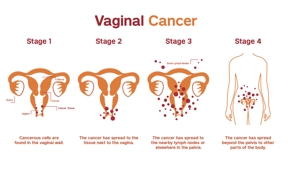

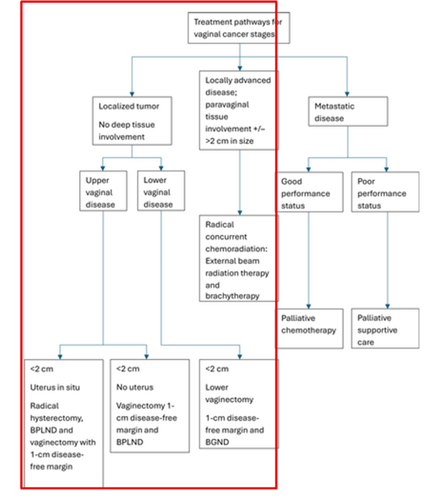
Source: Adams, T.S., Maluleke, J.C. and Cuello, M.A. (2025). Cancer of the vagina: 2025 Update. International Journal of Gynecology & Obstetrics, 171(S1), pp.48–59. doi:10.1002/ijgo.70325.
Curative surgery: This is performed only if it can be completely removed. This is referred to as complete tumour resection (maximum 2 cm). There are clear margins. No radiotherapy is needed after surgery (adjuvant radiotherapy).
Why?
A combination of both increases iatrogenic morbidity (illness linked to treatment or side effects). However, either of them offers similar survival benefit.
However, surgery depends on the location of the cancer and the staging. It should also depend on the extent and site of the lymph node staging.
It should not be close to the urethra or rectum. – otherwise there is a risk of additional resections (more cutting).
Every tumour resection needs to be with partial or complete colpectomy to ensure free margins.
Colpectomy is the removal of the vagina.
What if larger parts need to be removed?
Surgery should not be done to avoid loss of function unless there are contraindications (the technique would cause harm) for radiotherapy.
This includes previous radiotherapy, inflammation in the intestines (inflammatory bowel disease) like Chron’s disease and ulcerative colitis, or if the kidneys will be affected.
The Z-plasty technique is one of the main ways to heal a large surgical wound to lower tension and improve how it looks.
High rates of major intra- and grade three postoperative morbidities have been described (16% and 30%, respectively) related to the surgical treatment of vaginal cancer.
There is a risk of complications like:
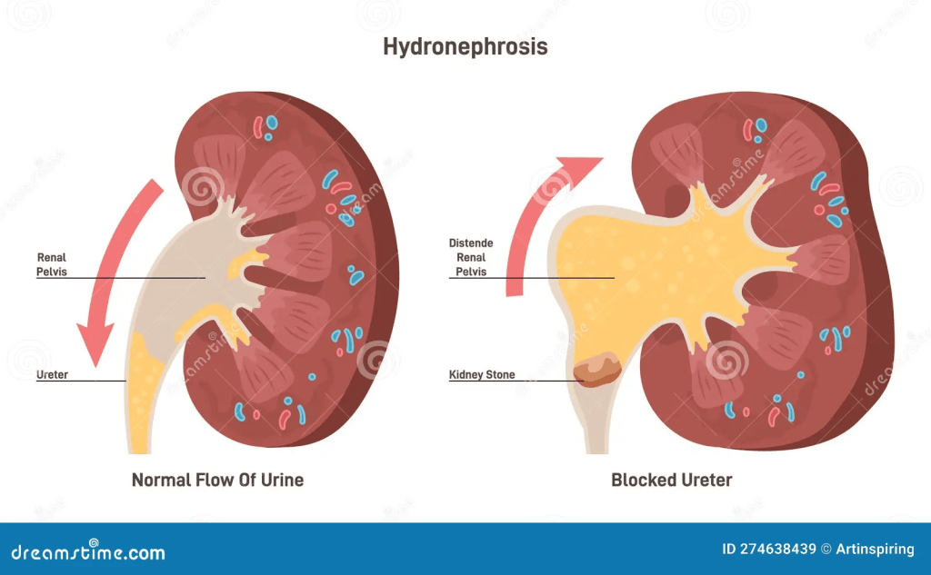
Hydronephrosis: This is where there is distension and can affect the obstruction of urine flow in the kidneys in acute stages. However, if this arises in the neck of the bladder, it can affect both kidneys. In chronic stages, it may impact the renal artery.
Fistula: This is when there is abnormal communication between hollow organs.
The first lymph node that is affected due to cancer (sentinel lymph node) is used to detect small metastasis and avoid any arising morbidity (with or without inguinal femoral lymphadenectomy. However, in vaginal cancer, the role of sentinel nodes has not been established and cannot be recommended.
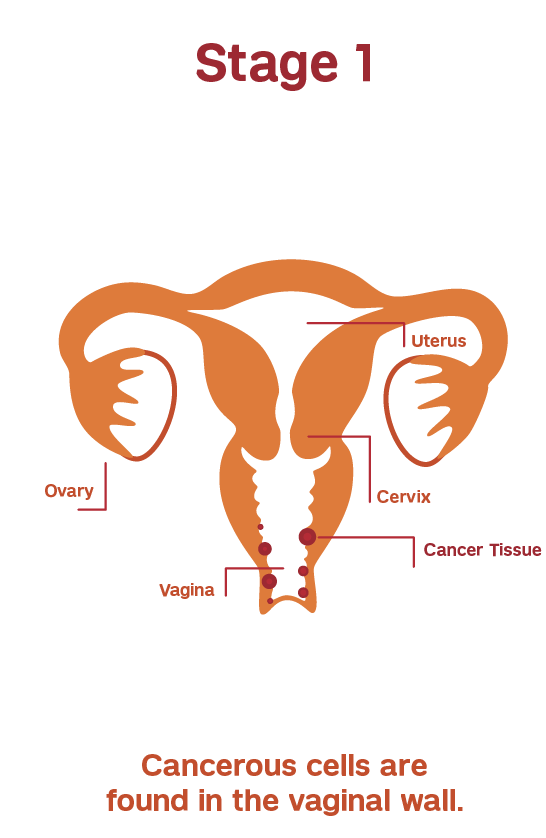
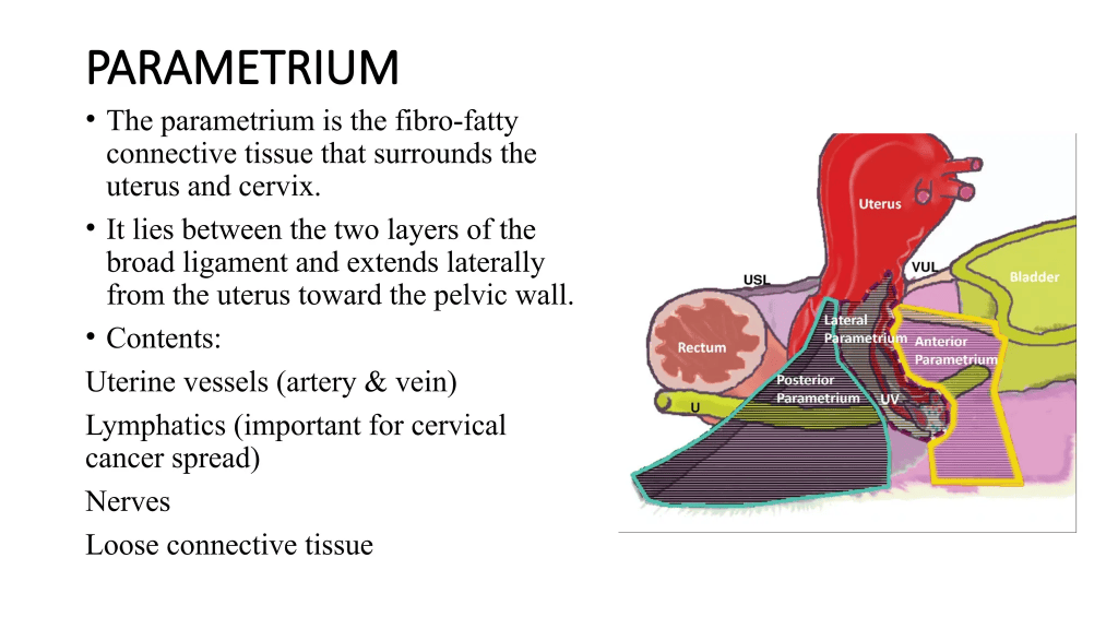
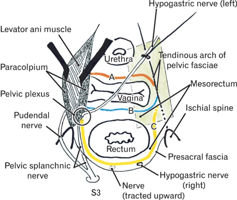
Hysterectomy (removal of the womb) and parametrial/paracolpia resection (removal of the parametrium and the vascular and nerve tissue along the vagina) and partial removal of the vagina (partial colpectomy) to ensure free margins.
The most common chemotherapies that are applied in brachytherapy (internal radiotherapy) and positive surgical margins are cisplatin or carboplatin (DNA alkylating agents to stop the growth of cancer)
Radiotherapy can also be given alone.
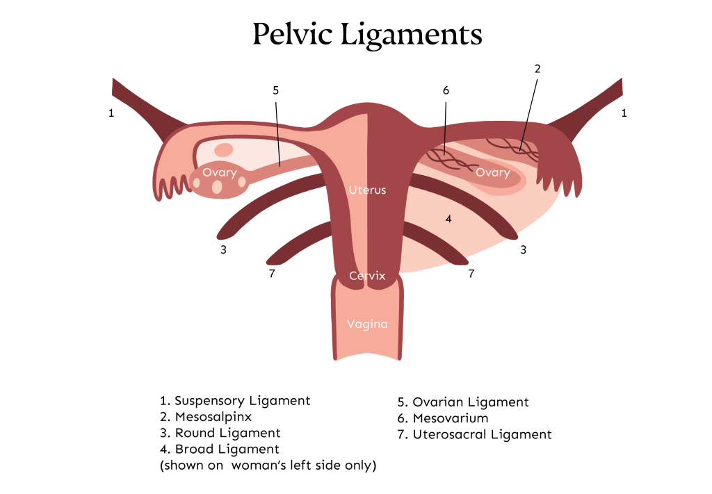
In premenopausal women, fertility preservation and ovarian transposition should be discussed up front. Ovarian transposition is where the ovaries are transposed or moved above the pelvic brim and to the side (lateral) to protect against radiotherapy. Key examples of lateral locations are the base of the round ligament in the uterus, the lower kidney pole, and the paracolic gutters. They are also protected with a lead shield centrally on the abdomen.

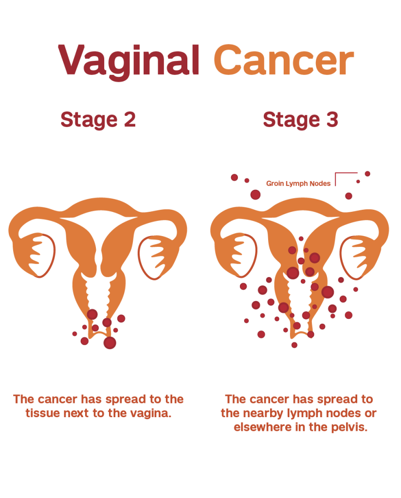
In locally advanced or node-positive vaginal cancer:
Chemoradiotherapy.
Cisplatin or another platinum-based chemotherapy is recommended along with radiotherapy.
This is followed by brachytherapy, especially in cases of T4 where there is a large fistula or cloacal malformation, or for patients with contraindications to radiotherapy such as previous pelvic radiotherapy, ulcerative bowel conditions like Crohn’s disease and ulcerative colitis, or pelvic kidneys.
What is cloacal formation?
Cloacal malformation can occur during early pregnancy (after five to six weeks from conception) when the rectum (organ that stores poo), urethra (the structure that allows urine to leave the body), and vagina do not separate and become a common channel that opens in the perineum. The perineum is where the anus and vagina are found. This is commonly characterised by having an:
- An absent anus (the structure that allows poo to leave the body)
- The external structures of the female reproductive system (external genitalia) are small.
- The clitoris is big
- An opening in the perineum can be found on routine ultrasound where both kidneys have hydronephrosis. It can also affect the development of the food pipe (oesophagus), heart, and lower spine (sacrum). Abnormal heart structure can be found on the echocardiogram. This is commonly treated with a nasogastric tube that goes down the throat and drains off the fluid to prevent damage to the kidneys; a stoma or colostomy (artificial opening to the bowel) can help remove the faeces, and breast or bottle feeding is introduced.
Reconstruction can then take place to create a new urethra, vagina and anus opening, but before this, additional scans such as a loopogram where contrast agent helps to view the large intestine, kidney function scans and also evaluating the bladder (cystoscopy).
Please visit the following link for further information:
Great Ormond Street Hospital for Children NHS Foundation Trust (2026) Cloacal Malformation. Available at: https://www.gosh.nhs.uk/conditions-and-treatments/conditions-we-treat/cloacal-malformation/ (Accessed: 14th November 2026)
The recent updates have indicated that external beam radiotherapy and brachytherapy with a total dose of 70 to 80 Gy with cisplatin chemotherapy is recommended.
Pre-menopausal women can also preserve fertility to prevent exposure to radiotherapy by applying ovarian transposition.
Surgical procedures to remove lymph nodes (lymphadenectomy) can help with symptom control and improve the quality of life for patients with palliative care.
Curative surgery helps to microscopically clear margins, and this depends on the size of the tumour.

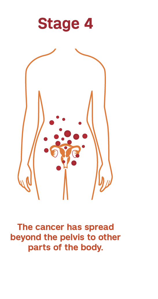
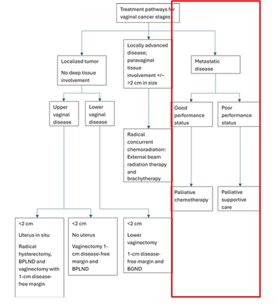
Source: Adams, T.S., Maluleke, J.C. and Cuello, M.A. (2025). Cancer of the vagina: 2025 Update. International Journal of Gynecology & Obstetrics, 171(S1), pp.48–59. doi:10.1002/ijgo.70325.
This stage is advanced, where there is limited evidence on chemotherapy, and it will be applied as in cervical cancer.
If there is cancer affecting many locations, it is referred to as oligometastatic disease.
This involves a combination of the following treatments:
Chemoradiotherapy à chemotherapy and radiotherapy.
Brachytherapy à internal radiotherapy.
Systemic therapy.
Personalised treatment is needed and depends on the size, location, symptoms, performance status, and how the tumour grows.
Other types of radiotherapy can be used: stereotactic radiotherapy, radiofrequency ablation.
Surgery can also be useful to include a stoma bag or stents to prevent obstruction.
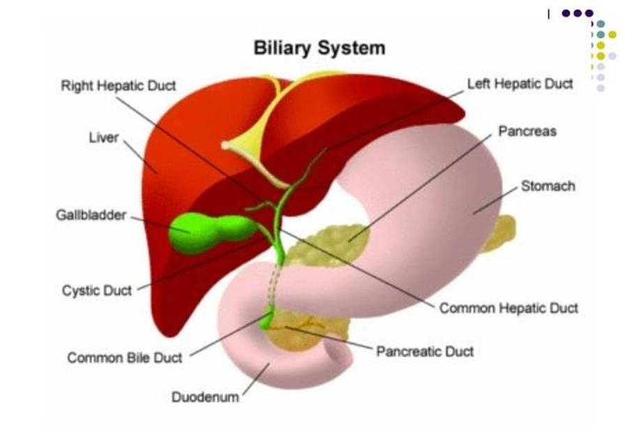
Palliative radiotherapy aims to help improve the quality of life psychologically, spiritually, socially, and clinically, and minimise the distress from active care to palliative care and to help them live alongside their cancer. Through patient assessment, the aim is to minimise the symptoms that cause the pain, and this requires pain management to identify whether the pain is acute (sudden onset) or chronic, related to the malignancy. Does the patient have somatic, visceral, or neuropathic pain? If it is somatic, it may be localised and persistent where inflammation may occur or presence of bone metastases. If it is visceral, it is commonly poorly localised with different modes of intensity and associated with symptoms like bleeding, discharge, or pelvic pain, nausea, abnormal lymph nodes (lymphadenopathy), or smooth muscle spasm that causes colic-linked symptoms, for instance, in the intestines, the bladder, or the biliary system. If it is neuropathic, it may be associated with compression of the spinal nerve root, like S3, giving a shooting or burning pain. The most important factor to consider is the pain perception of the patient. This can be overcome by palliative radiotherapy, either as a single fraction or a short course.
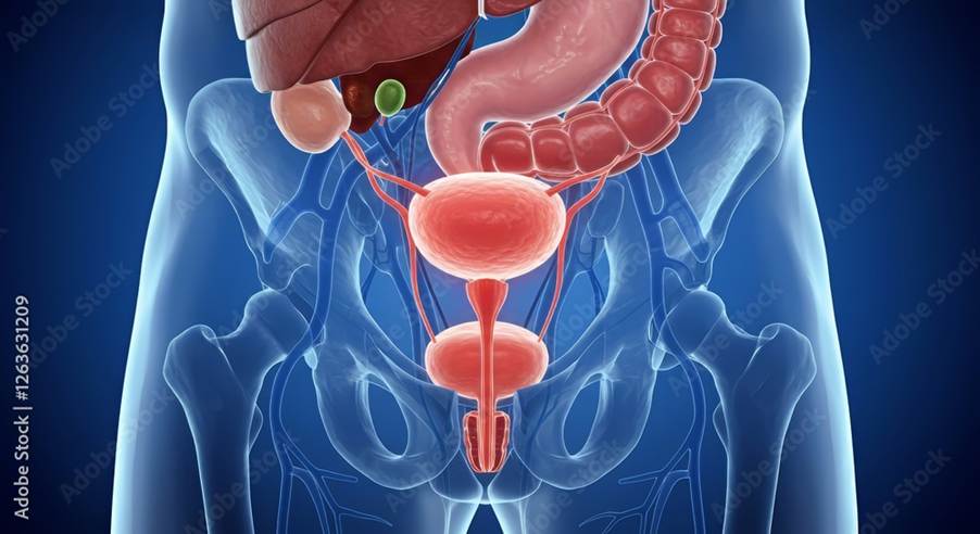
Below summarizes the main points for the histological classification, signs, symptoms, diagnosis and treatments in accordance to the European Society of Gynaecological Oncology (ESGO), European Society for Radiotherapy and Oncology (ESTRO) and the European Society of Paediatric Oncology (SIOPe) 2023 guidelines. A 2025 update of Vaginal cancer has been provided by experts from the following reference:
Adams, T.S., Maluleke, J.C. and Cuello, M.A. (2025). Cancer of the vagina: 2025 Update. International Journal of Gynecology & Obstetrics, 171(S1), pp.48–59. doi:10.1002/ijgo.70325.
Please read the guidelines attached for further details.
References
Adams, T.S., Maluleke, J.C. and Cuello, M.A. (2025). Cancer of the vagina: 2025 Update. International Journal of Gynecology & Obstetrics, 171(S1), pp.48–59. doi:10.1002/ijgo.70325.
Great Ormond Street Hospital for Children NHS Foundation Trust (2026) Cloacal Malformation. Available at: https://www.gosh.nhs.uk/conditions-and-treatments/conditions-we-treat/cloacal-malformation/ (Accessed: 14th November 2026)
Hinata, N., Hieda, K., Sasaki, H., Kurokawa, T., Miyake, H., Fujisawa, M., Murakami, G. and Fujimiya, M. (2014). Nerves and fasciae in and around the paracolpium or paravaginal tissue: An immunohistochemical study using elderly donated cadavers. Anatomy & Cell Biology, 47(1), p.44. doi:10.5115/acb.2014.47.1.44.
Lee, J.H., Jeong, Y.K., Park, J.K. and Hwang, J.C. (2003). “Ovarian Vascular Pedicle” Sign Revealing Organ of Origin of a Pelvic Mass Lesion on Helical CT. American Journal of Roentgenology, 181(1), pp.131–137. doi:10.2214/ajr.181.1.1810131.
Nout, R.A., Calaminus, G., Planchamp, F., Chargari, C., Lax, S., Martelli, H., McCluggage, W.G., Morice, P., Pakiz, M., Schmid, M.P., Stunt, J., Timmermann, B., Vokuhl, C., Orbach, D. and Fotopoulou, C. (2023). ESTRO/ESGO/SIOPe Guidelines for the management of patients with vaginal cancer. International Journal of Gynecologic Cancer, [online] 33(8). doi:10.1136/ijgc-2023-004695.
Sai, K. (n.d.) Cervical Cancer Anatomy Up Till Workup. Available at: https://www.slideshare.net/slideshow/anatomy-of-cervix-and-cervical-cancer-workup/283474012 (Accessed: 14th November 2026)
Tulandi, T. (2026) Ovarian transposition before pelvic radiation. Available at: https://www.uptodate.com/contents/ovarian-transposition-before-pelvic-radiation#H30824440 (Accessed: 8th October 2026)
Updated: November 2026. Next Review September 2028


Leave a Reply