
The Function Of The Female Reproductive System

Polite notice: This page contains diagrams of the reproductive organs of males and females (private parts). It is for educational purposes on cancers that arise in those areas. Please ensure that when visiting the page, there is a sense of manners. We understand that in some cultures, there would be discomfort, but it is important to be aware of information about cancer.
The Function Of The Reproductive System
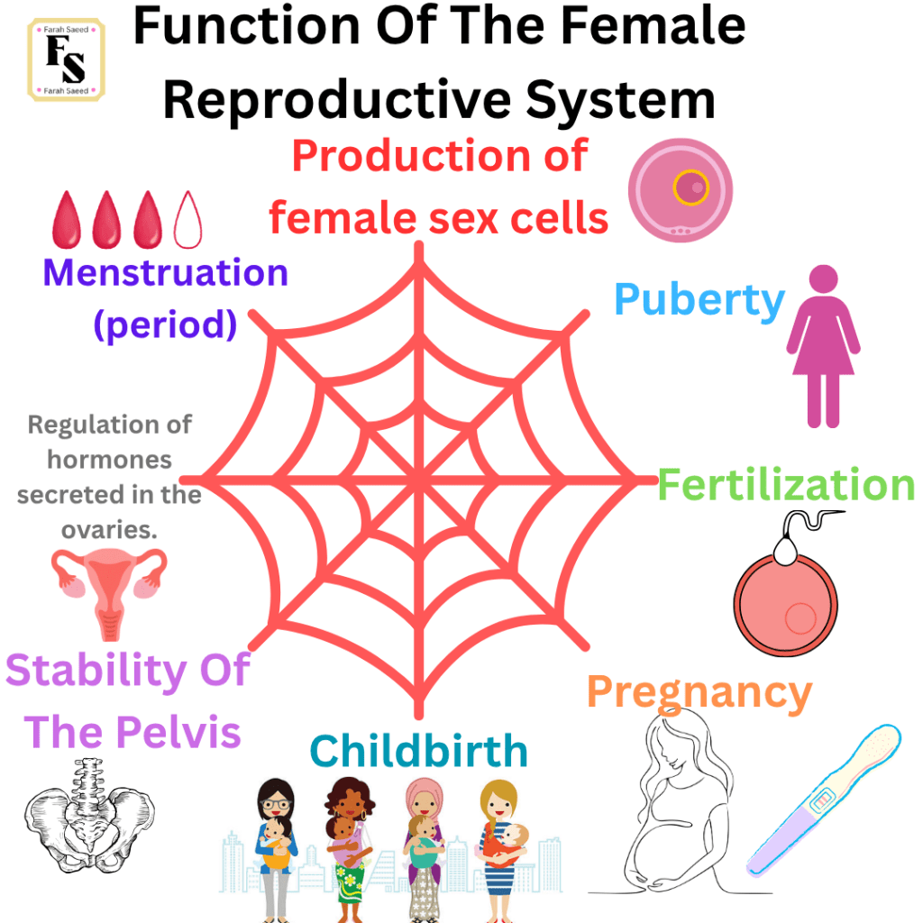
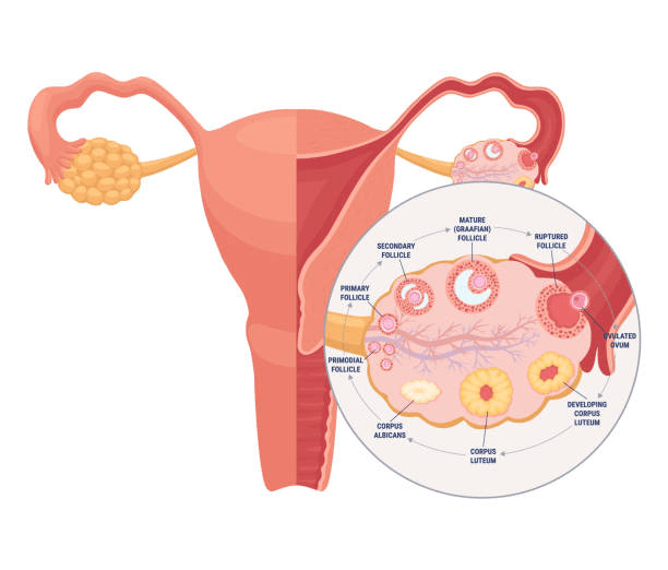
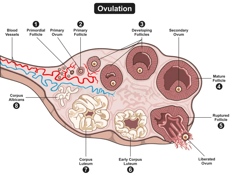
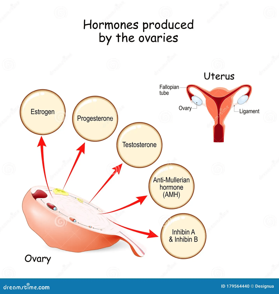
Function One: Production Of Female Sex Cells
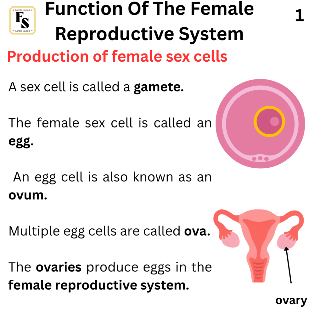
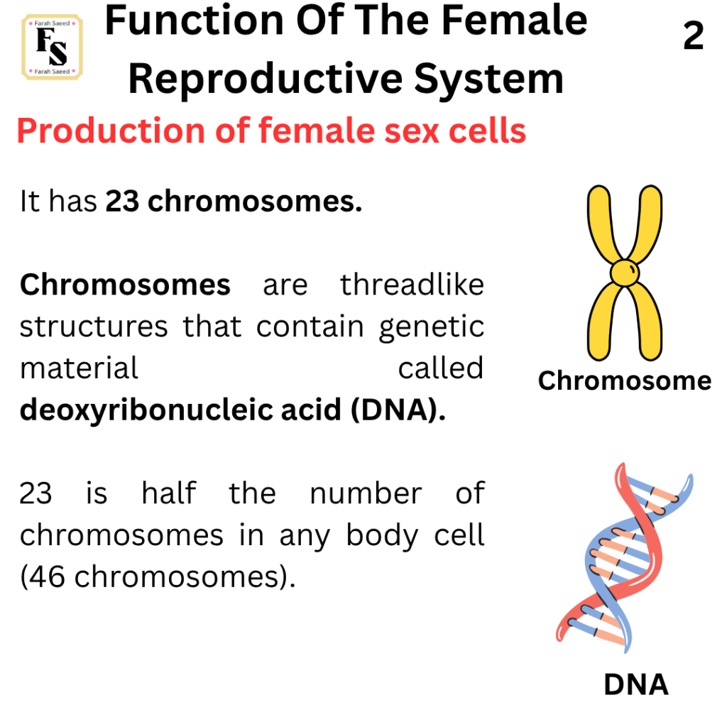
Supporting Images Of Function One
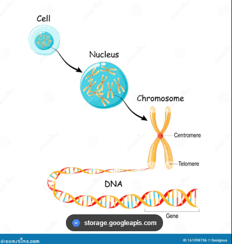
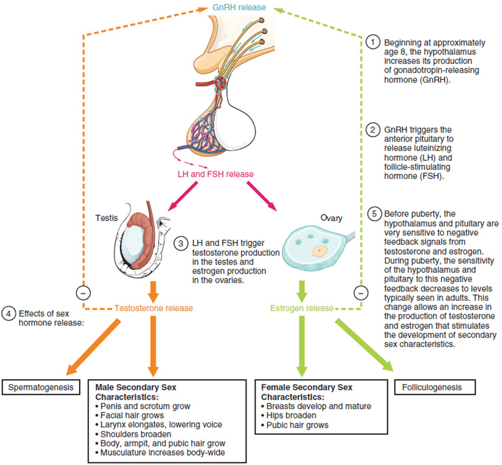
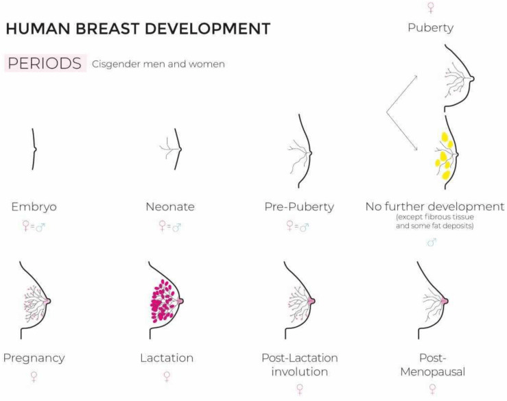
Function Two: Puberty Changes
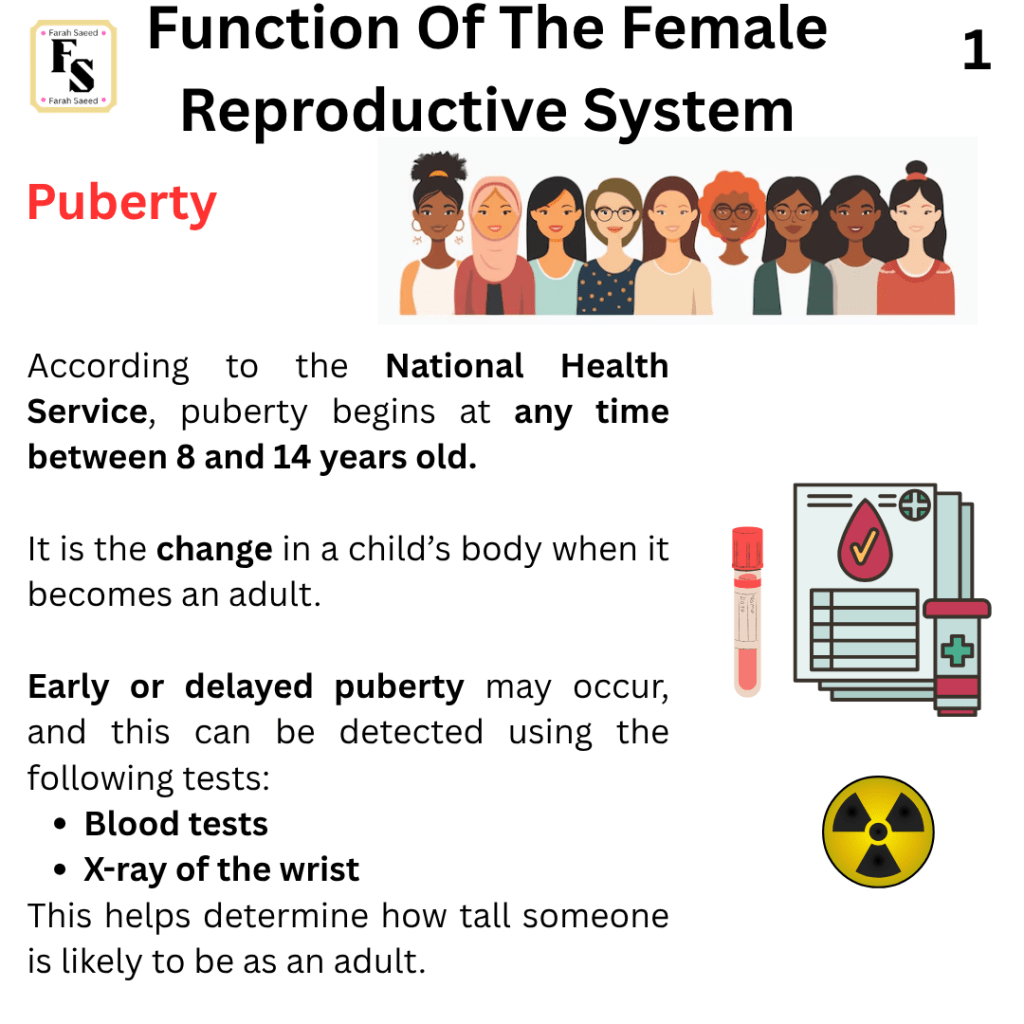
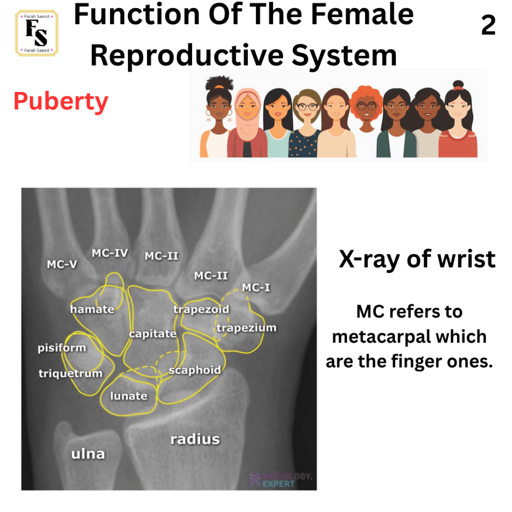
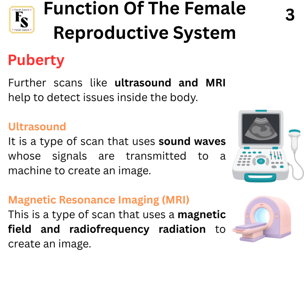
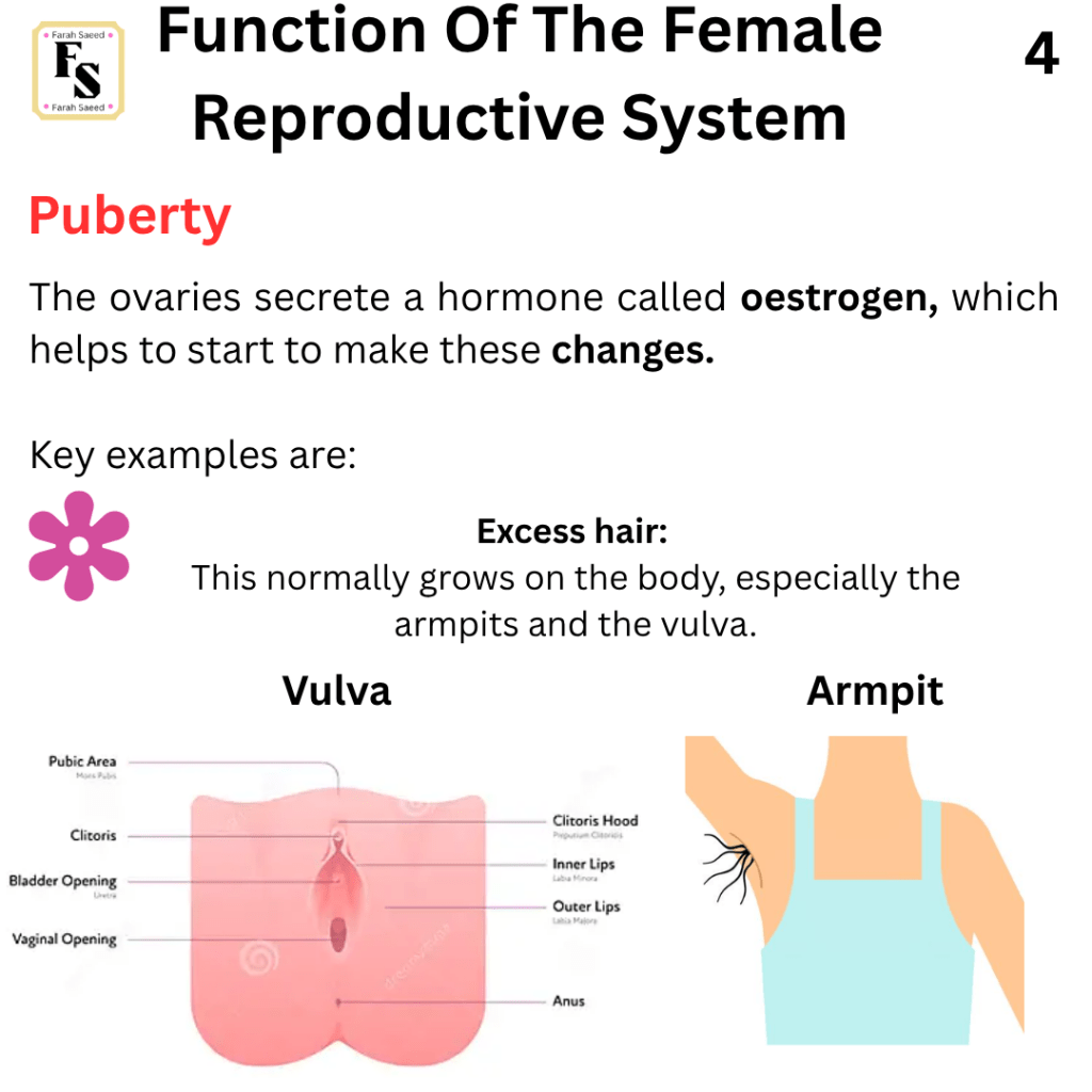
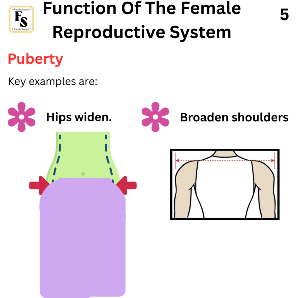
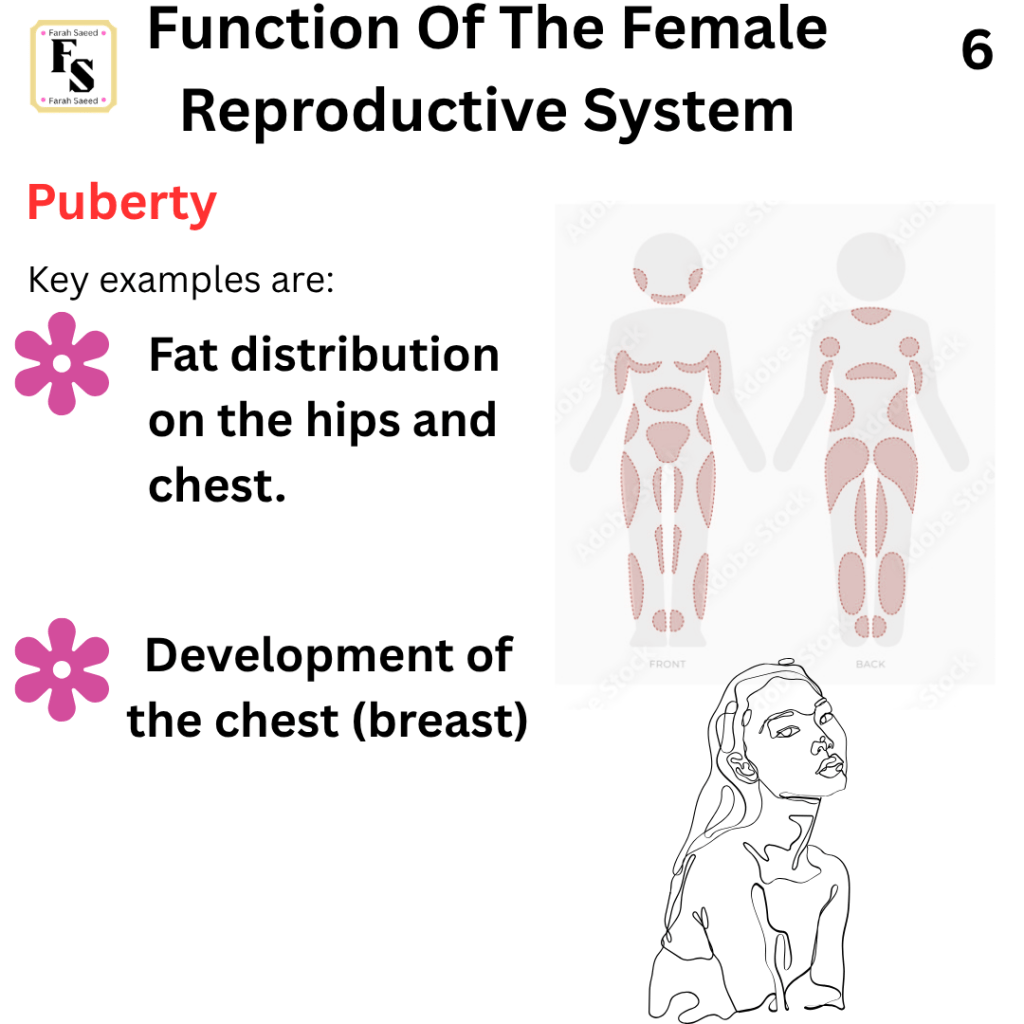
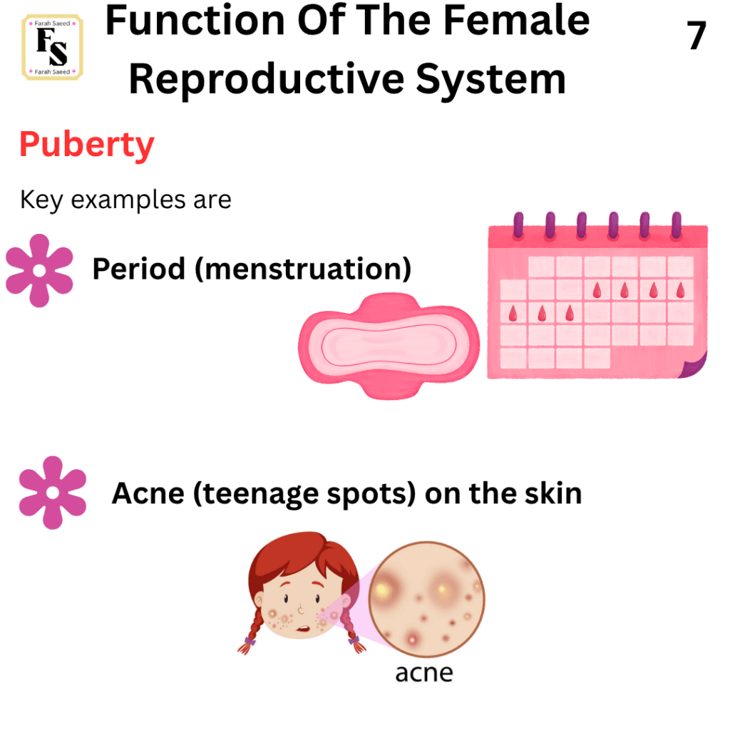
Supporting Images For Function Two
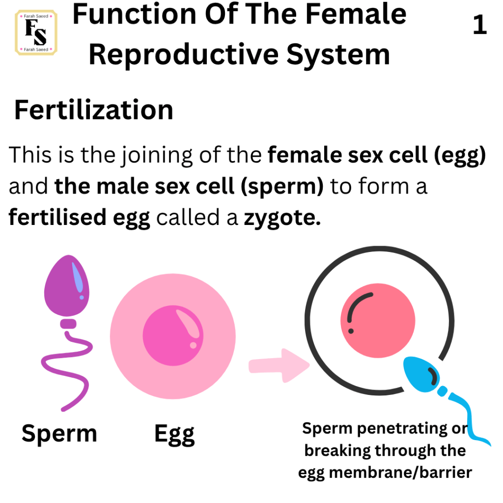
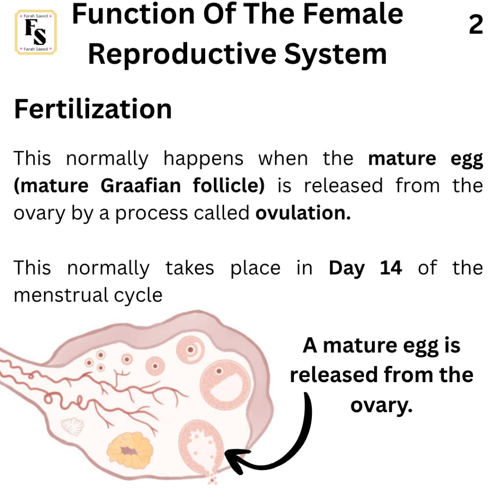
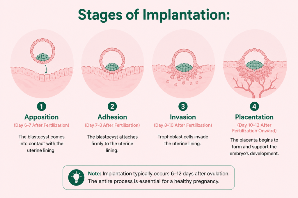
Function Three: Fertilization
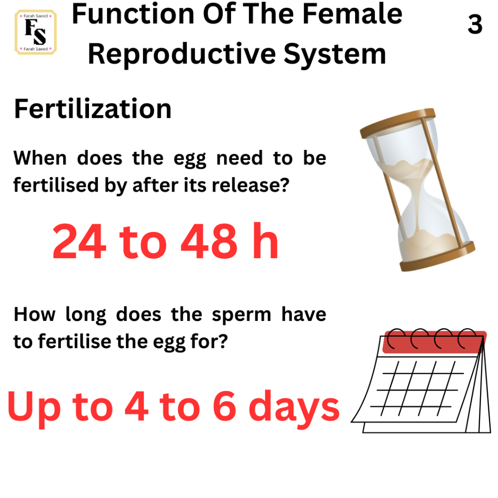
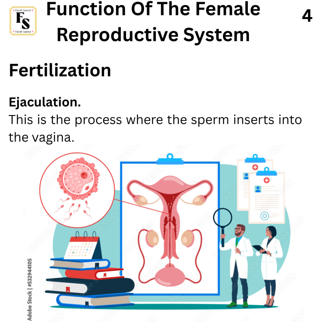
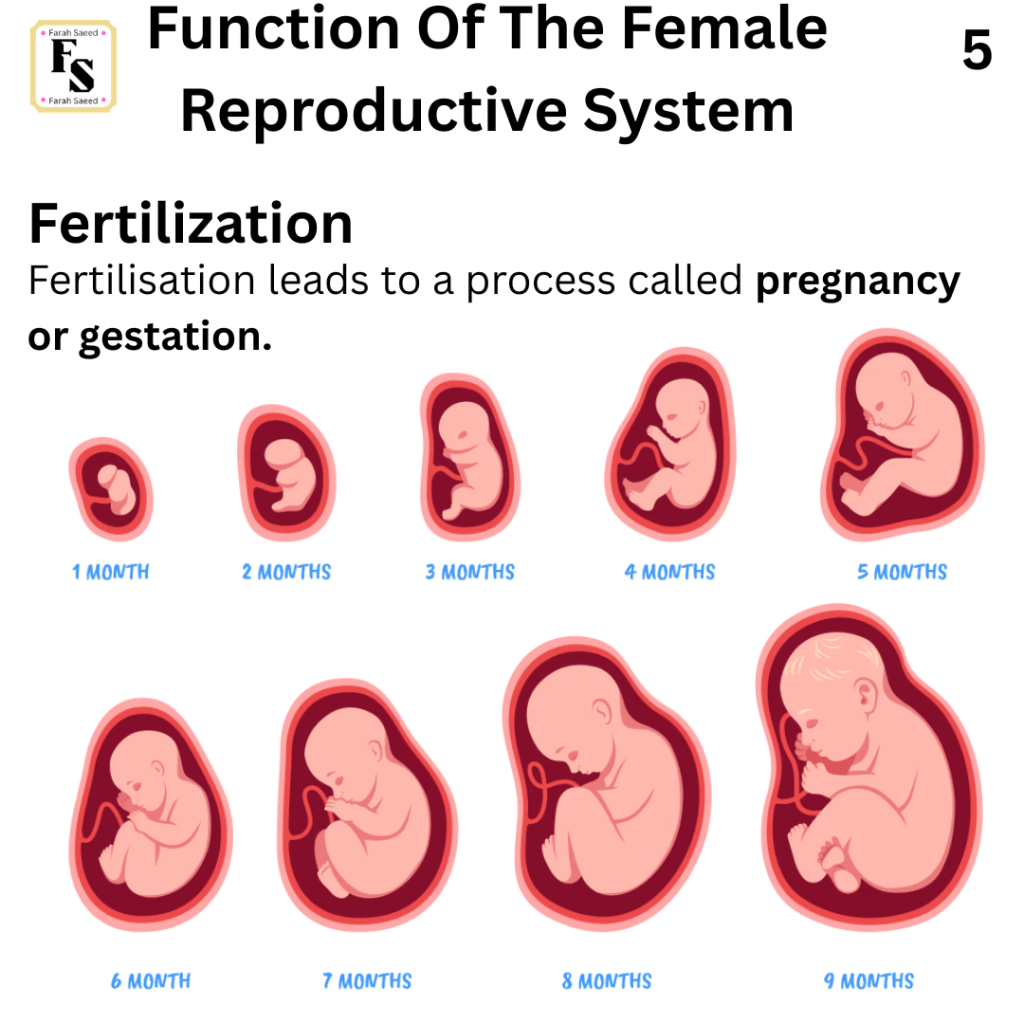
Supporting Images And Video Resources For Function Three
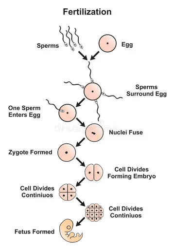
How Fertilization Happens By Dr. Paulien Moyaert
Fertilization explained – sperm and egg fusion by Henrik’s Lab
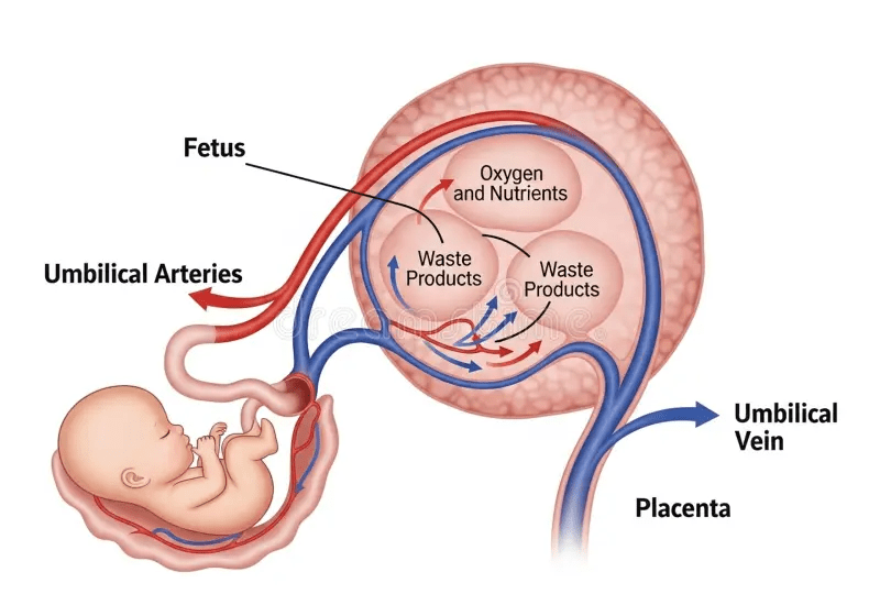
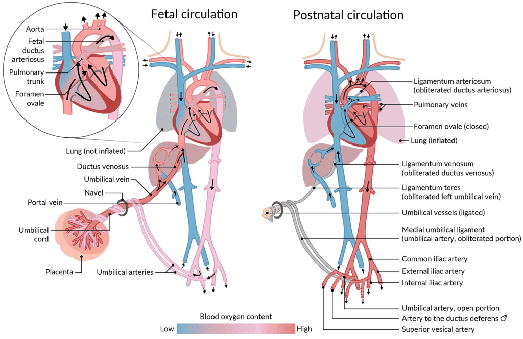
Function Four: Pregnancy
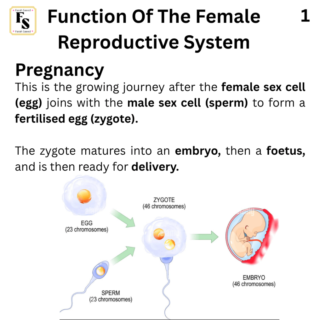
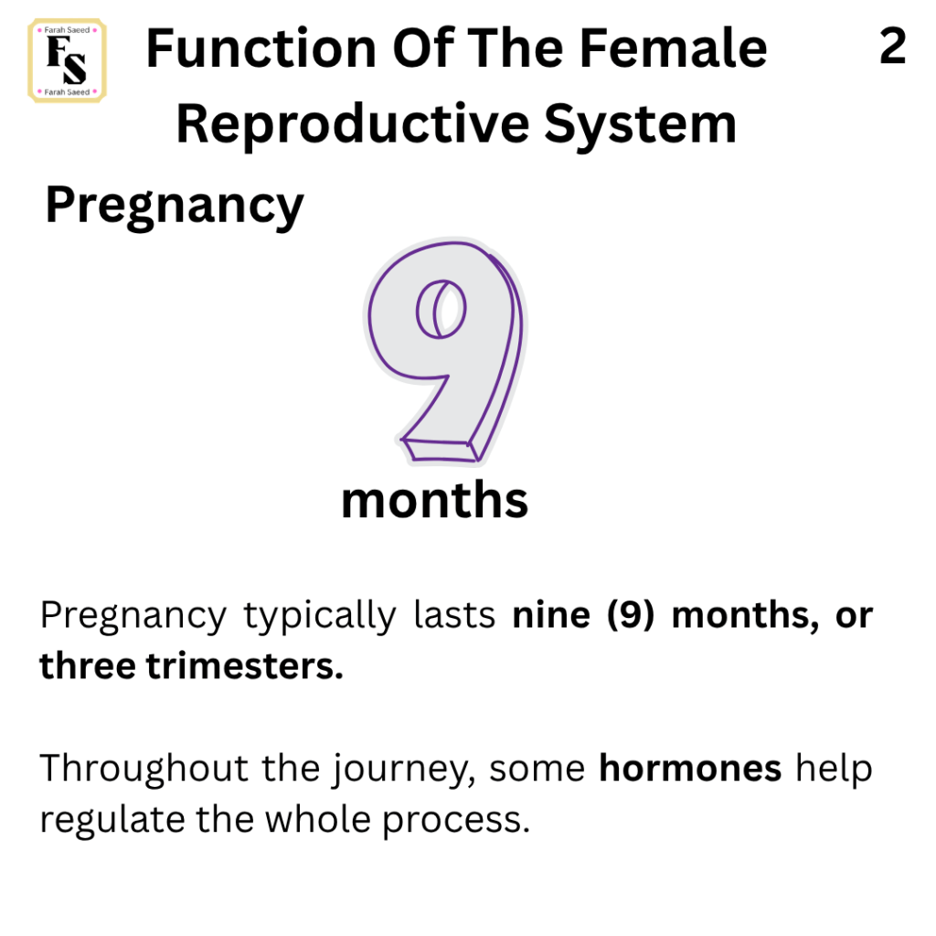
Supporting Images and Video Resources For Function Four.
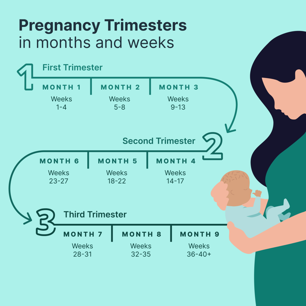
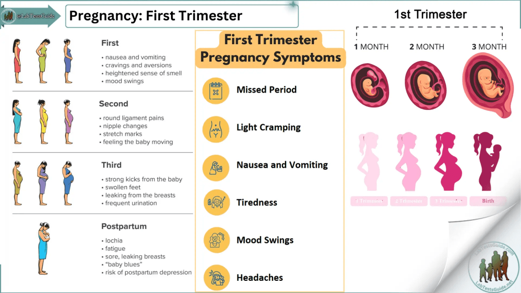
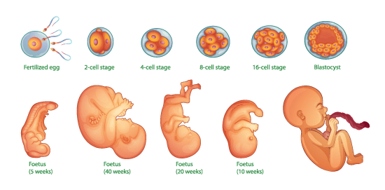
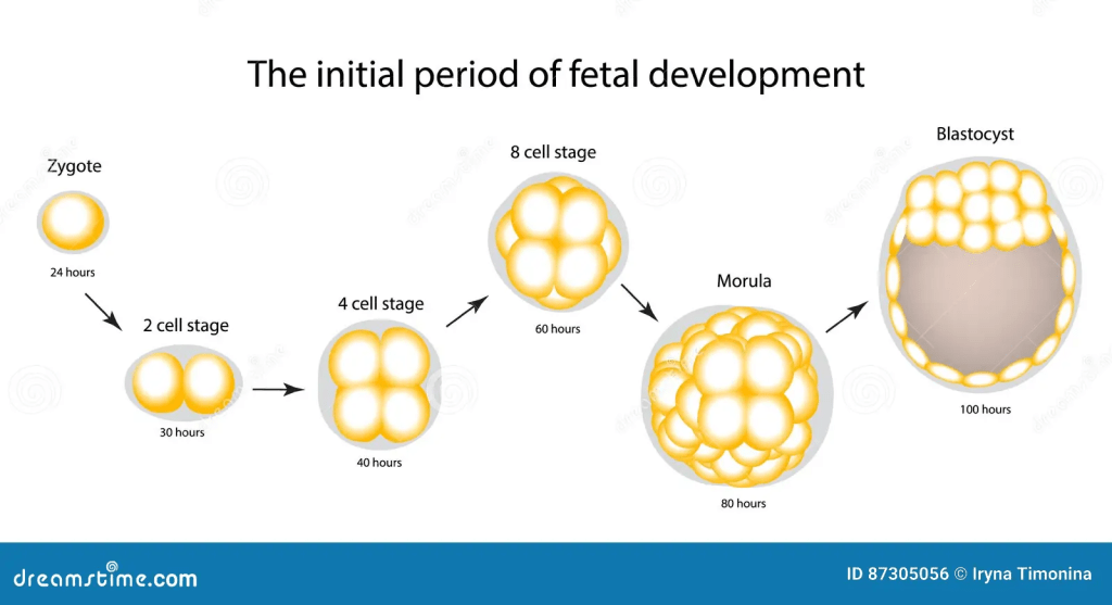
The development journey for the baby by Bupa Health
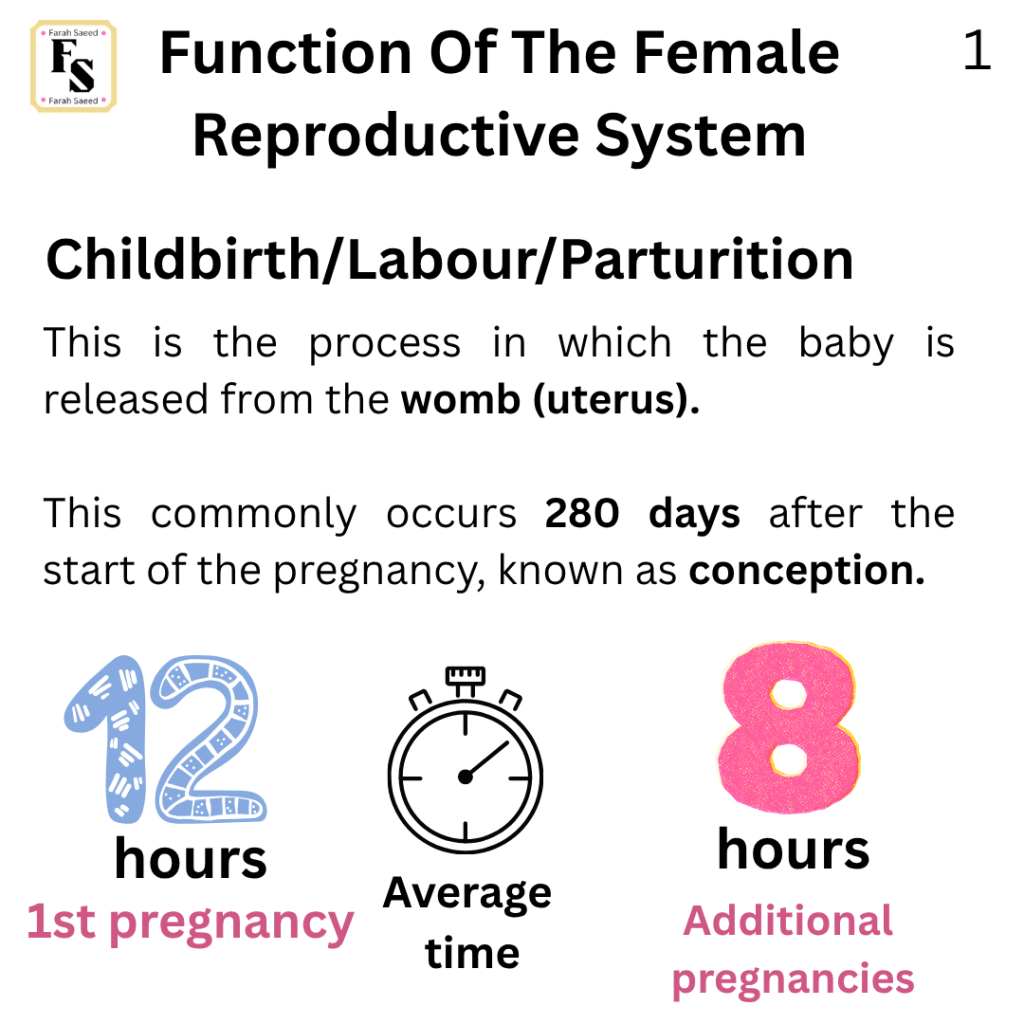
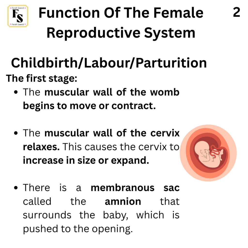
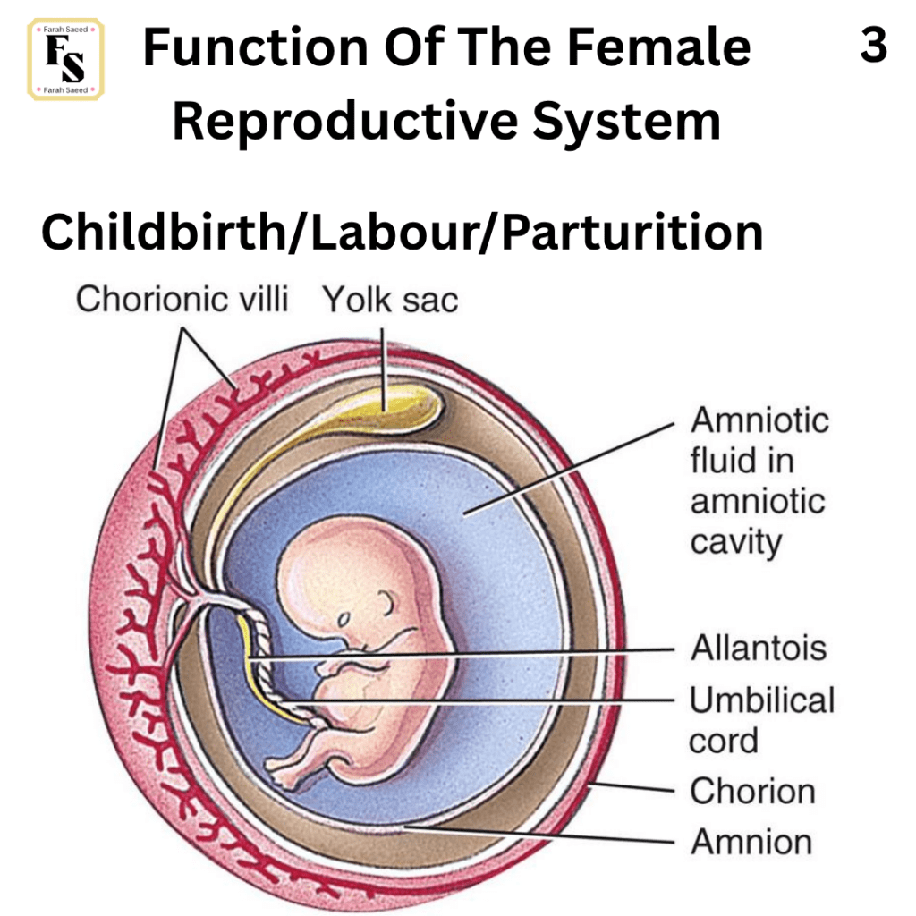
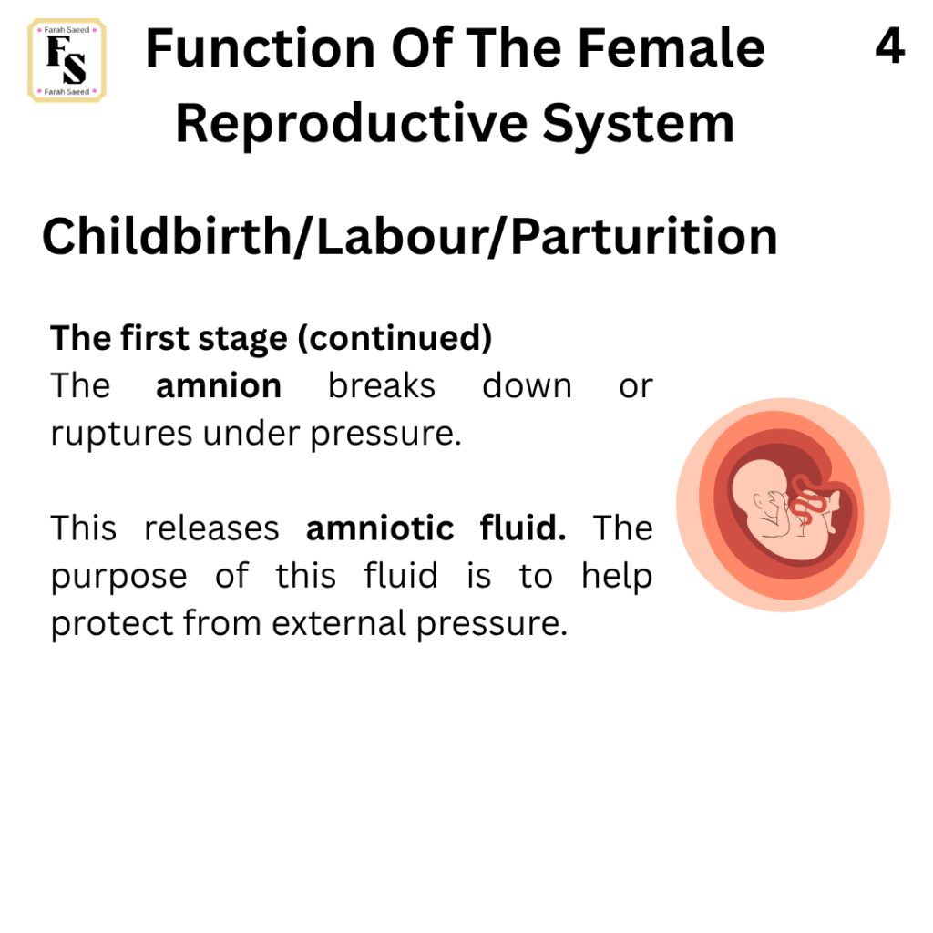
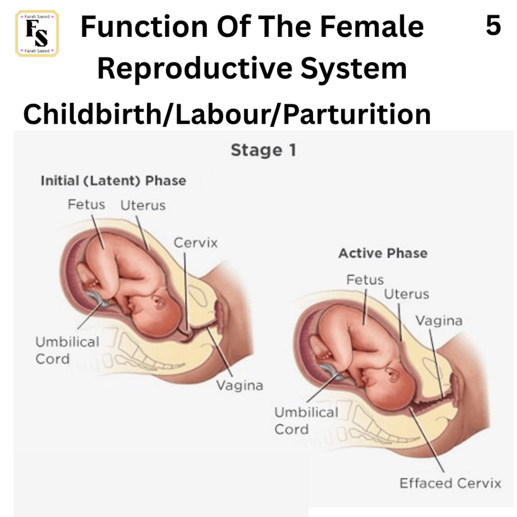
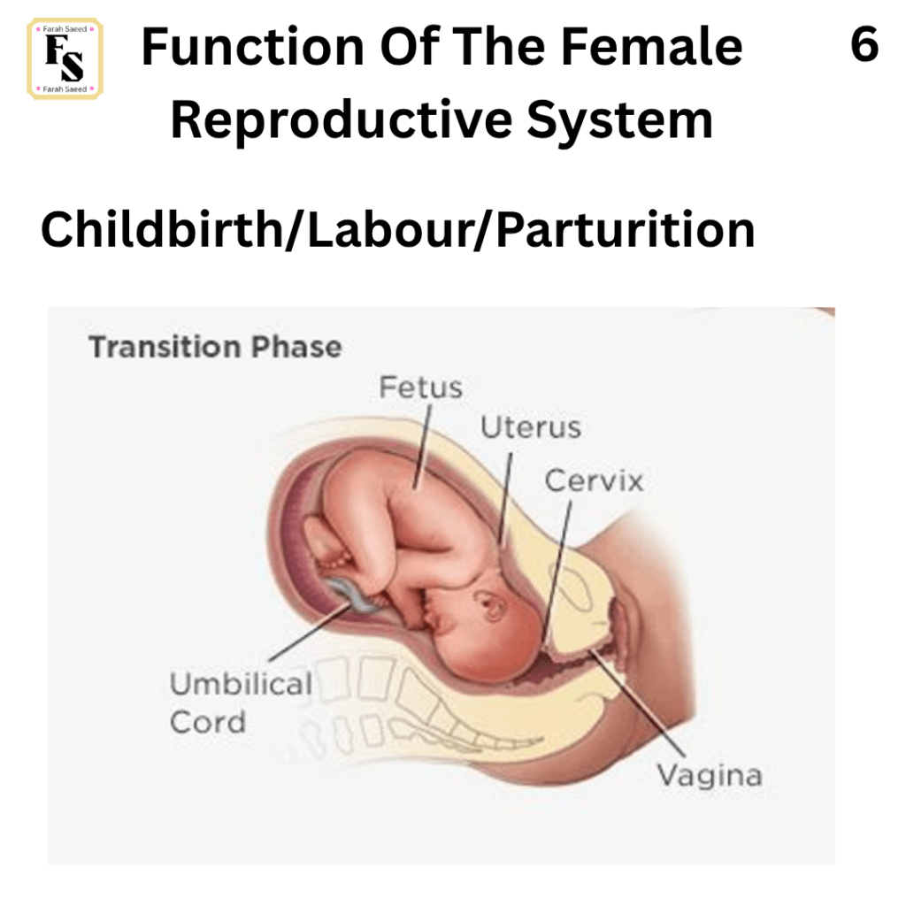
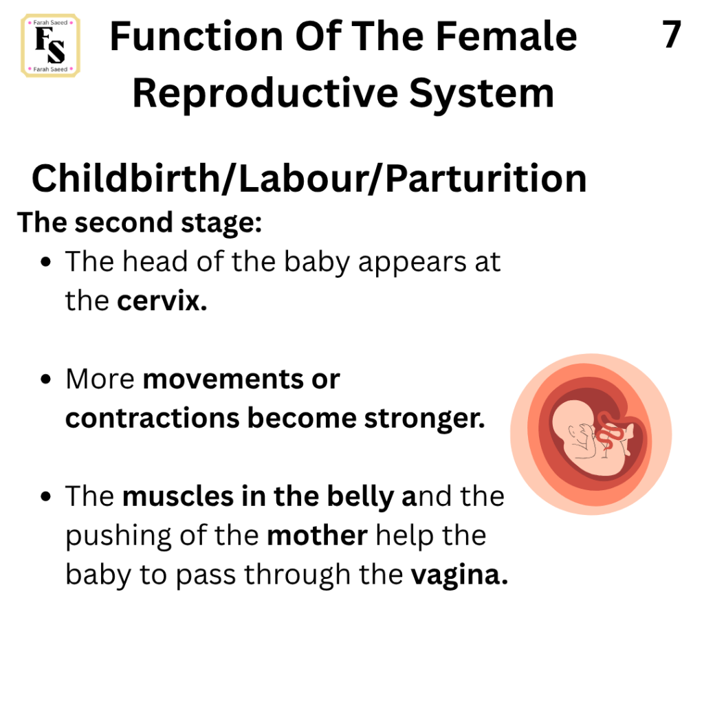
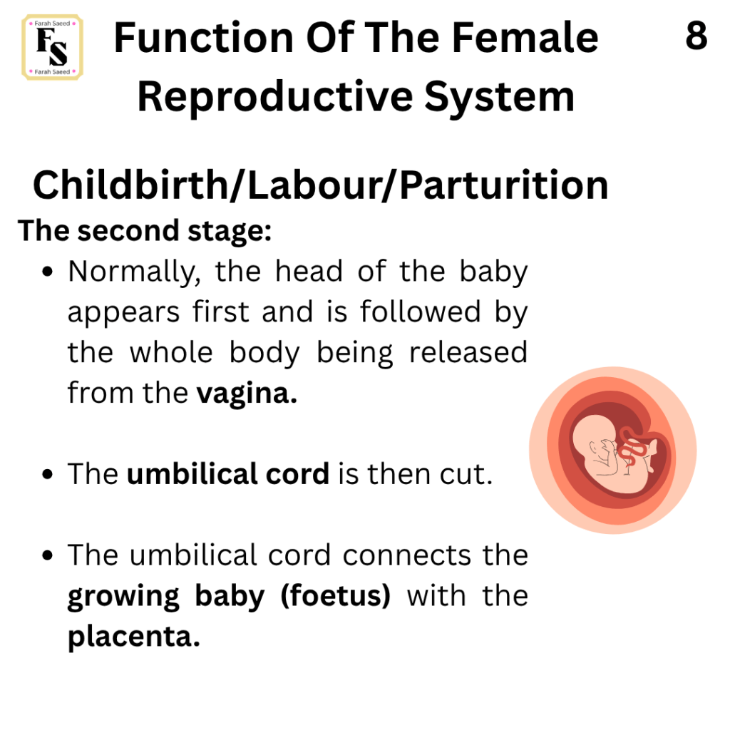
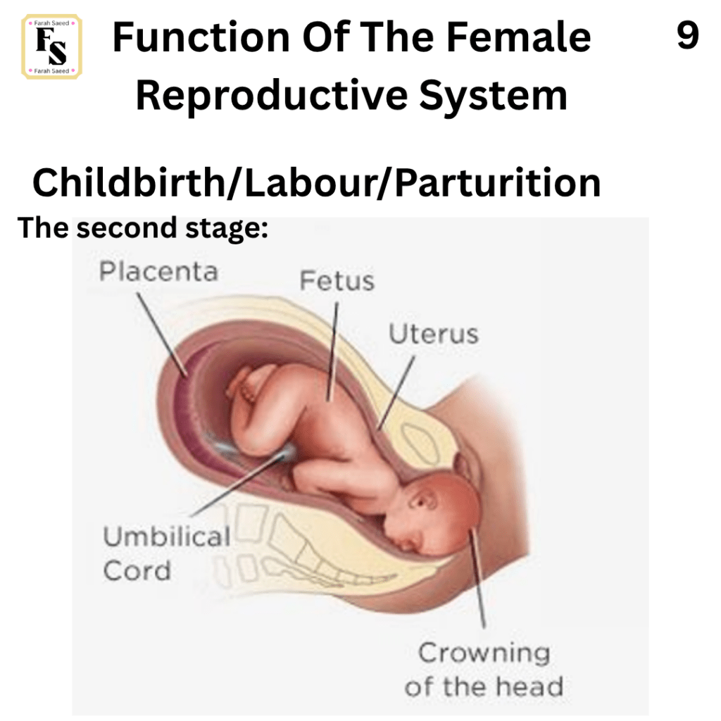
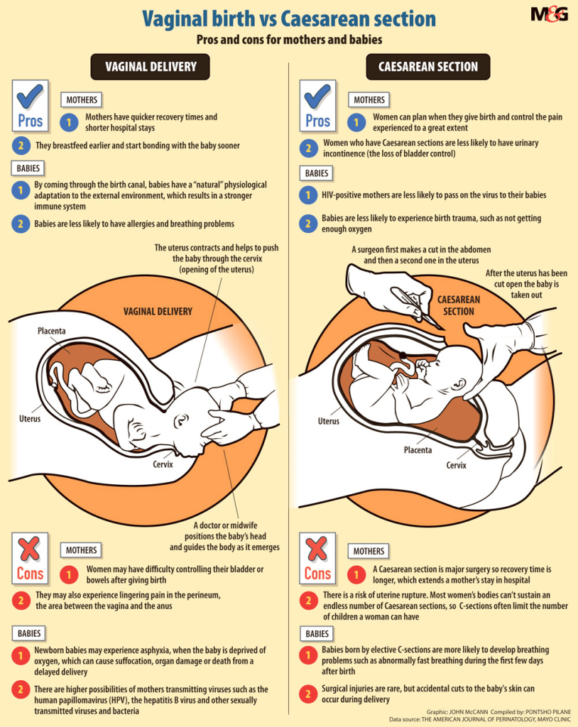
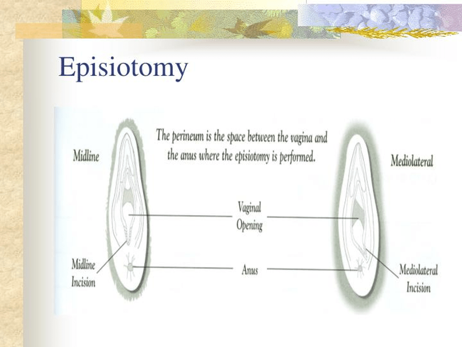
Function Five: Childbirth/Labour/Parturition
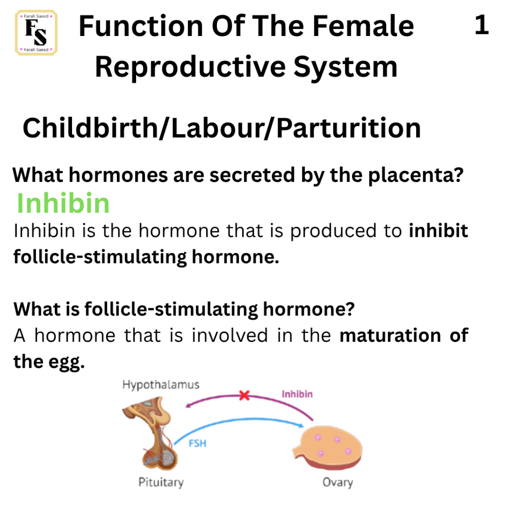
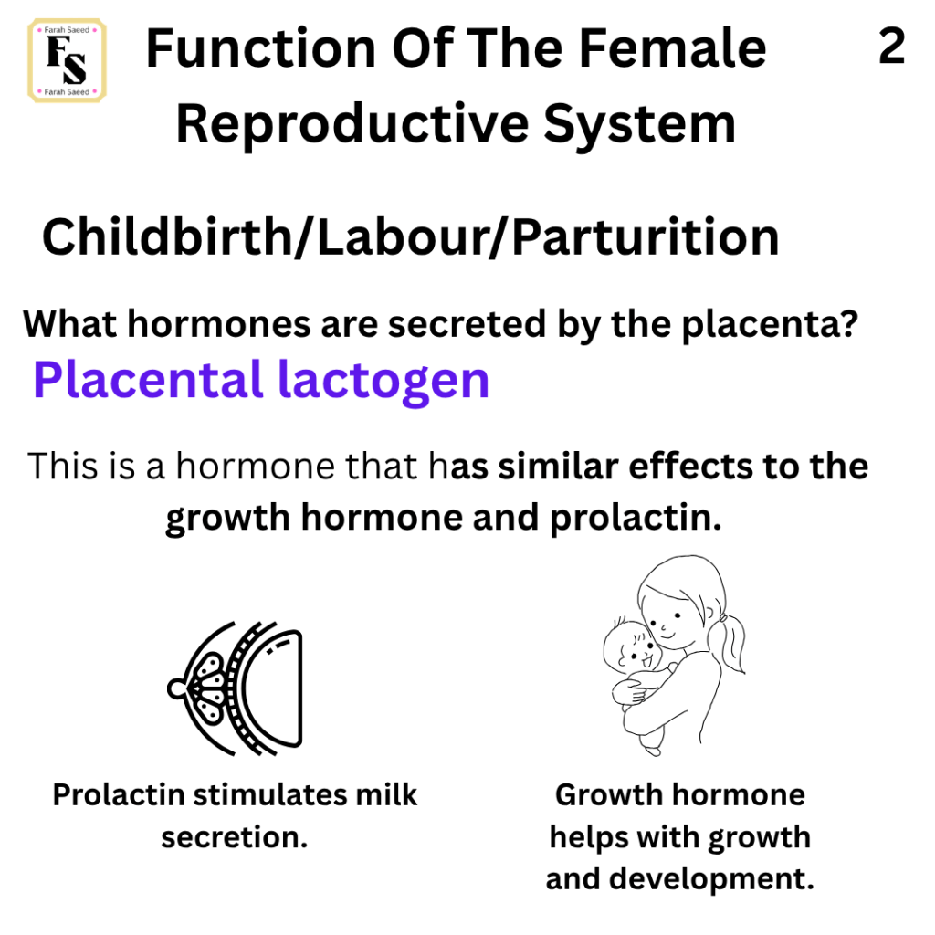
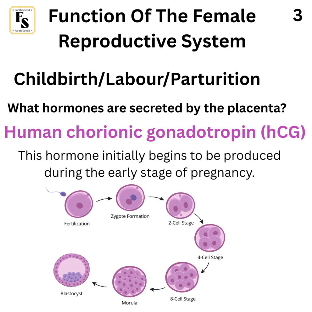
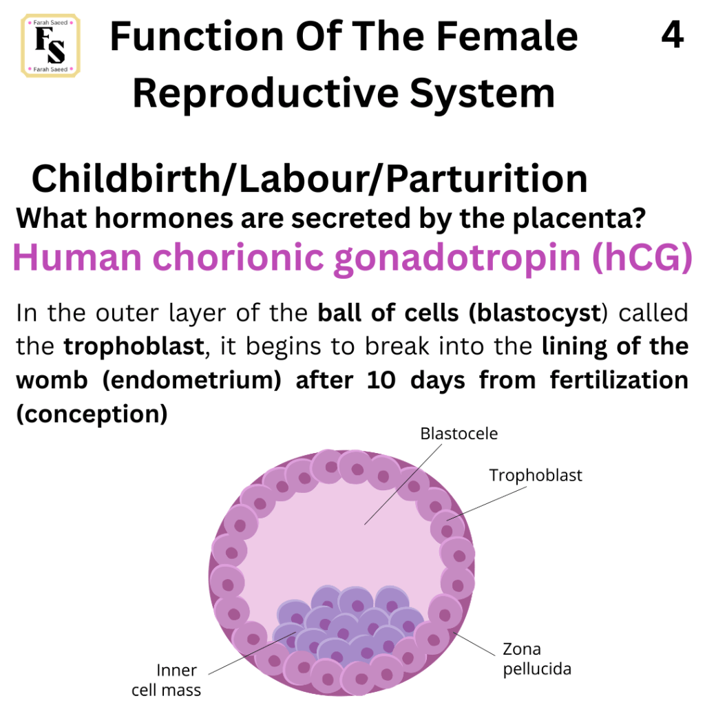
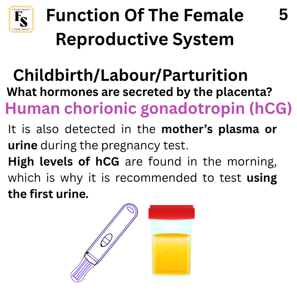
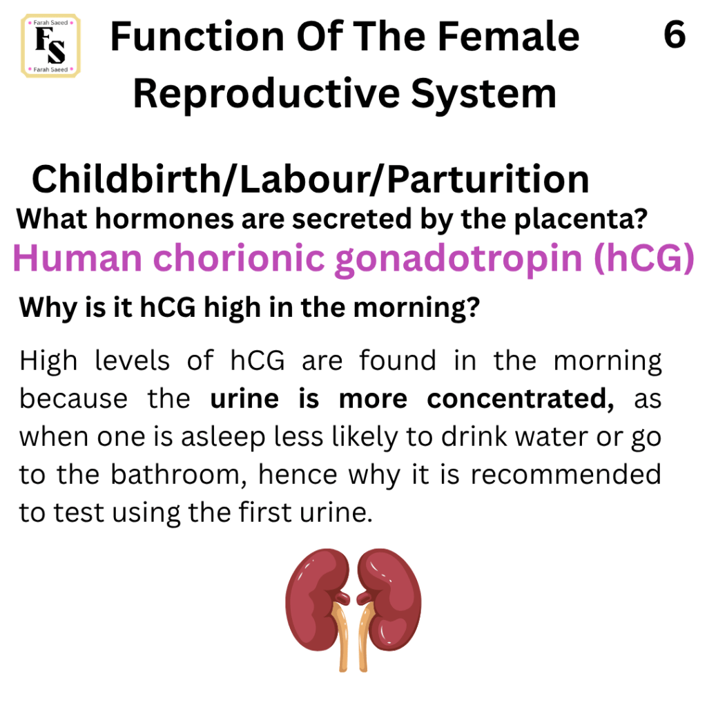
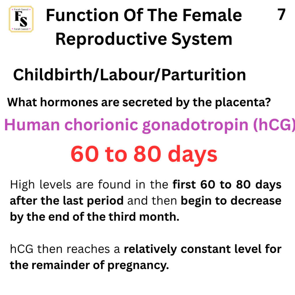
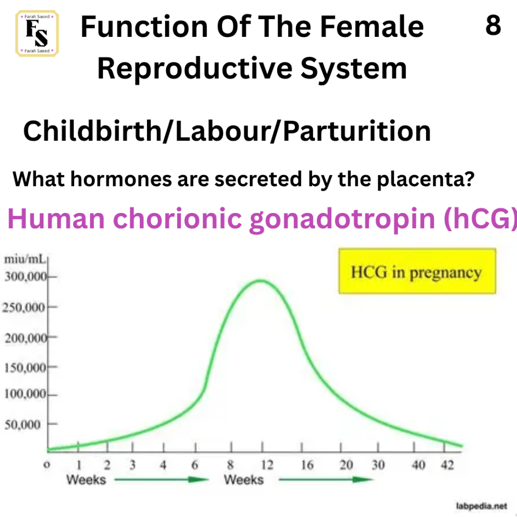
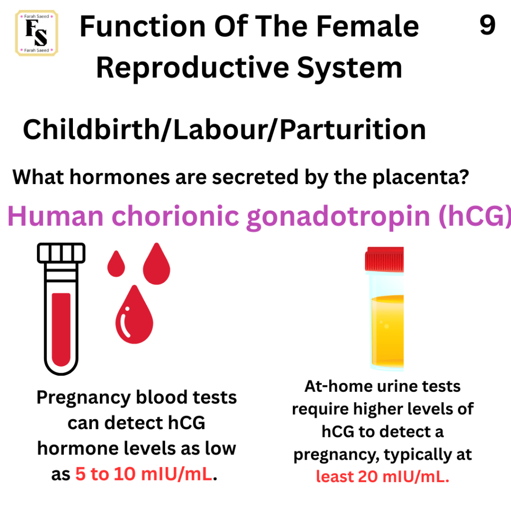
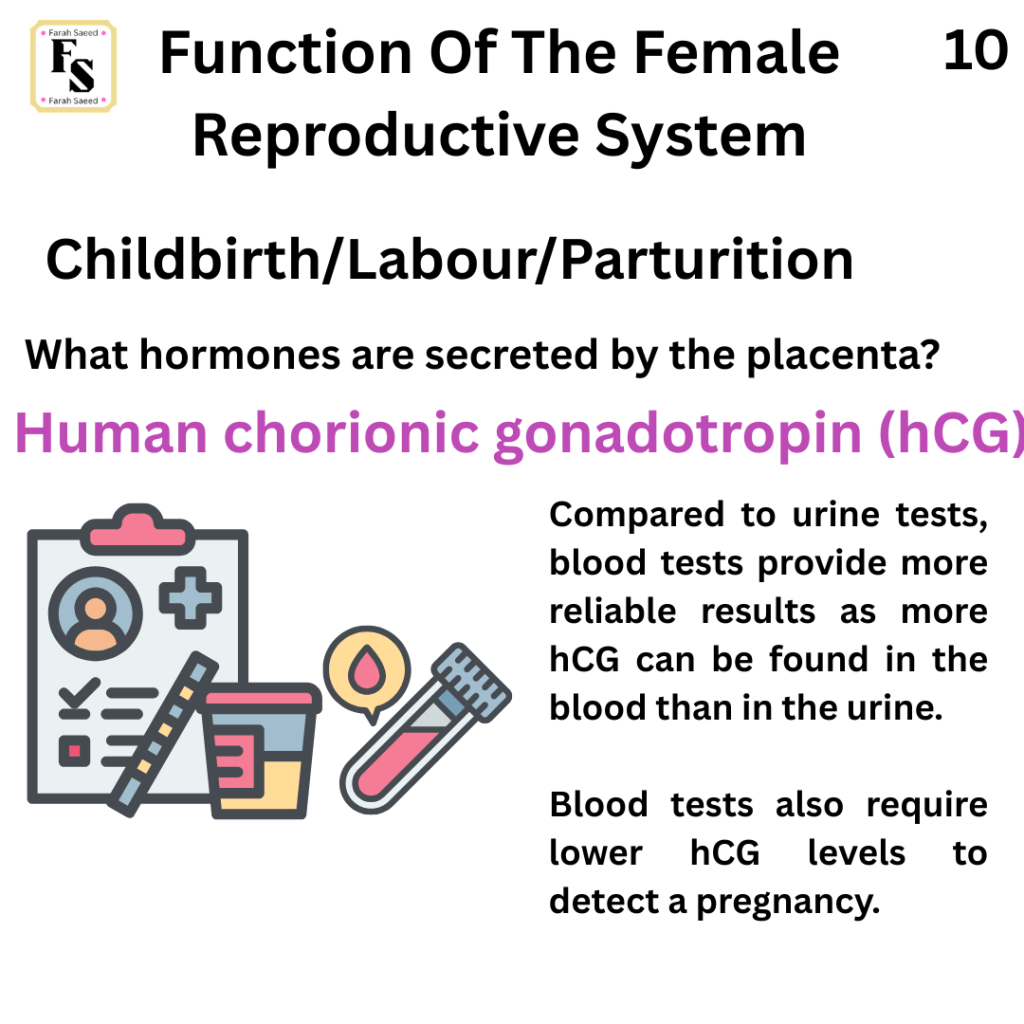
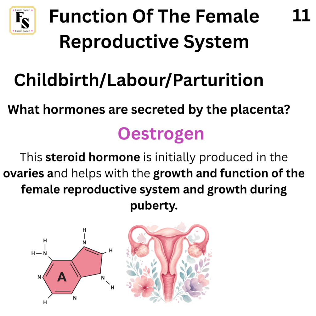
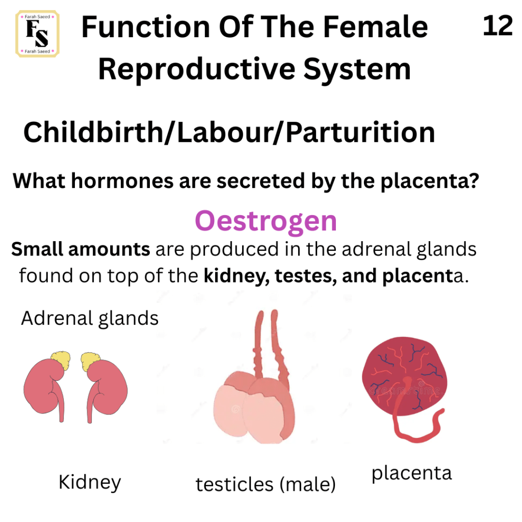
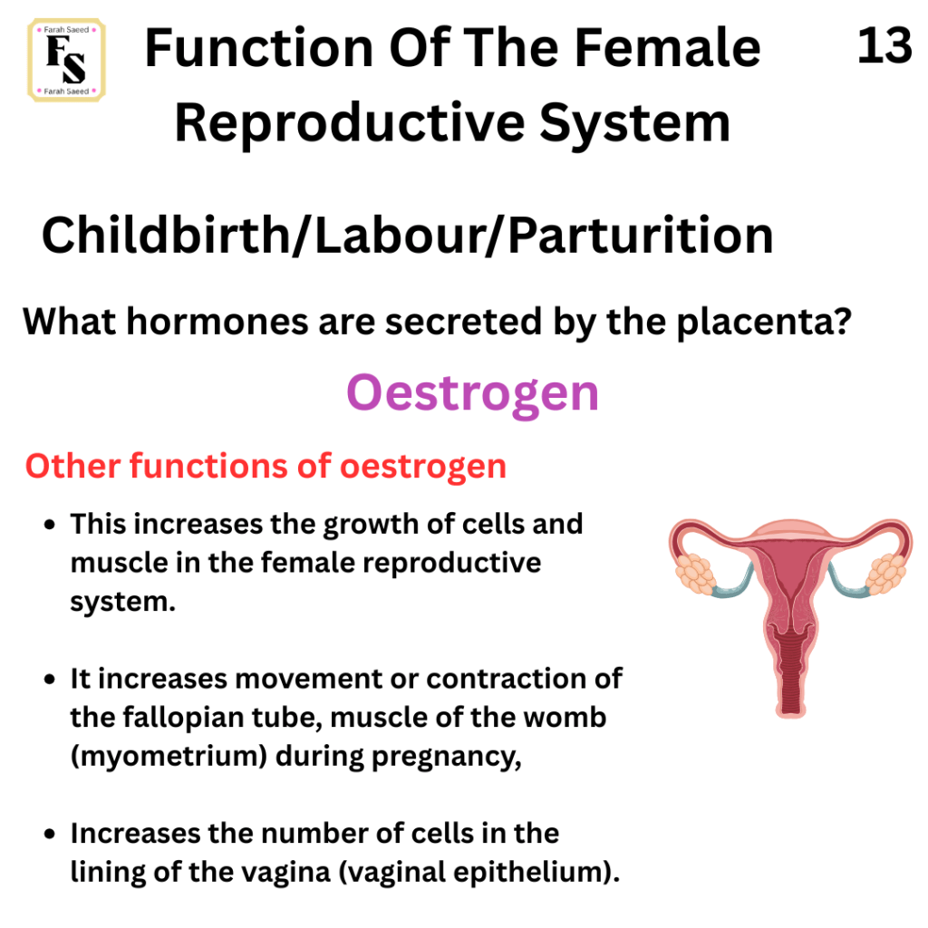
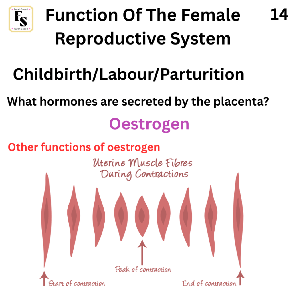
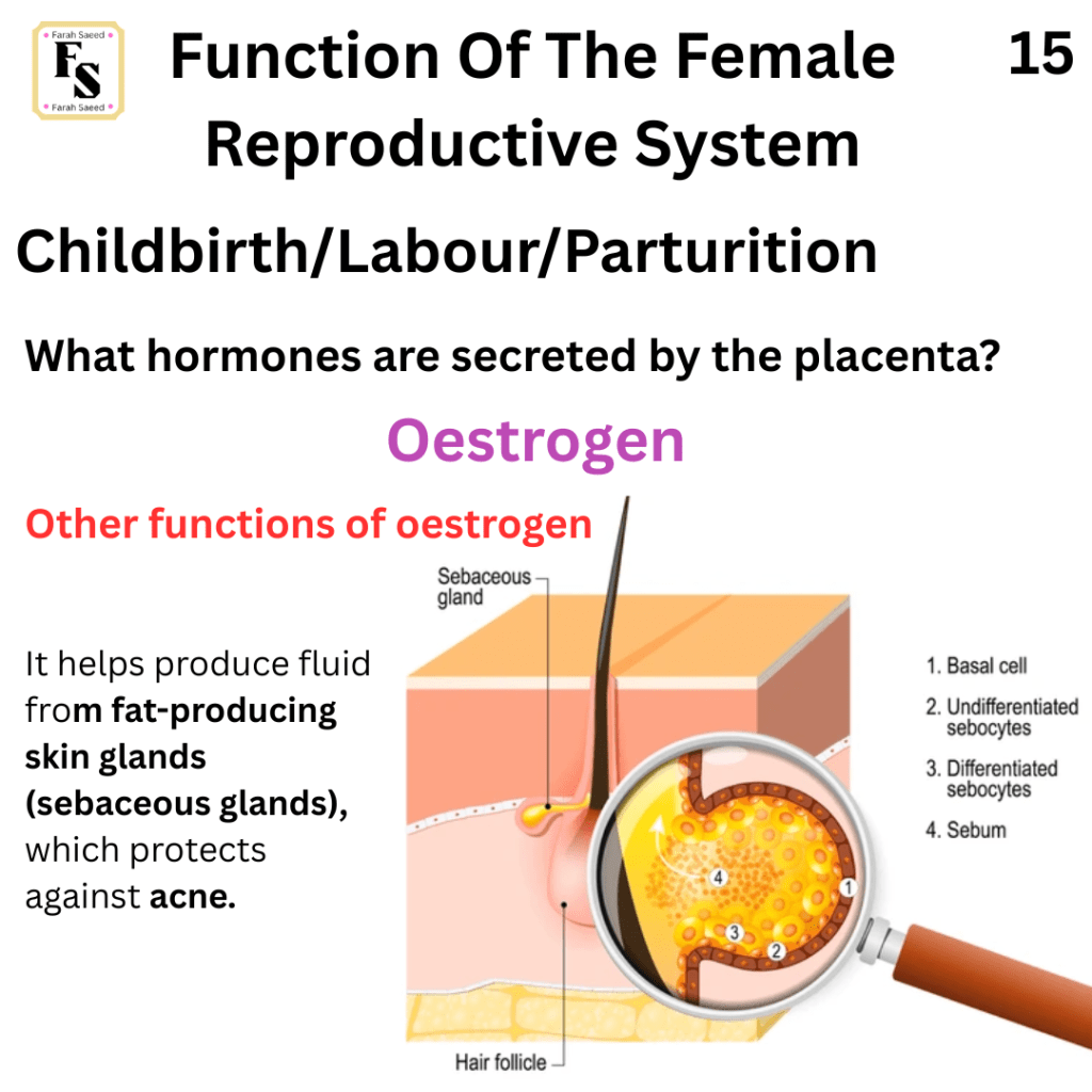
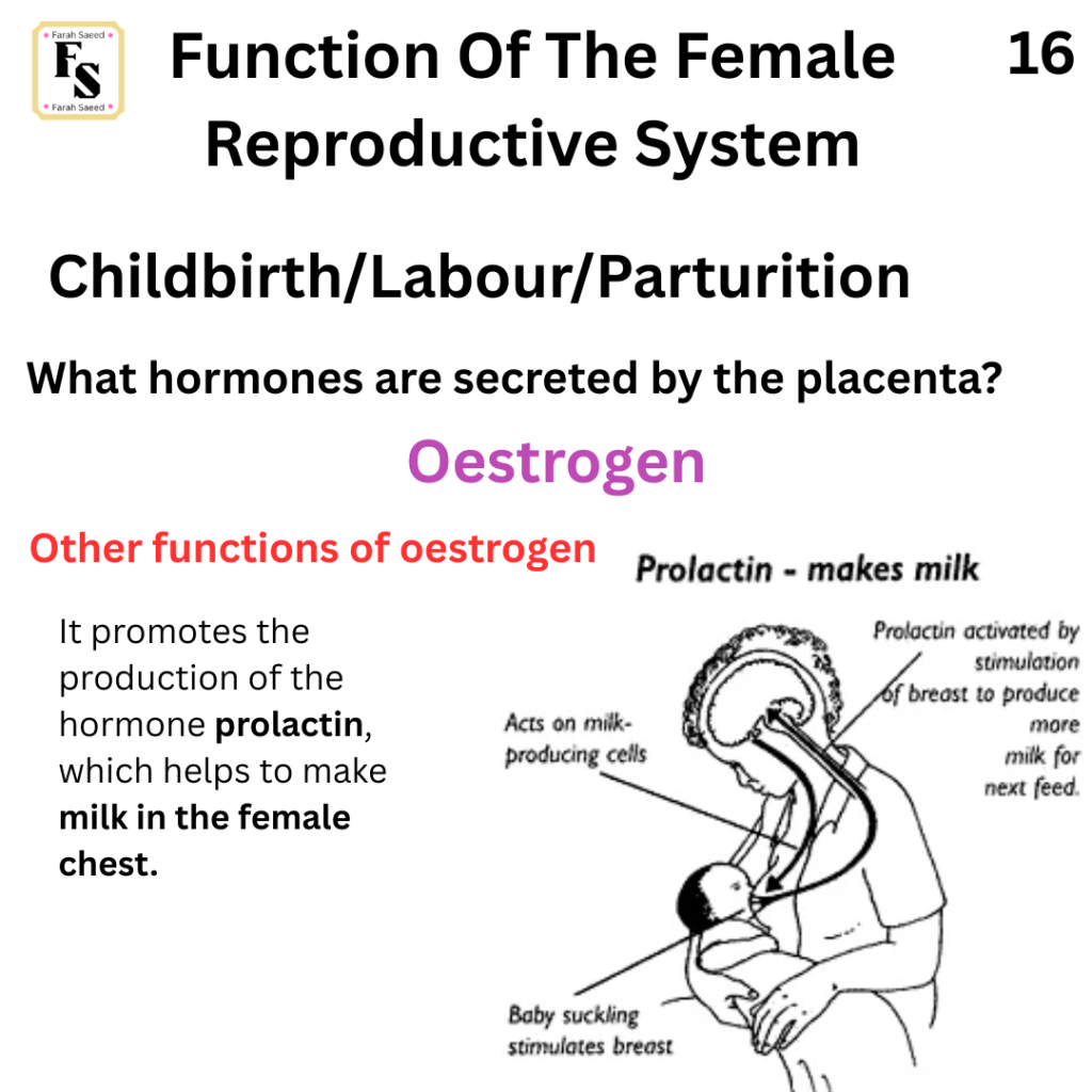
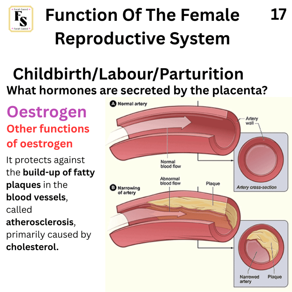
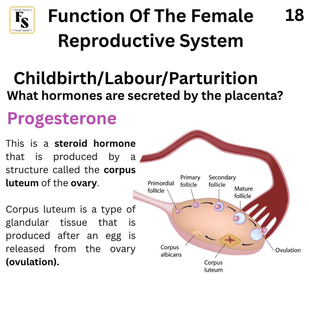
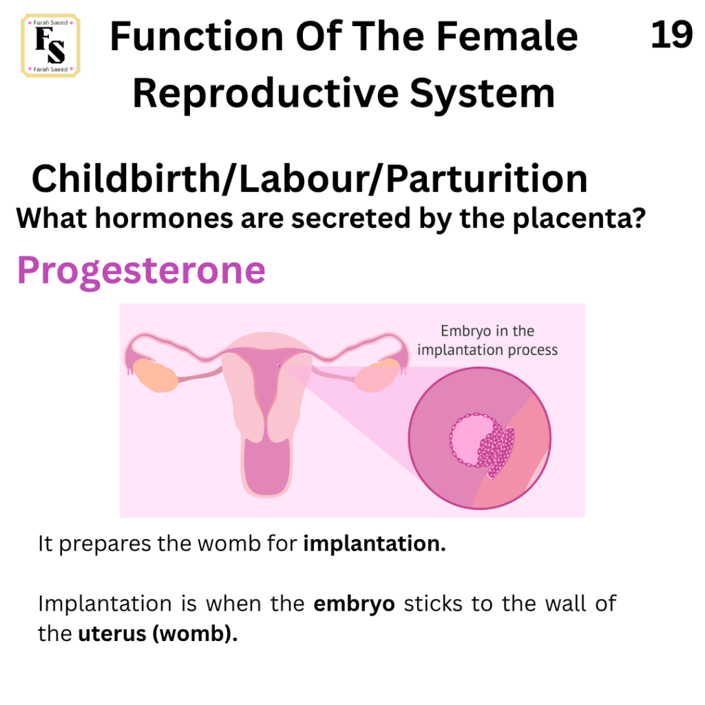
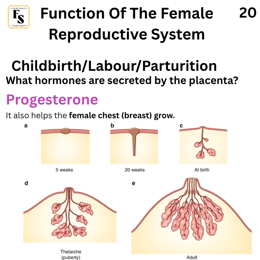
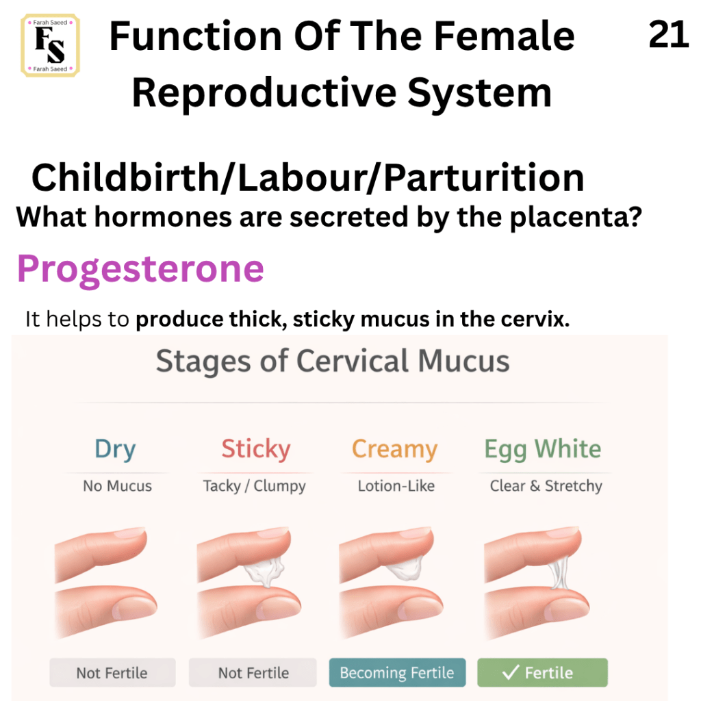
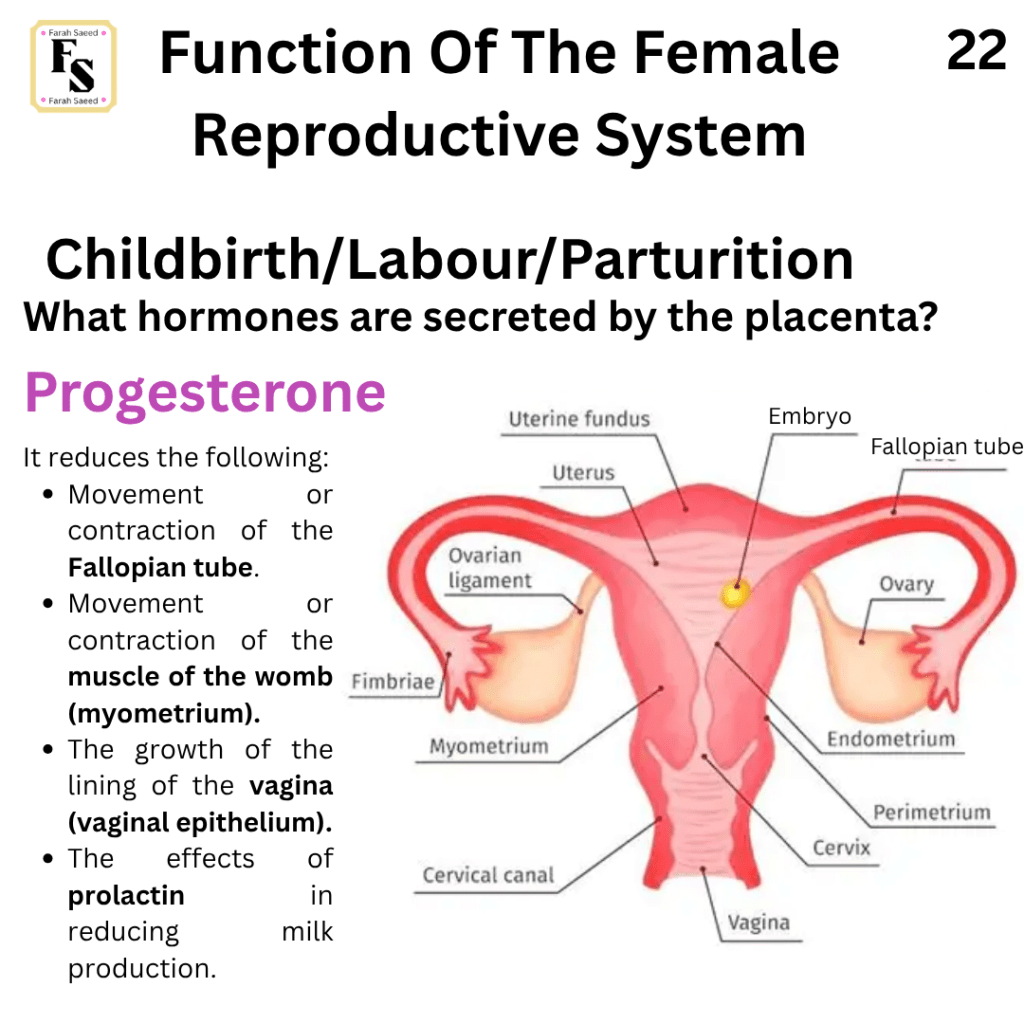
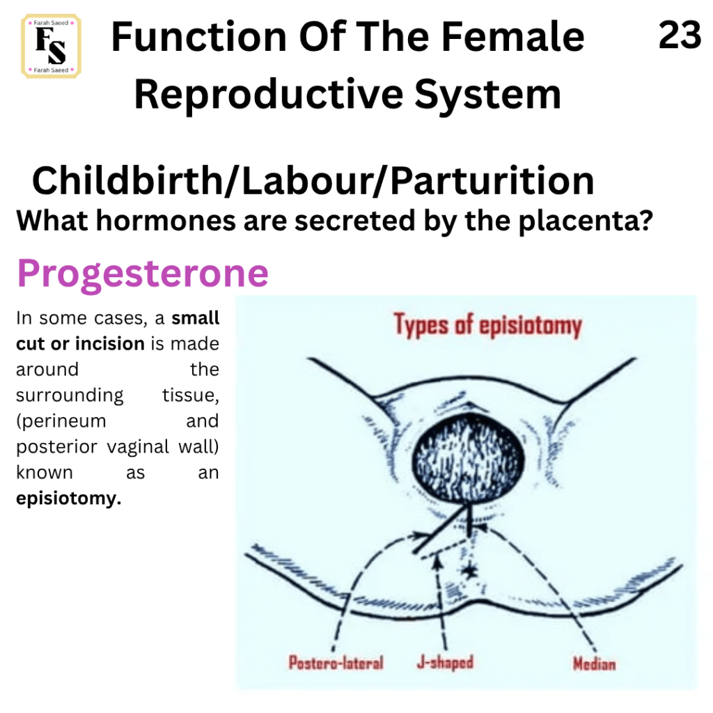
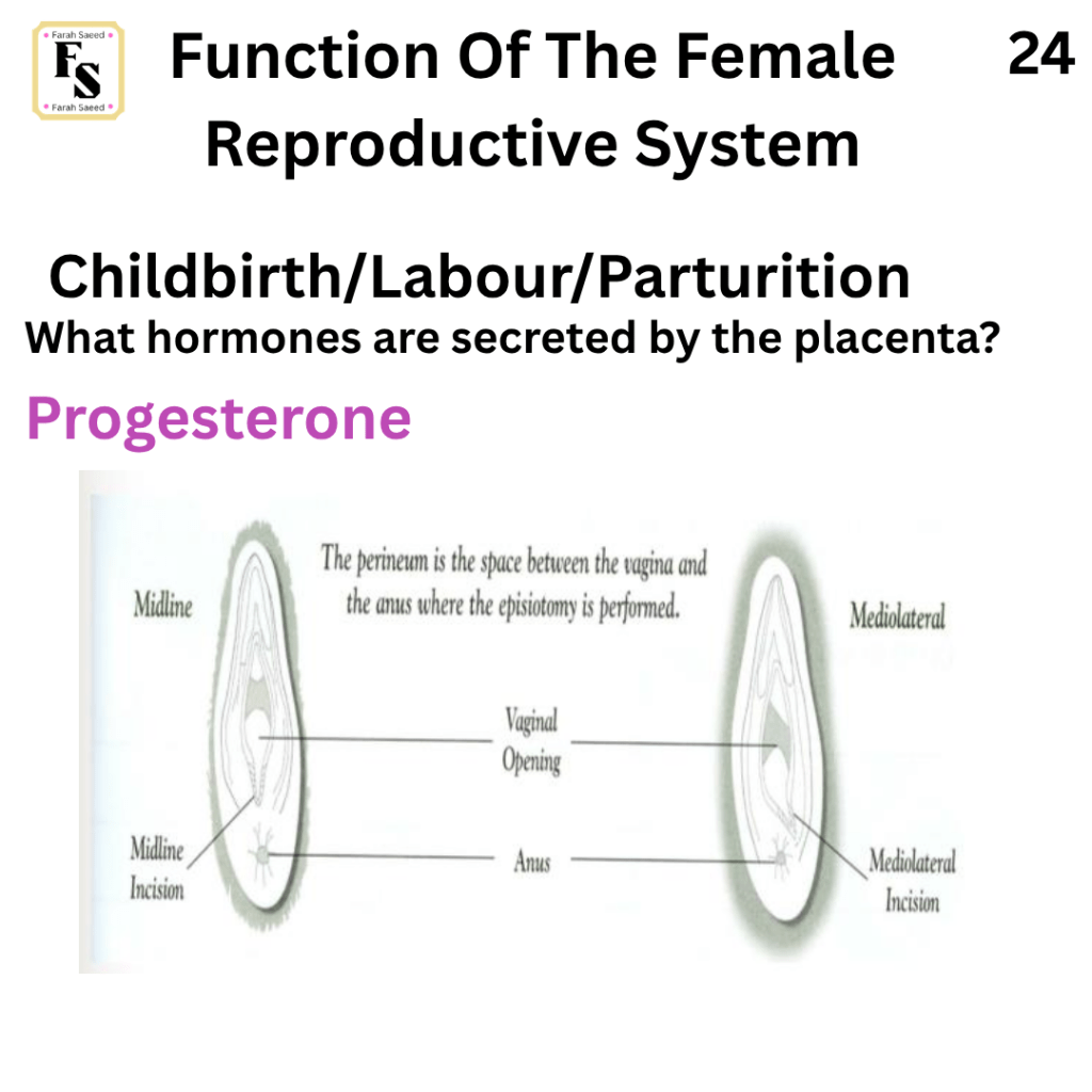
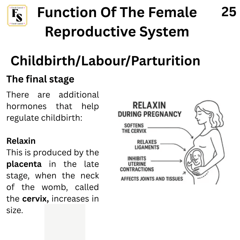
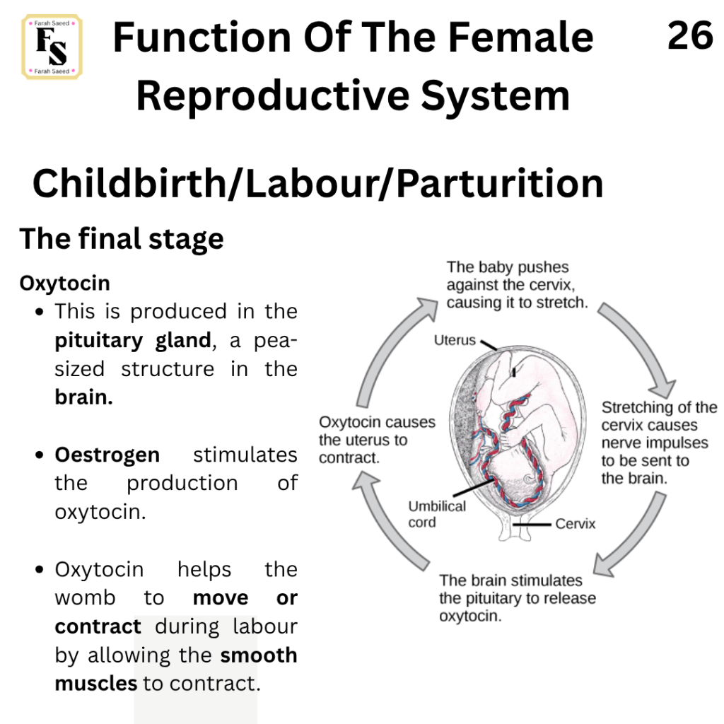
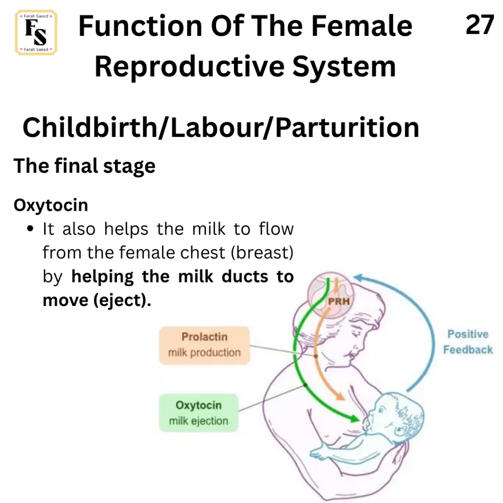
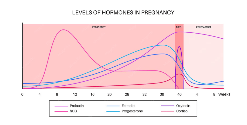
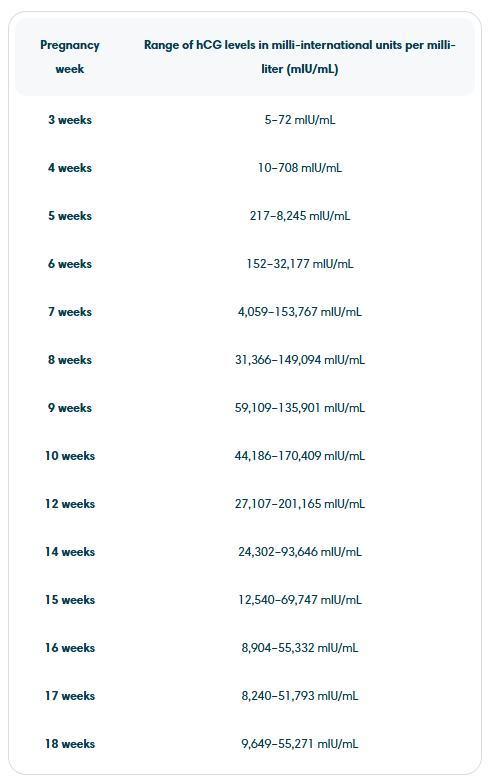
What Hormones Are Secreted By The Placenta?
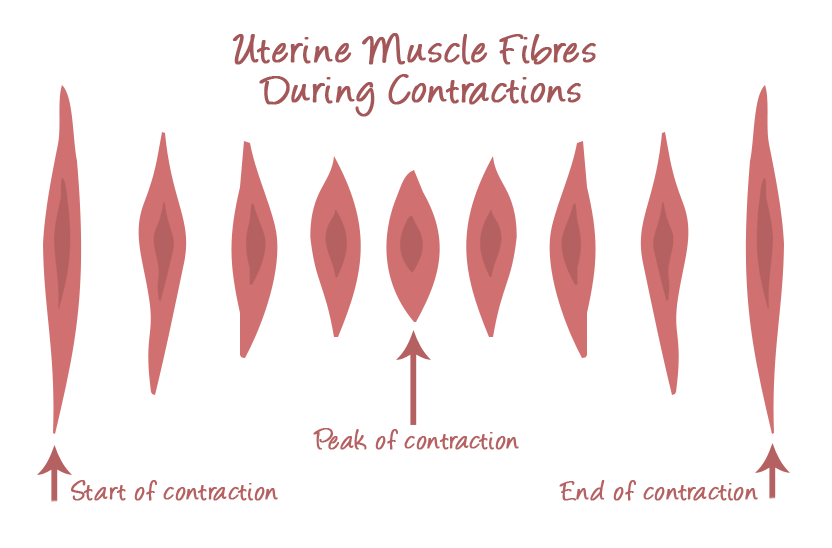
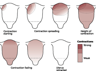
Video and Imaging Resources For Function Five

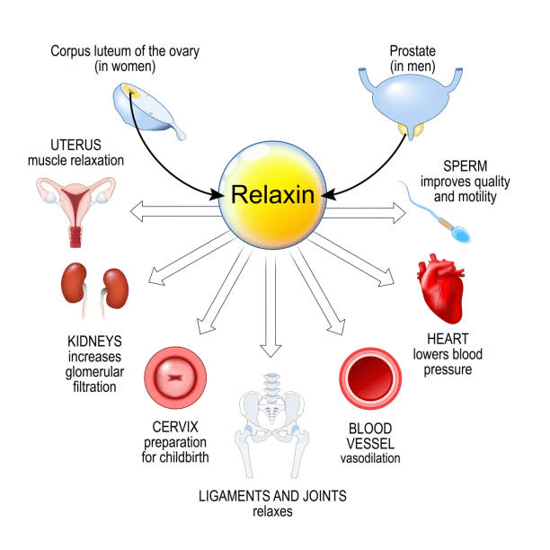
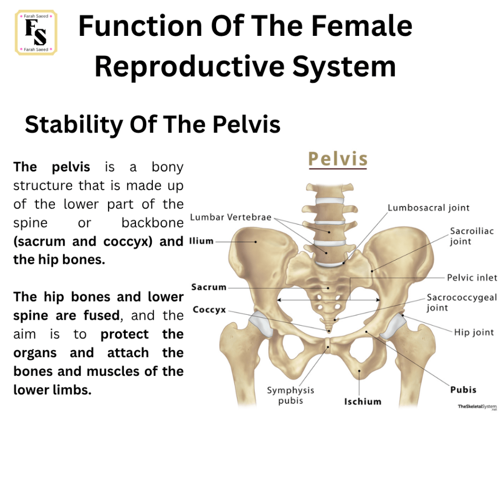
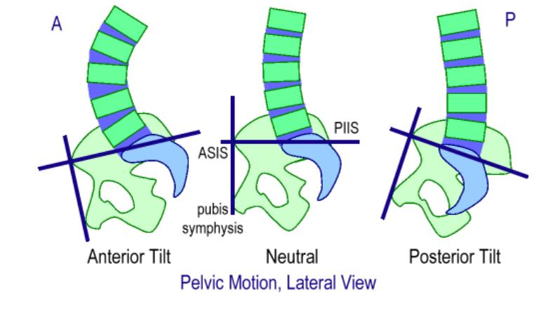
Function Six: Stability Of The Pelvis
Supporting Images For Function Seven.
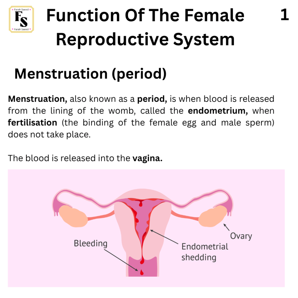
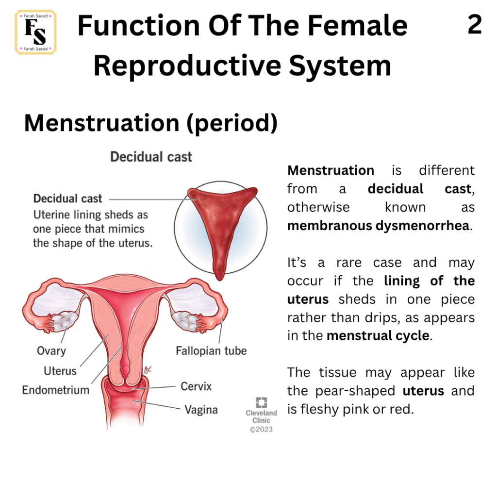
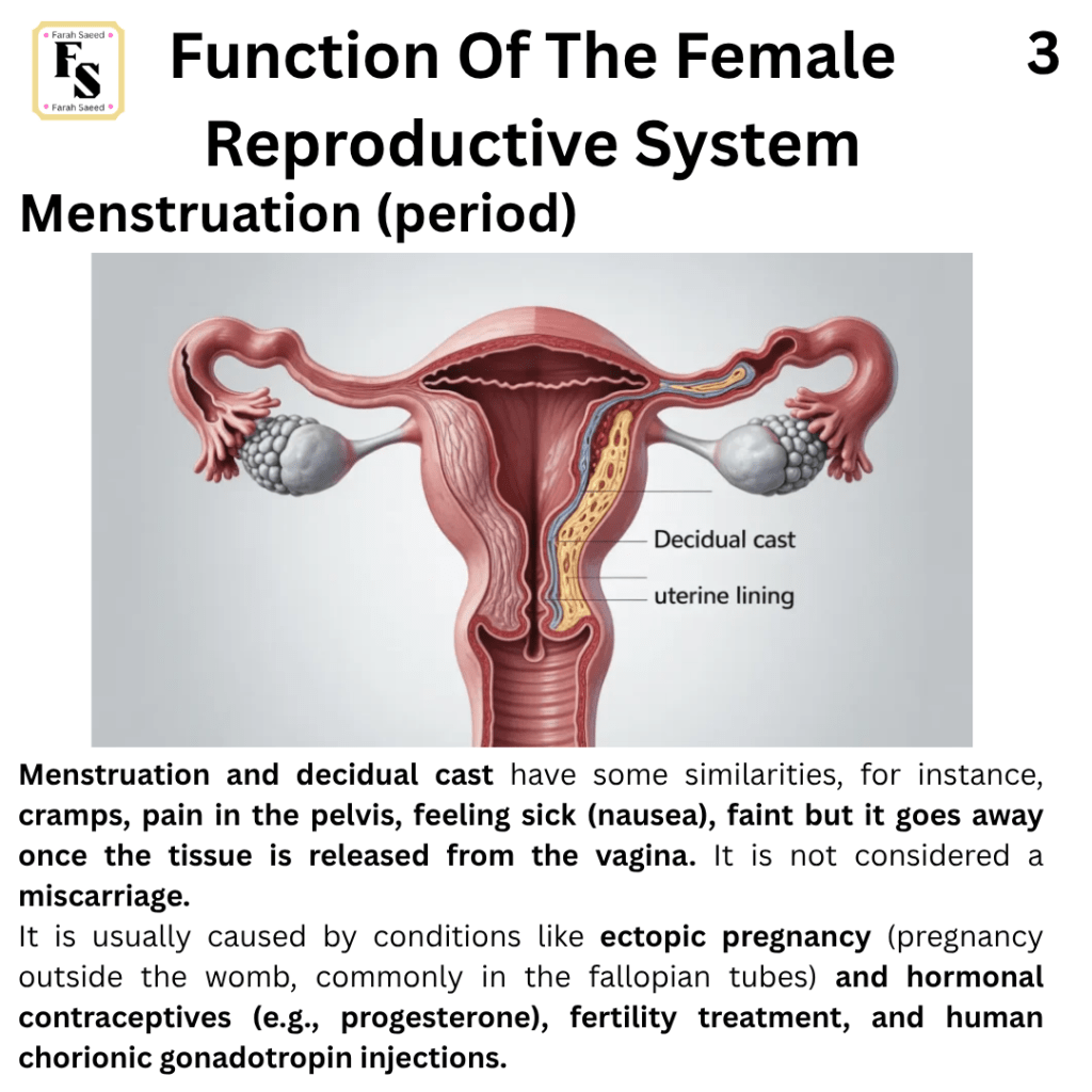
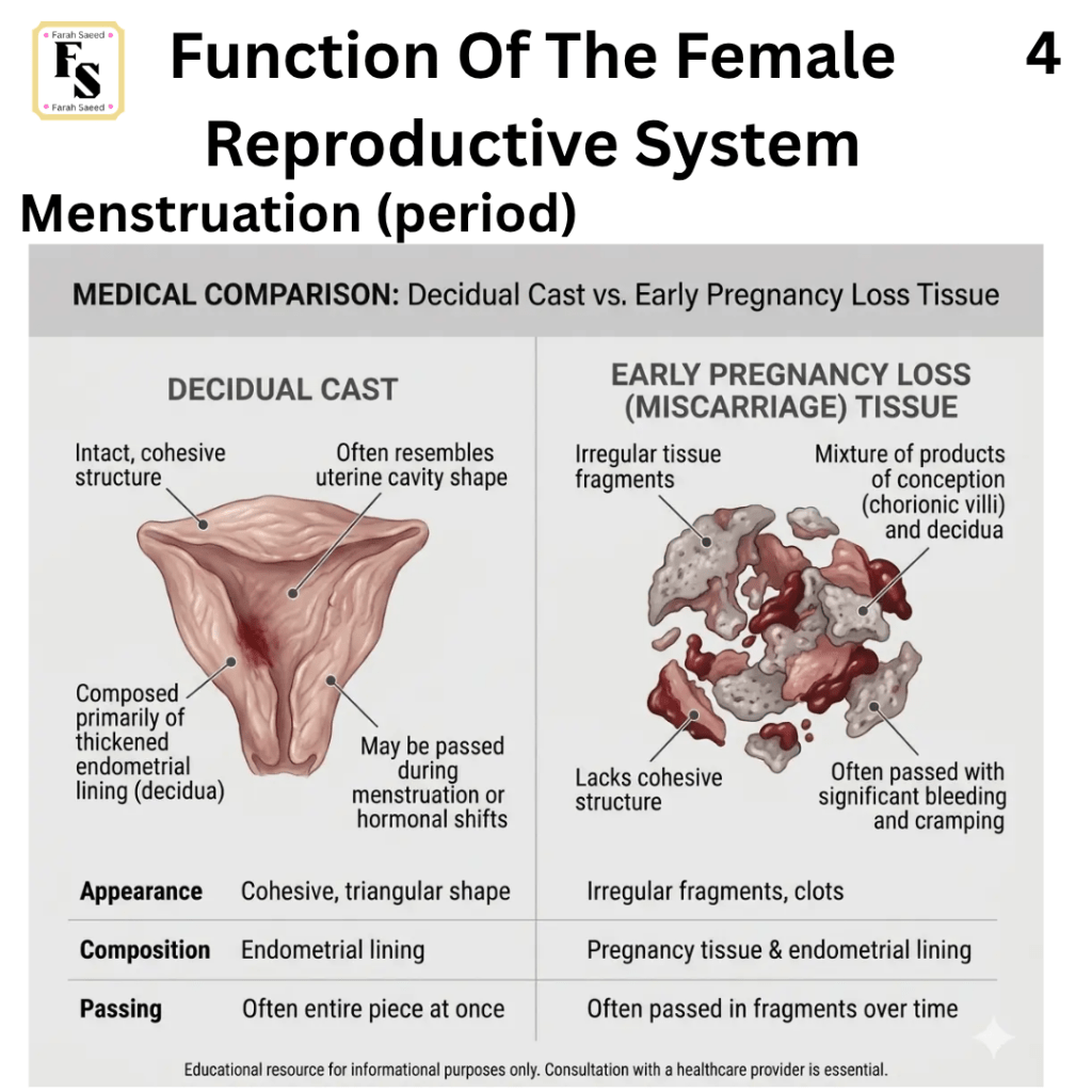
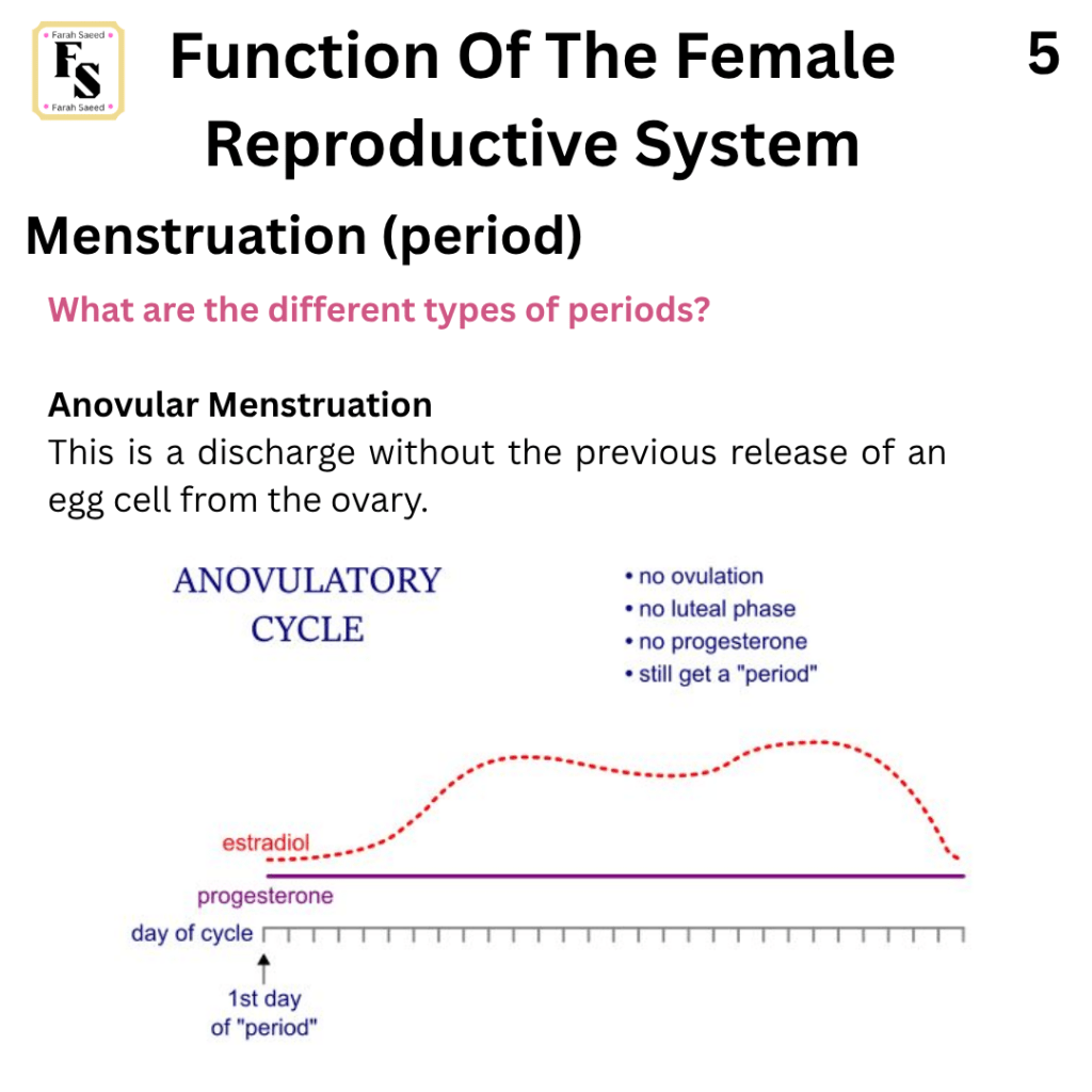
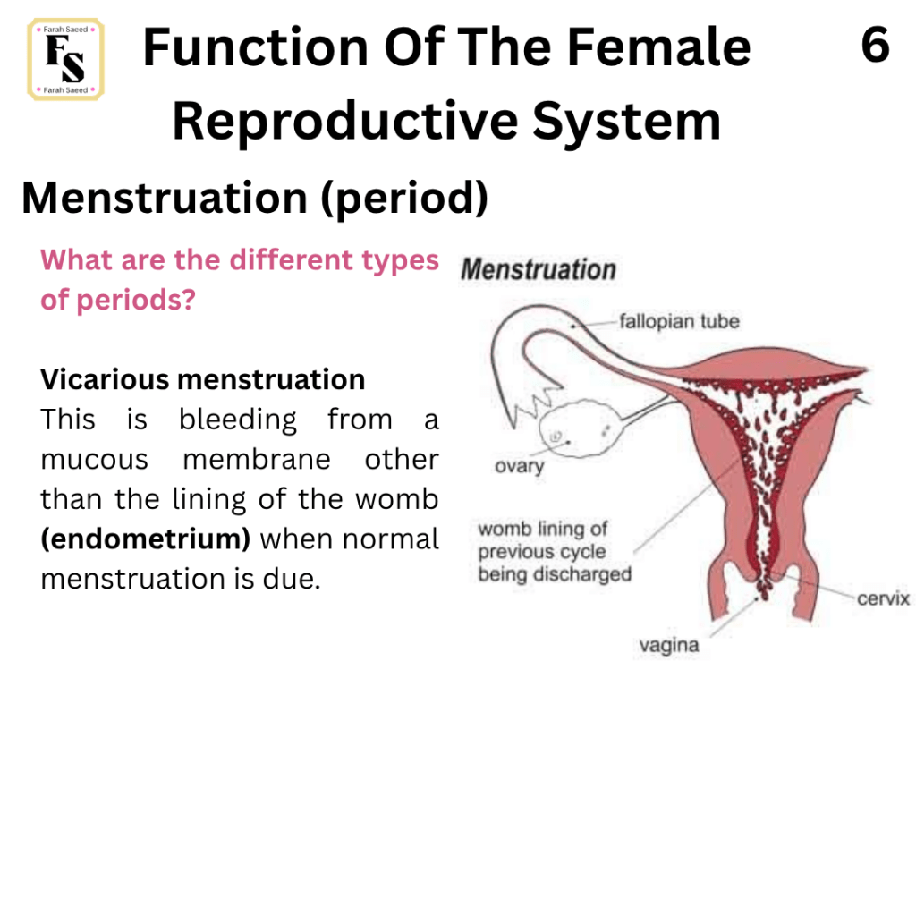
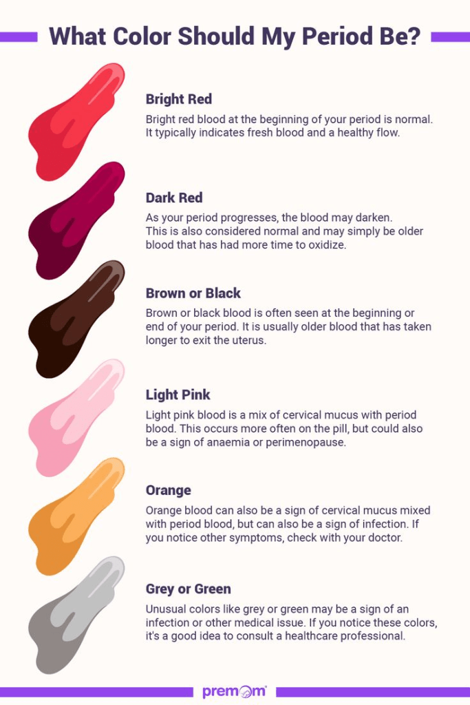
Function Seven Menstruation
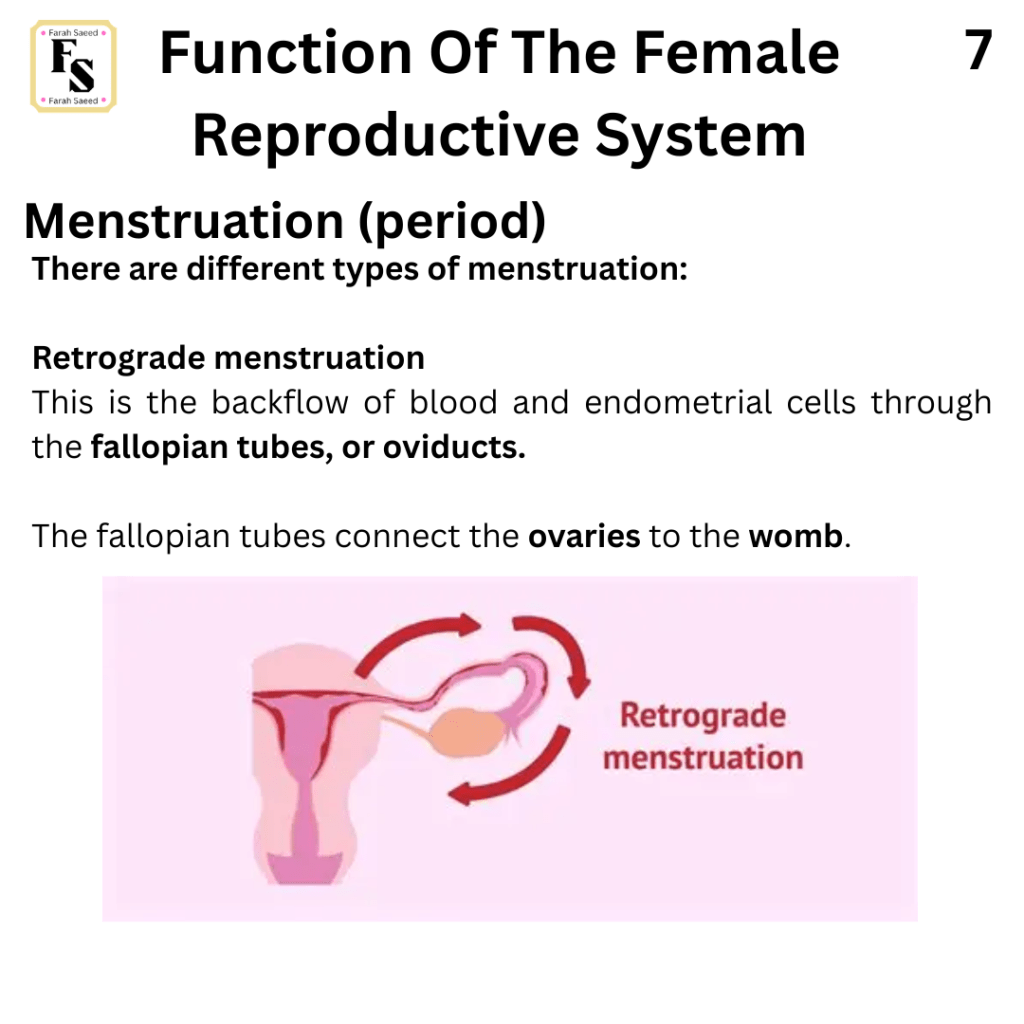
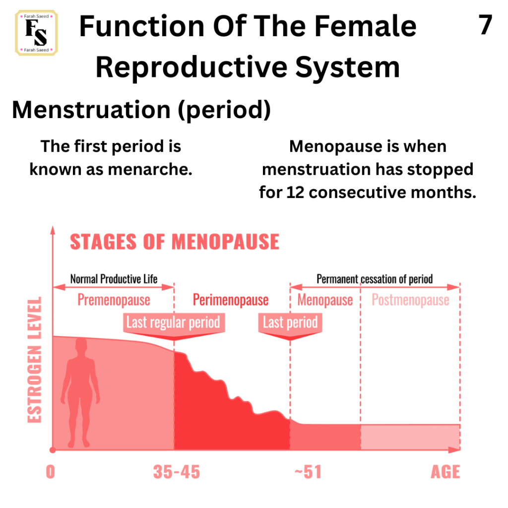
Supporting Images of Function Seven
Function Eight: The Hypothalamic-Pituitary-Ovary (HPO) Axis
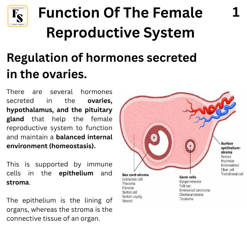
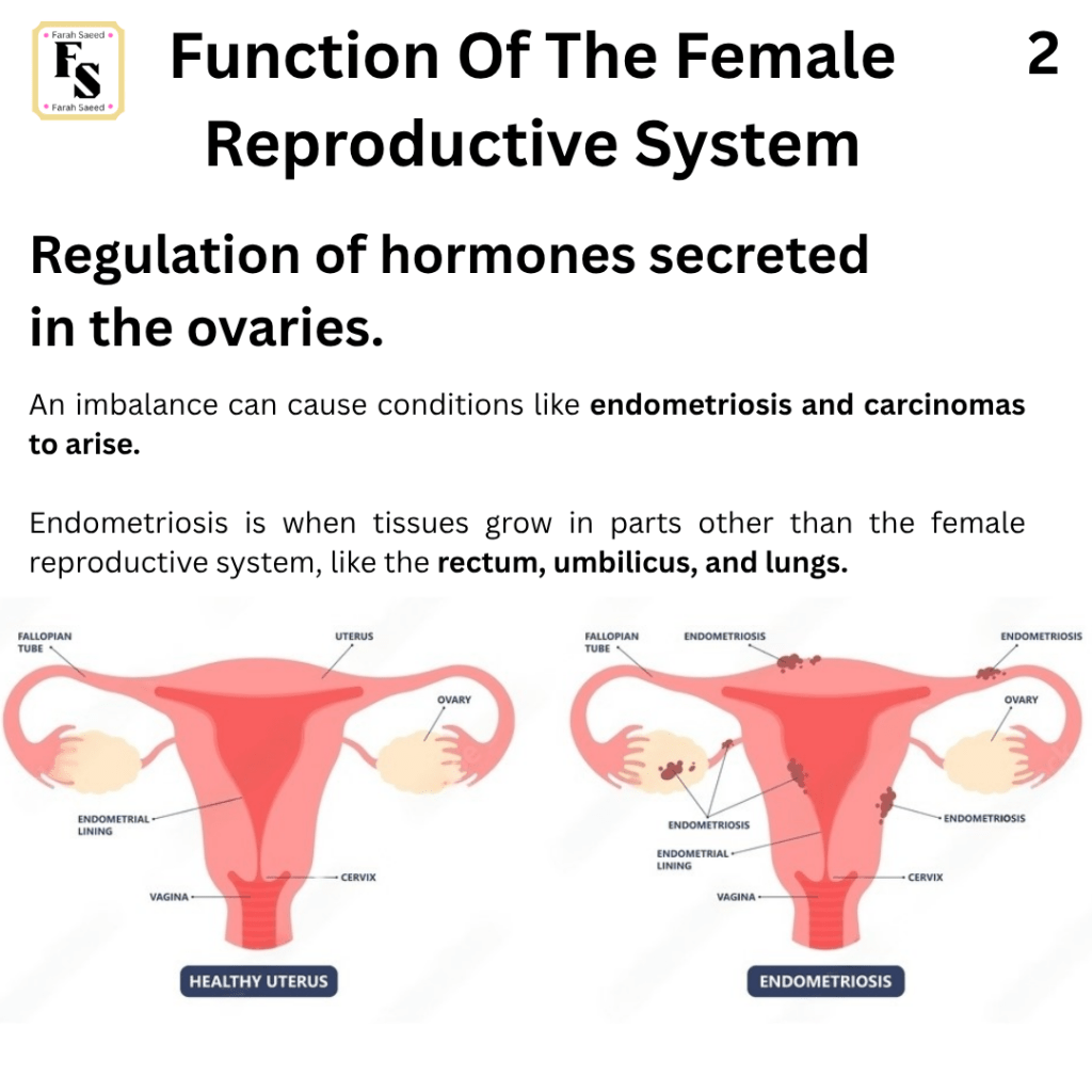
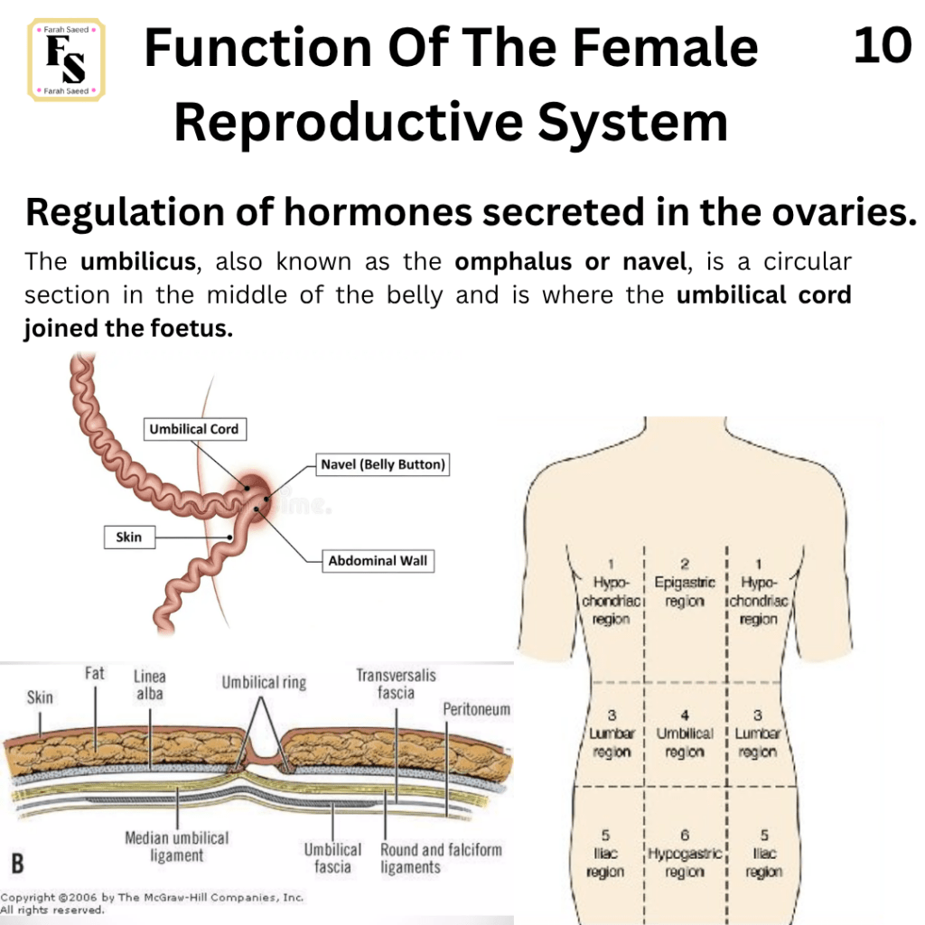
Supporting Images For Function Eight
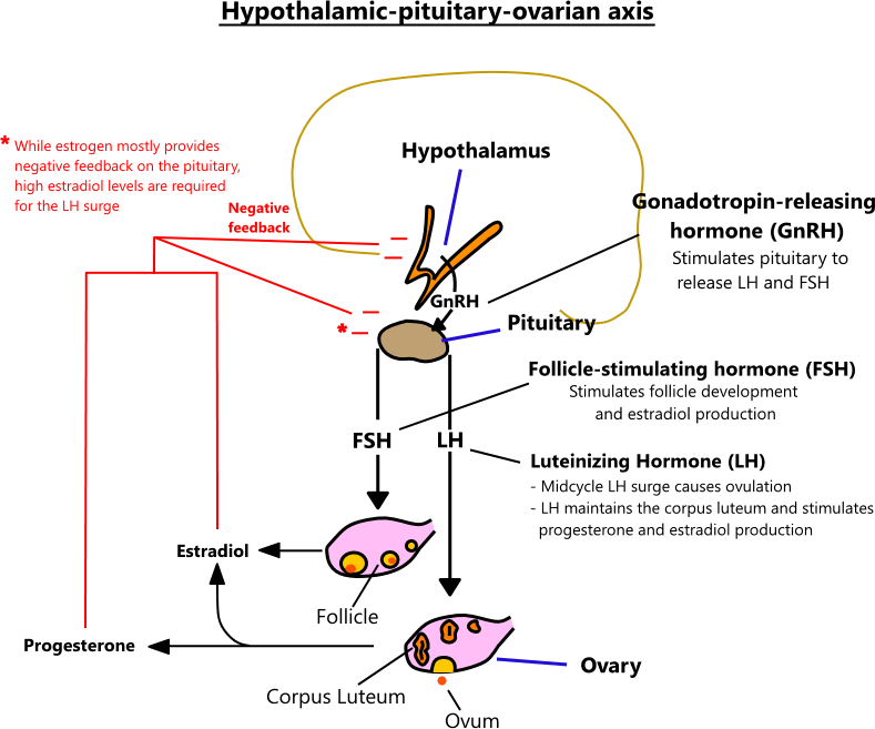
References
Biology Insights (2025) Why Are HCG Levels Higher in the Morning? Available at: https://biologyinsights.com/why-are-hcg-levels-higher-in-the-morning/ (Accessed: 25th August 2026)
Chumduri, C. and Turco, M.Y. (2021). Organoids of the female reproductive tract. Journal of Molecular Medicine, 99(4), pp.531–553. doi:10.1007/s00109-020-02028-0.
Cleveland Clinic (2023) Decidual Cast. Available at: https://my.clevelandclinic.org/health/diseases/24742-decidual-cast (Accessed: 25th August 2026)
National Health Service (2026) Early or delayed puberty. Available at: https://www.nhs.uk/conditions/early-or-delayed-puberty/ (Accessed: 22nd August 2026)
Pampers (2022) hCG Levels in Early Pregnancy. Available at: https://www.pampers.co.uk/pregnancy/healthy-pregnancy/article/hcg-levels (Accessed: 22nd August 2026)
Teach Me Anatomy (n.d.) The female reproductive tract. Available at: https://teachmeanatomy.info/pelvis/female-reproductive-tract/ (Accessed: 7th August 2026)
The Eve Appeal (2026) Gynaecological cancers. Available at: https://eveappeal.org.uk/information-and-advice/gynaecological-cancers/ (Accessed: 7th August 2026)
An Overview: The Supporting Tissues Of The Female Reproductive System.
The role of the female reproductive system is supported by its organs, supporting tissues, hormones, prostaglandins, blood vessels, and nerves.
Organ: These are different types of tissues that join together to form a structure and have a specific function.
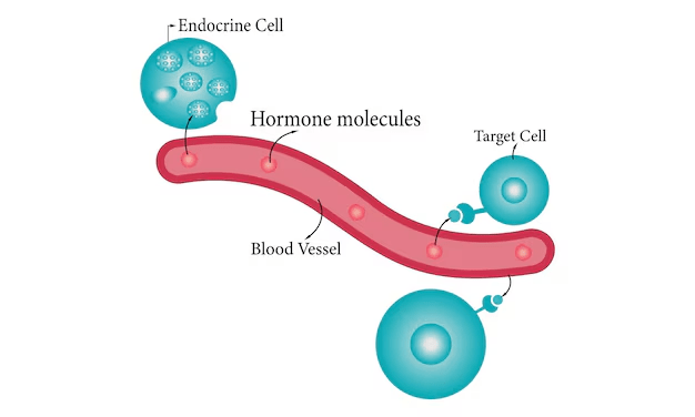
Hormone: This is a chemical messenger produced by structures called glands and is released into the blood to target organs and start a response.
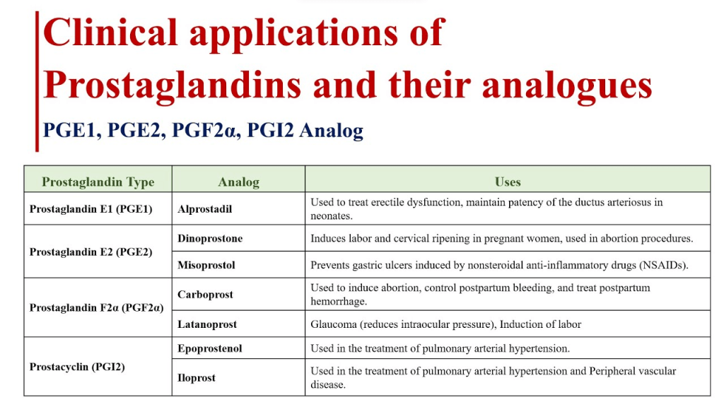
Prostaglandins: This is a type of fatty acid (eicosanoid) that helps organs to function. In the case of the female reproductive system, especially after the release of the egg from the ovary, during menstruation (period), after the egg has been fertilised and is ready to be stuck on the wall of the womb, pregnancy, and childbirth.
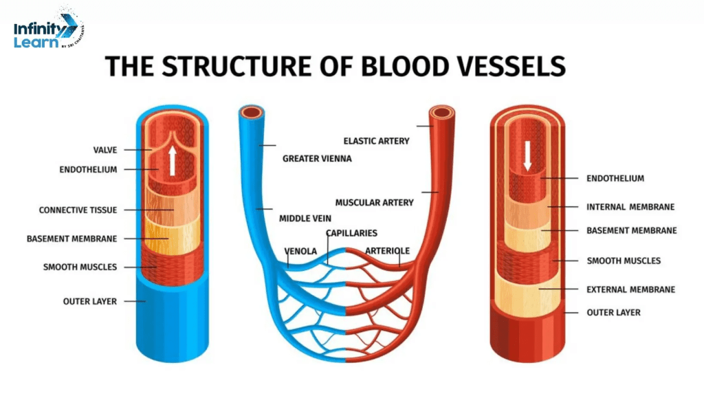
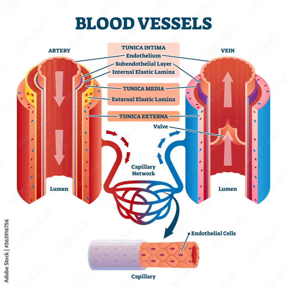
Blood vessels: These are tubes that carry blood around the body to organs and tissues.
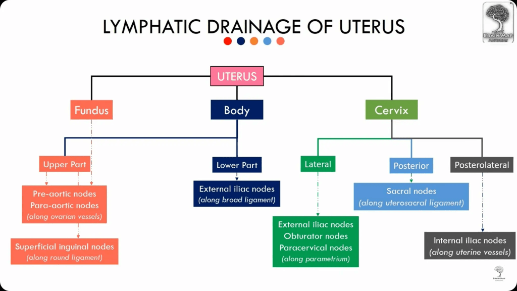
Lymphatic vessels. There are vessels that carry fluid called lymph and passes through filtering structures called lymph nodes. Its purpose is to boost immune system and bath tissues.
Nerves: These consist of nerve fibres that start, integrate, and conduct signals.
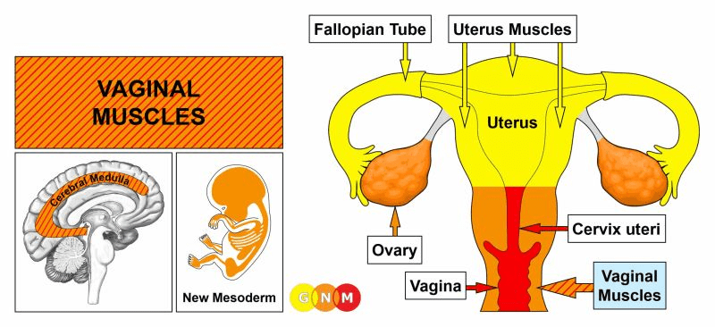
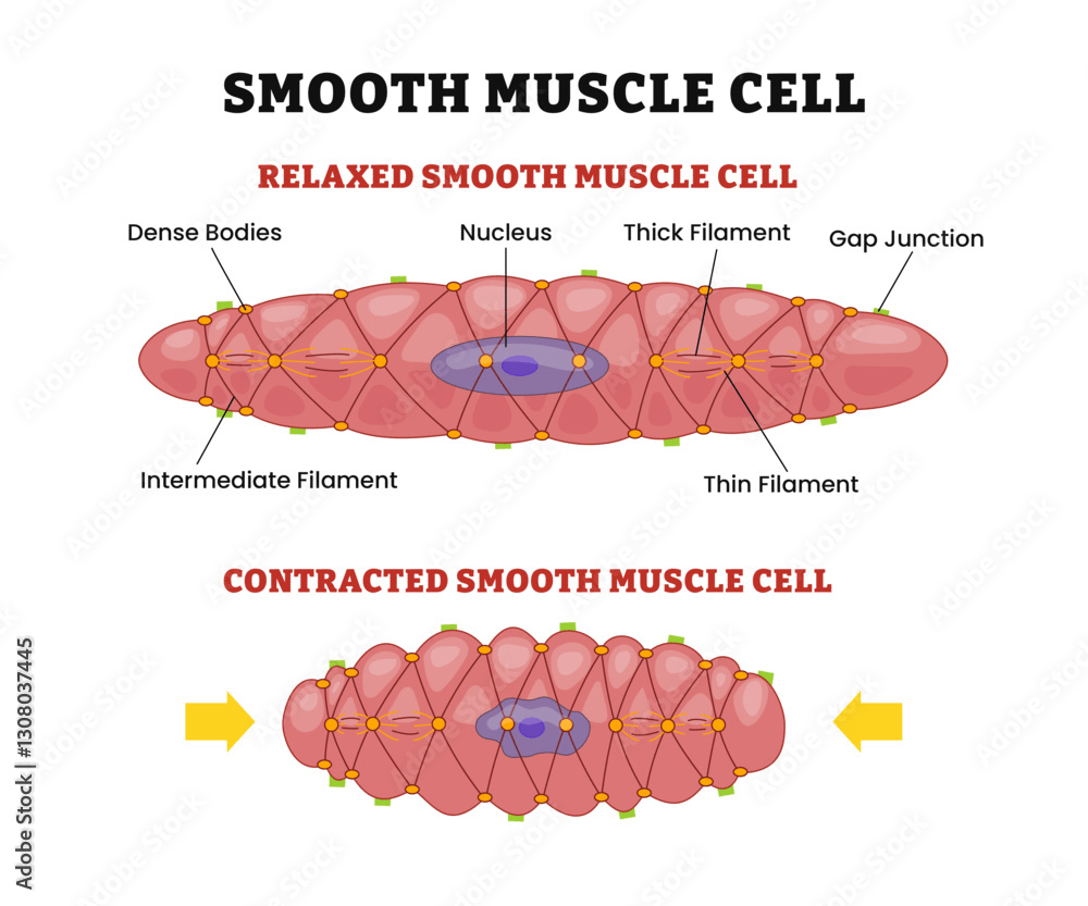
Muscles: They are muscle fibres containing proteins (actin, myosin and intermediate filaments) and connected together to help with movement.
Supporting Images
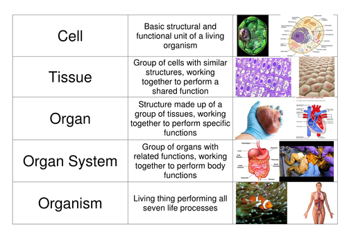
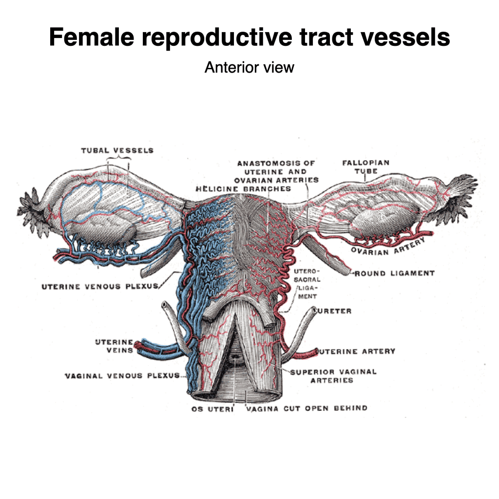
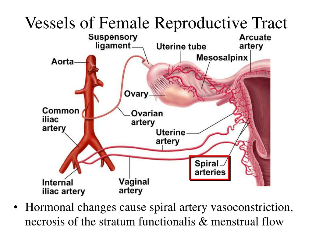
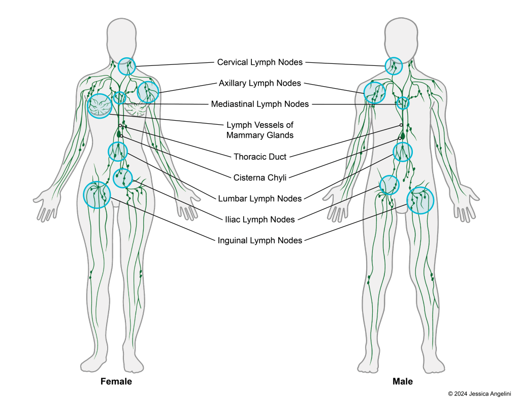
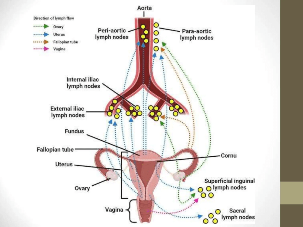
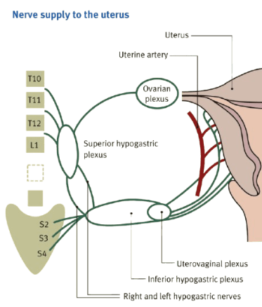
Please see images to support your understanding of the following explanation.
There are several groups of nerves that go through the belly wall (hypogastric nerve plexus) to supply the female reproductive system and connect with the spine/backbone.
Specific sections of the spine: the lower end of the thoracic (T11 – T12), lumbar (L1) and sacrum (S2-S4).
Superior hypogastric plexus
It has two or three nerves.
One nerve accompanies the ovarian artery and supply nerve fibers to the ovary and outer parts of the uterine tube.
The end of parts of the nerves (terminal fibers) interacts with the fibers present in the uterine (womb) to stimulate the uterine tube.
The nerves then send a signal to the spinal cord through the spinal roots present at the back (posterior). It is aimed at the 10th and 11th nerve in the thoracic segment of the spine. Some are able to enter the 9th and 12th position of the thoracic nerves.
The ovarian plexus is stimulated by the sympathetic nerves that are activated during fight or flight response.
The middle hypoglastic plexus.
This is subdivided into right and left hypogastric nerves.
The middle hypoglastic plexus helps to supply the ovaries, the uterine tube and blood vessels and gives off bundle of nerves (fascicles) to the common and external iliac arteries.
Inferior hypogastric plexus
The nerves have an effect on the womb (uterus), cervix and vagina.
How?
- The blood vessels namely arteries of the vagina, uterus and spiral arteries.
- The nerves on the upper part of the vagina has structures called ganglia. They are subdivided into smaller nerve fibres that affect the arteries in the vagina and also affect the muscle in the vagina, urethra and the vulva.
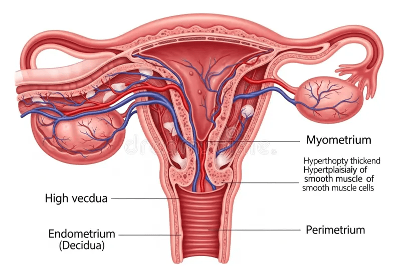
- The other nerves near the uterine artery affects the body and fundus/gravis of the womb as well the muscle of the womb (myometrium).
- The end part of the nerves (terminal fibres) supplies the narrow part of the uterine tube called the isthmus and the end part of the womb. They interact with superior and middle hypogastric plexus.
Neupsy Key (n.d) Innervation of Female Reproductive Organs. Available at: https://neupsykey.com/innervation-of-female-reproductive-organs/ (Accessed: 22nd August 2026)To find out more about nerve plexuses, please visit: Plexuses


Leave a Reply