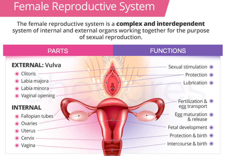
Brauer and Smith (2015) discovered that female oestrogen can affect the nerves around the uterus and vagina of the female reproductive system. The structure of the female reproductive system is illustrated in Figure 1. The uterus, otherwise known as the womb, is where the baby grows, beginning in its outermost layer known as the endometrium. The endometrium is also the layer that is shed during menstruation (period). Other functions include its contraction during labour/childbirth, known as parturition. The vagina is a muscular tube where the male reproductive organ (penis) enters through the opening of the vagina and releases the semen containing the male sex cell (sperm) during a process called ejaculation. It is lined with mucous membrane from the neck of the cervix (cervical uteri) to the outer part/exterior.
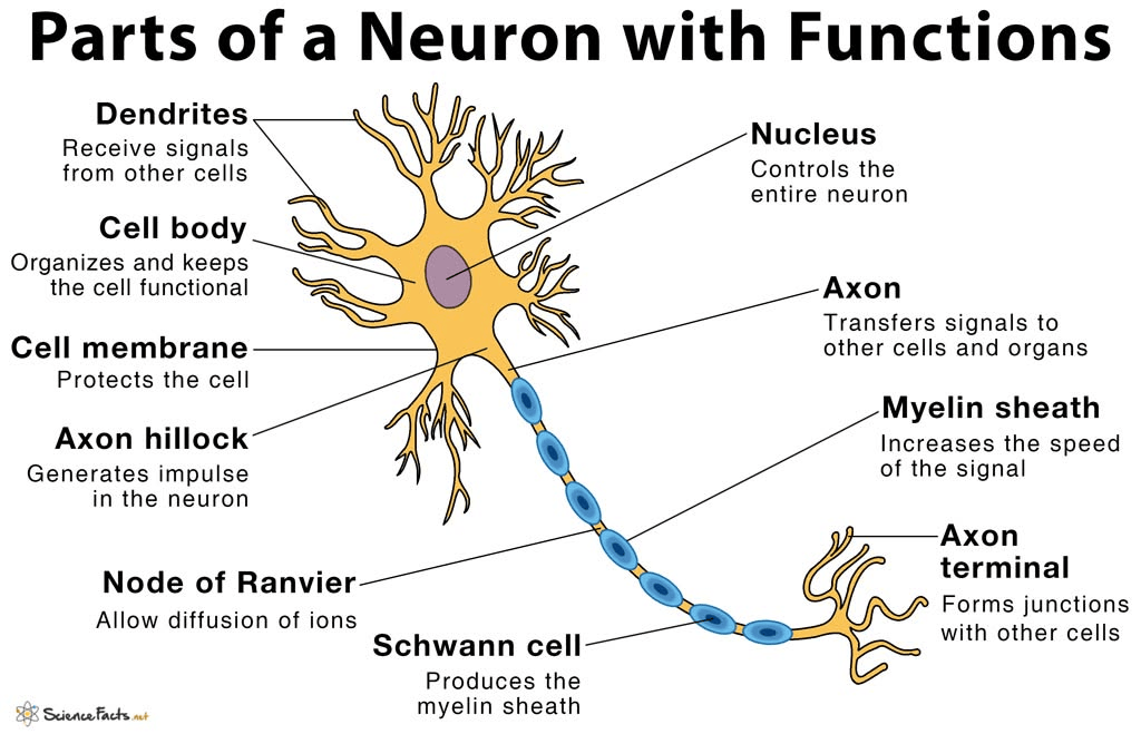
In the uterus, sympathetic nerve endings (terminal axons) in the uterine muscle (myometrium) break down or degenerate. The structure of the neuron is presented in Figure 2.
The release of the following proteins that are stimulated by the presence of oestrogen, achieves this:
Brain-derived neurotrophic factor (BDNF)
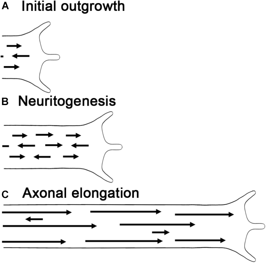
Neuropeptide is a type of peptide and a member of the neurotrophin growth factor family that regulates neurite growth and survival. Neurites are part of the growth of the peripheral nervous system, where it extends the axon and dendrites. Other members of the BDNF family are nerve growth factor (NGF), neurotrophin-3 (NTF3), and neurotrophin-4/5 (NTF5). BDNF cooperates with neurotrophin receptor tyrosine kinase 2 (Ntrk2) and can facilitate several cellular functions: adhesion to surfaces (adhesion), formation of new blood vessels (angiogenesis), cell death (apoptosis), and growth of cells (proliferation). BDNF and Ntrk2 can be found in the vascular smooth muscle (blood vessel), muscle of the uterus/womb (myometrium), and the lining (lumen and glandular tissue). Other studies have found that it can help differentiate into cytotrophoblasts; it is part of the trophoblast that maintains its structure and does not embed into the maternal tissues. Some studies discovered that BDNF can prevent the outgrowth of the neurite in the ganglion of the cervix and myometrium.
Neurotromin
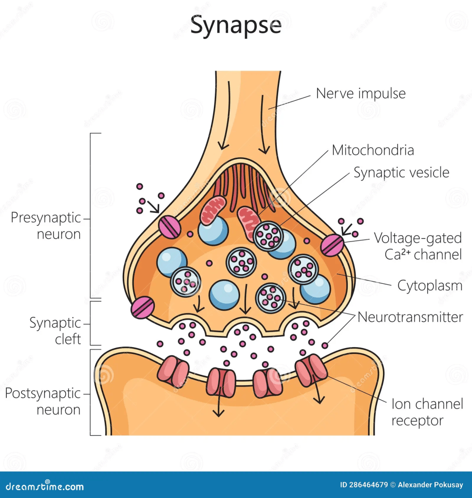
It is a molecule that is involved in cell adhesion, interaction with other cells, and neurite outgrowth by facilitating the recognition and regulation of the neurone. It is found on the plasma membrane, especially in the postsynaptic, presynaptic, and extracellular regions. Please see Figure 3.
Semaphorins
They are members of a protein family that helps with the growth of the axons (the structures where the signal passes through in the nerves) and the dendrites (branches of the nerves). It also helps in the formation of nerve bundle fibres, movement of the nerve cells, transport, and cell death (apoptosis). Other functions is the initiation of the immune response, acute inflammation, and the cardiovascular system. For instance, SEMA3A is expressed by a type of immune cell called dendritic cells, and it prevents the activation of T immune cells. On the other hand, SEMA4A associates with Tim-2 on T cells, and this leads to the activation and growth of T cells. SEMA4D indirectly co-stimulates the activity of T cells.
Pro-Nerve Growth Factor (proNGF)
It is a precursor of the nerve growth factor (NGF) that promotes the growth and survival of nerve cells (neurons) in the central and peripheral nervous systems. At first, NGF is translated as a precursor, proNGF, and this results in three states: it can remain as proNGF, or proNGF can be cleaved intracellularly (inside the cells) by a protein called furin, or proNGF can be cleaved extracellularly (outside the cells) by proteins called plasmin or metalloproteinases.
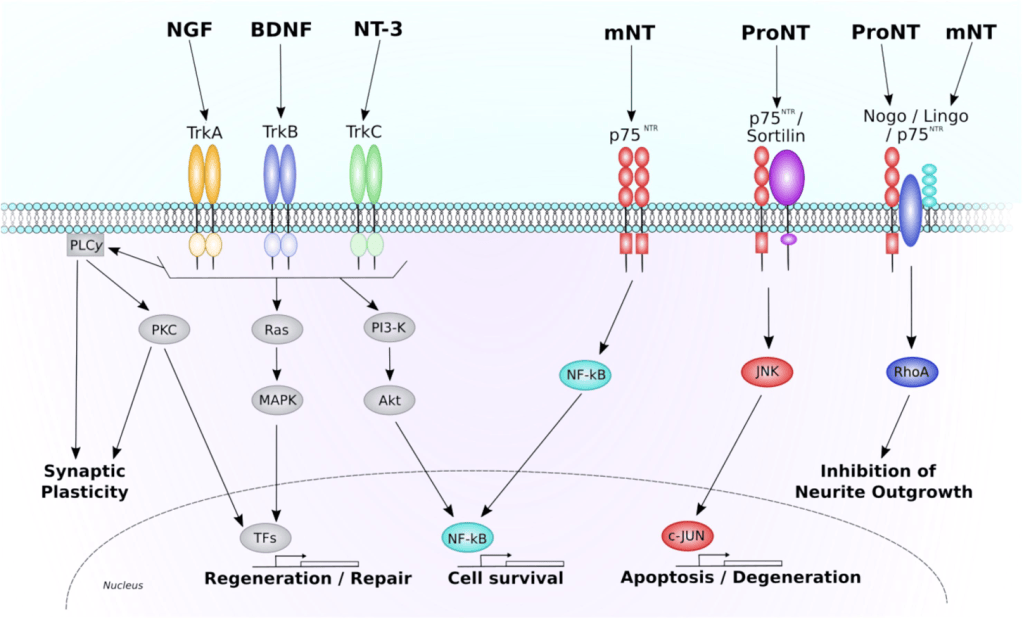
There are two main cell signalling pathways: phosphatidylinositol-3-kinase (PI3K)/Akt and Ras/Extracellular signal-regulated kinase (ERK). The PI3K/Akt pathway is involved in the survival of cells mediated by neurotrophins. The ERK pathway helps to maintain cell survival following stress and also to grow neurites (neuritogenesis). Figure 4 presents some of the signalling pathways that are mediated during the effects on the neurons. There are several receptors that bind to NGF and proNGF to initiate function: tropomyosin-related kinase A (TrkA), p75NTR pan-neurotrophin receptor, and sortilin. This is similar to a lock-and-key process where NGF and proNGF are keys, and they can bind to locks (receptors) on the cell surface.
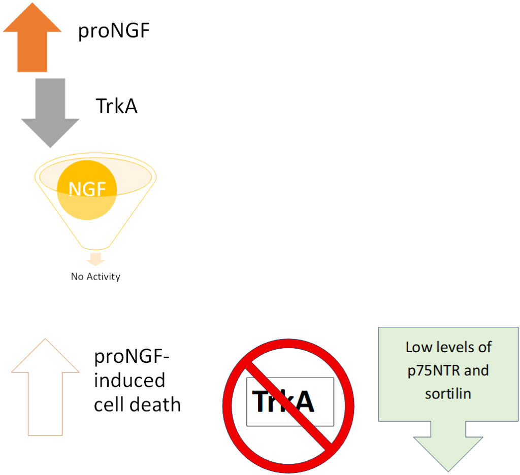
Lowering the levels of p75NTR lowers the ability of NGF to mediate survival of sensory neurones of the embryo (developing/early-staged baby). The neurones express both TrkA and p75NTR.
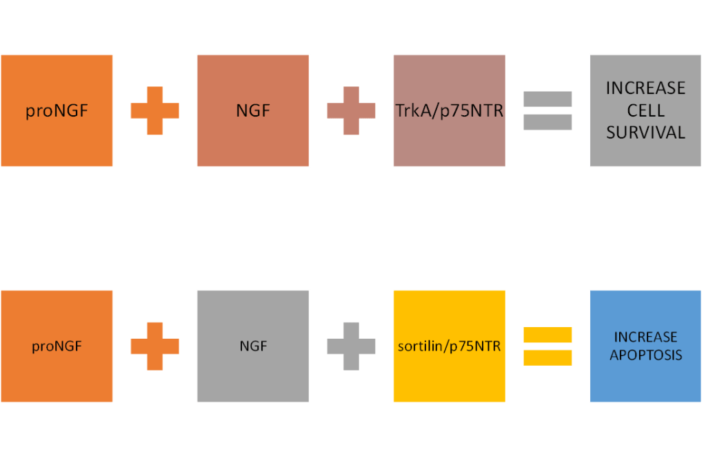
In the absence of TrkA, P75NTR further helps to promote cell death with sortilin; this is achieved via the tumour suppressor gene p53, ceramide, and other pathways, such as c-Jun N-terminal kinase. On the other hand, when p75NTR is co-expressed with TrkA rather than sortilin, alternative signalling pathways are induced to mediate survival of neurones and neurite outgrowth. For example, NF-Kb and Rho signalling pathways.
One of the main factors that affect the binding between the receptor (lock) and protein/ligand (key) is the ability or strength to bind to the binding site. TrkA has a higher strength or affinity to bind to mature NGF than its precursor proNGF. Alternatively, p75NTR has higher affinity for proNGF than mature NGF.
The combination of sortilin and p75NTR receptors binds to neurotrophins and has higher affinity for proNGF but not NGF. Even when transporting proteins and other chemicals within the cells in membrane-bounded structures called vesicles, they vary in kinetics or rate of the reaction. Most vesicles that contain NGF have TrkA; on the contrary, Most vesicles that have proNGF also have TrkA and p75NTR.
One of the main factors affecting the binding between the receptor (lock) and protein/ligand (key) is the ability or strength to bind to the binding site. TrkA has a higher strength or affinity to bind to mature NGF than its precursor, proNGF. Alternatively, p75NTR has higher affinity for proNGF than mature NGF.
The combination of sortilin and p75NTR receptors binds to neurotrophins and has higher affinity for proNGF but not NGF. Even when transporting proteins and other chemicals within the cells in membrane-bounded structures called vesicles, they vary in kinetics or rate of the reaction. Most vesicles that contain NGF have TrkA; on the contrary, Most vesicles that have proNGF also have TrkA and p75NTR.
Other parts of the extracellular matrix

This is a mixture of proteins and minerals that are found in the fluid outside the cells.
However, oestrogen does not affect neurotrophins that increase the growth of axons, but it does affect the expression of neurotrophins that promote degeneration. Under low female oestrogen conditions, the sympathetic terminal axons can be remade again.
In the vagina, the breakdown of the autonomic and nociceptive axons (the nerve fibre endings that are associated with pain). The suppression of BMP4 (Bone morphogenetic protein 4) can be induced by oestrogen and can affect the nociceptive axons. BMP4 is a growth factor of the tumour growth factor (TGF)-Beta that is involved in the development of neurons, production of new blood vessels, and bone development. The receptors of BMP (BMPR1A and BMPR2) bind together and activate via phosphorylation (the addition of phosphate groups). BMPR1A then mediates signalling by phosphorylating SMAD1/5/8 to the nucleus and either switch on or off the transcription of the target genes.
Overall, there is a link between the female oestrogen and neuronal growth, and this is observed through the effect of the proteins released when there are high levels of oestrogen.
References
Brauer, M. and Smith, P.G. (2015). Estrogen and female reproductive tract innervation: Cellular and molecular mechanisms of autonomic neuroplasticity. Autonomic Neuroscience, 187, pp.1–17. doi:10.1016/j.autneu.2014.11.009.
Ioannou, M. and Fahnestock, M. (2017). ProNGF, but Not NGF, Switches from Neurotrophic to Apoptotic Activity in Response to Reductions in TrkA Receptor Levels. International Journal of Molecular Sciences, [online] 18(3), pp.599–599. doi:10.3390/ijms18030599.
Kanth, S.M., Gairhe, S. and Torabi-Parizi, P. (2021). The Role of Semaphorins and Their Receptors in Innate Immune Responses and Clinical Diseases of Acute Inflammation. Frontiers in Immunology, 12. doi:10.3389/fimmu.2021.672441.
LifeMap Sciences (2026) NTM gene Neurotrimin. Available at: https://www.genecards.org/card/NTM (Accessed: 6th Septemer 2026)
UniProt (n.d.) P12644 · BMP4_HUMAN. Available at: https://www.uniprot.org/uniprotkb/P12644/entry (Accessed: 6th September 2026)
Wessels, J.M., Wu, L., Leyland, N.A., Wang, H. and Foster, W.G. (2014). The Brain-Uterus Connection: Brain Derived Neurotrophic Factor (BDNF) and Its Receptor (Ntrk2) Are Conserved in the Mammalian Uterus. PLoS ONE, 9(4), p.e94036. doi:10.1371/journal.pone.0094036.


Leave a comment