
The Structure And Function Of Each Part Of The Female Reproductive System

Polite notice: This page contains diagrams of the reproductive organs of males and females (private parts). It is for educational purposes on cancers that arise in those areas. Please ensure that when visiting the page, there is a sense of manners. We understand that in some cultures, there would be discomfort, but it is important to be aware of information about cancer.
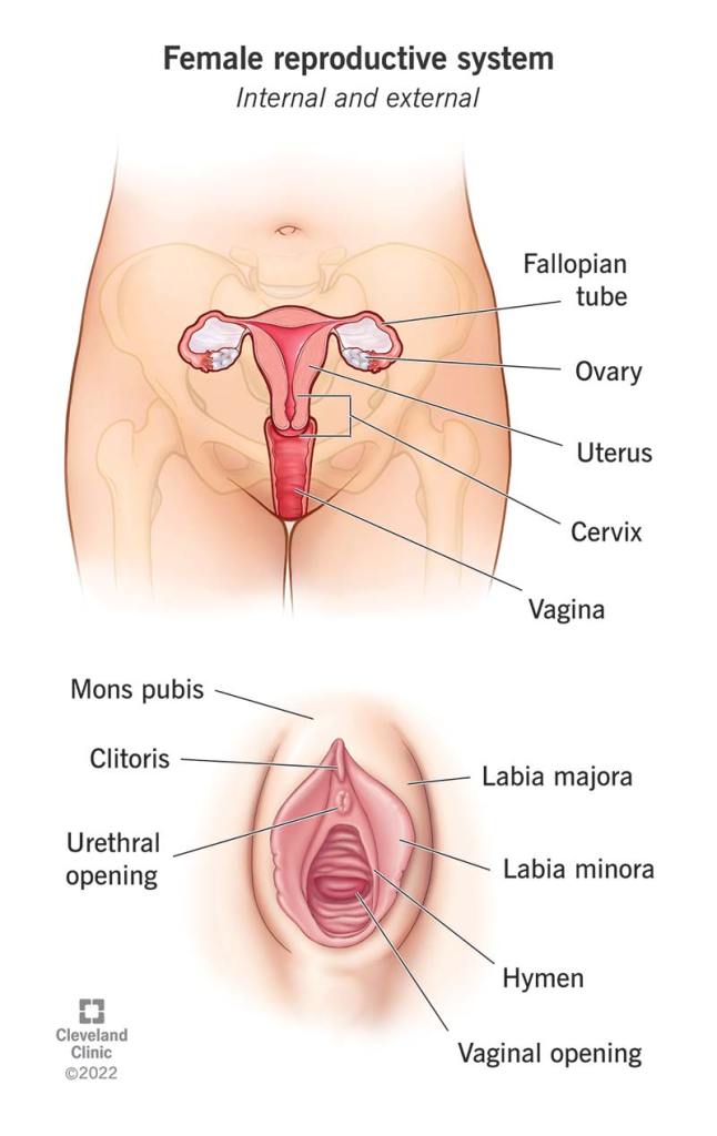
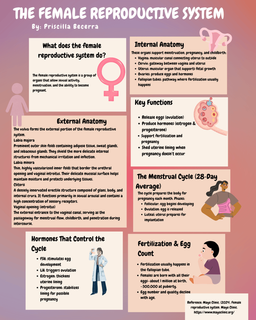
The female reproductive system is divided into INTERNAL GENITALIA and EXTERNAL GENITALIA.
Genitalia – This is in reference to the reproductive organs.
Internal – Deep Inside
External – Outside or Outer structures
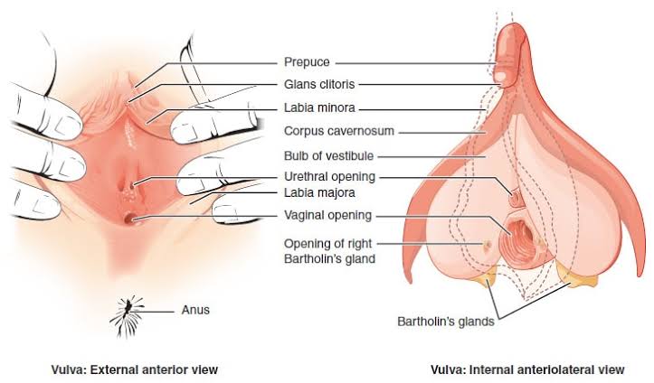
External Genitalia
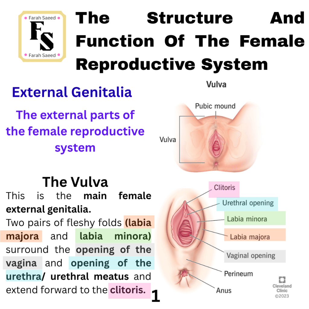
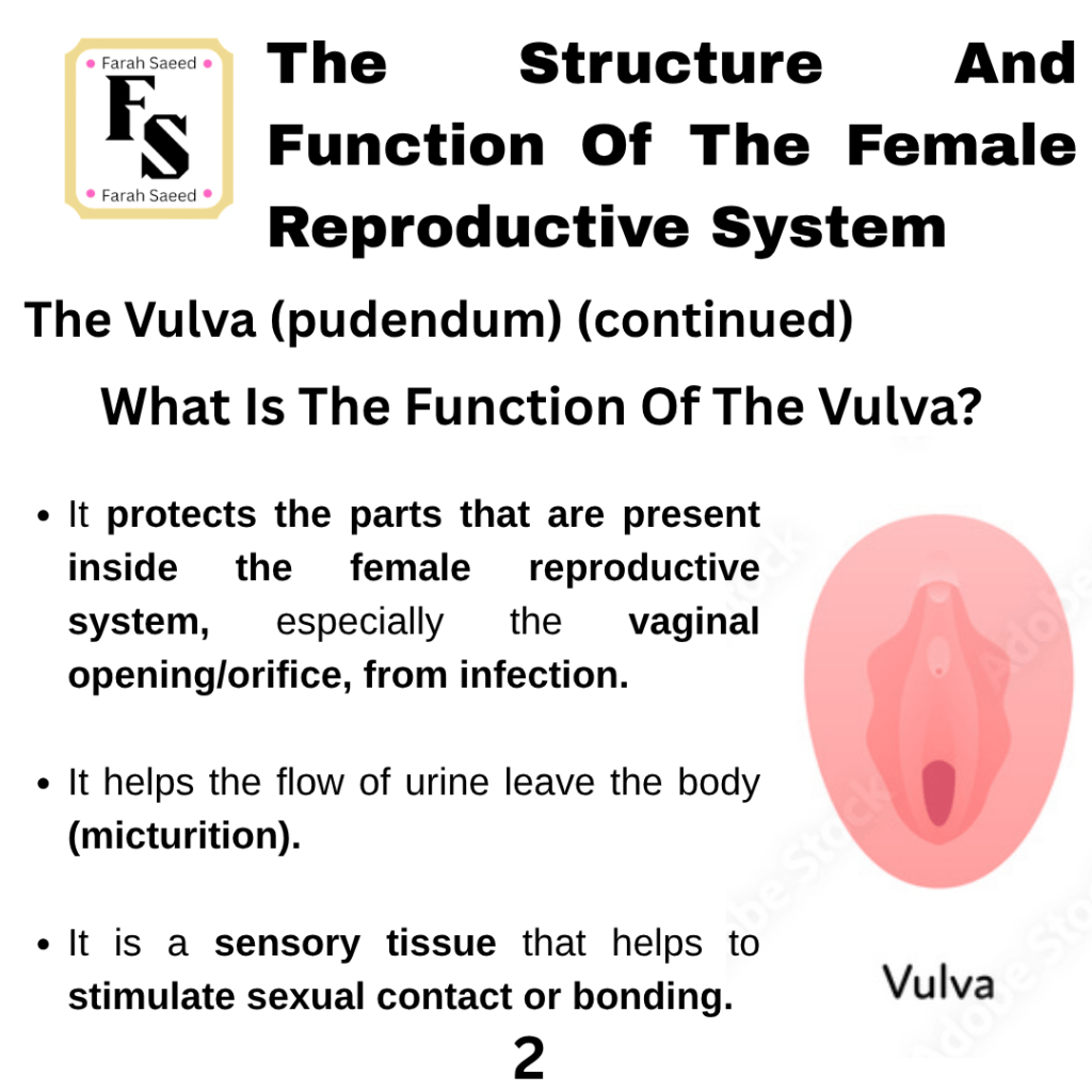
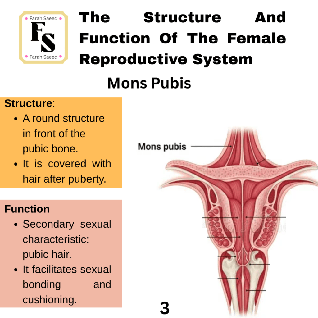
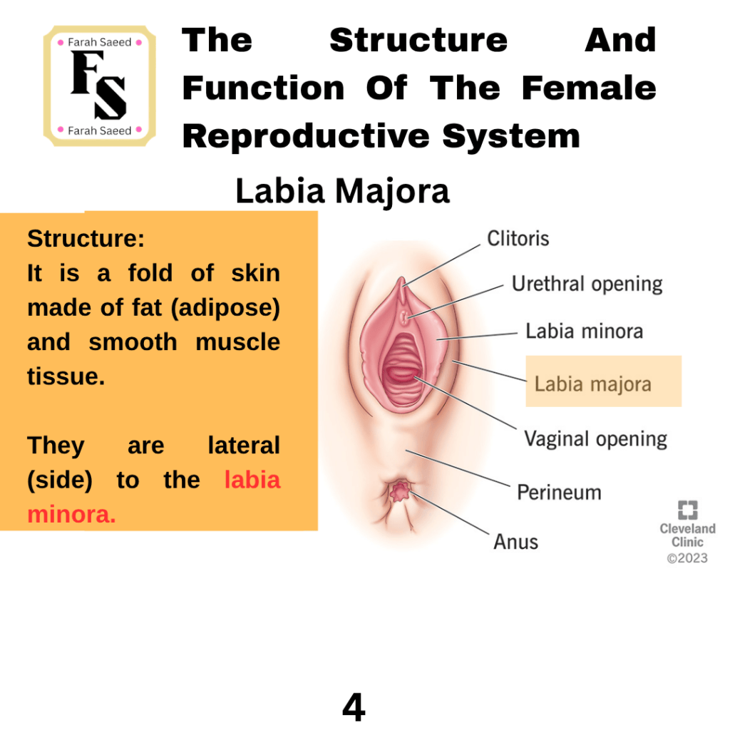
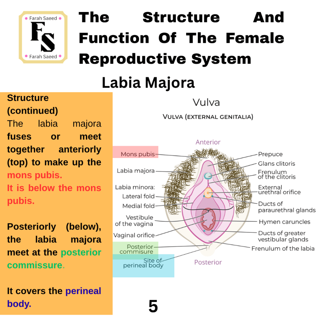
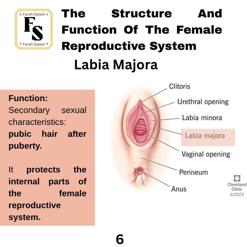
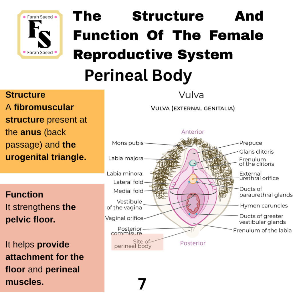
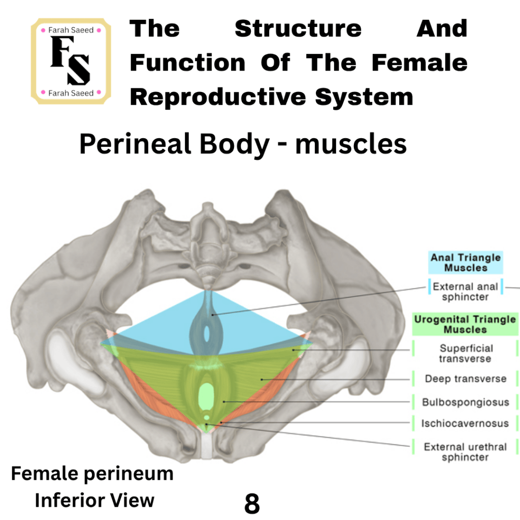
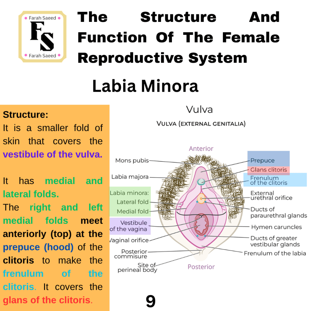
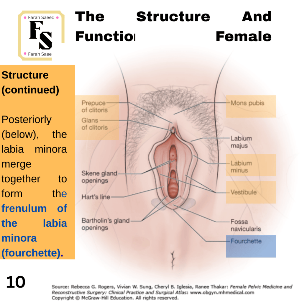
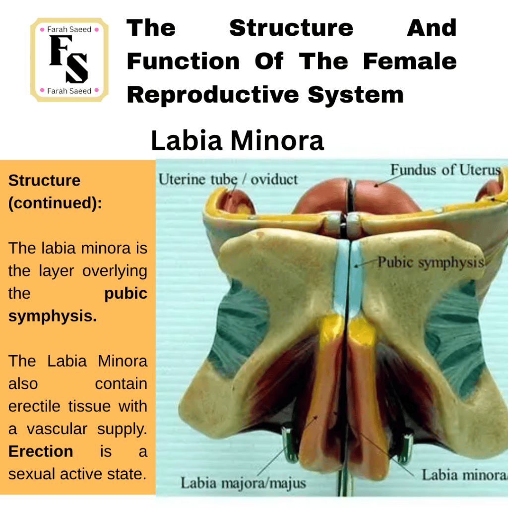
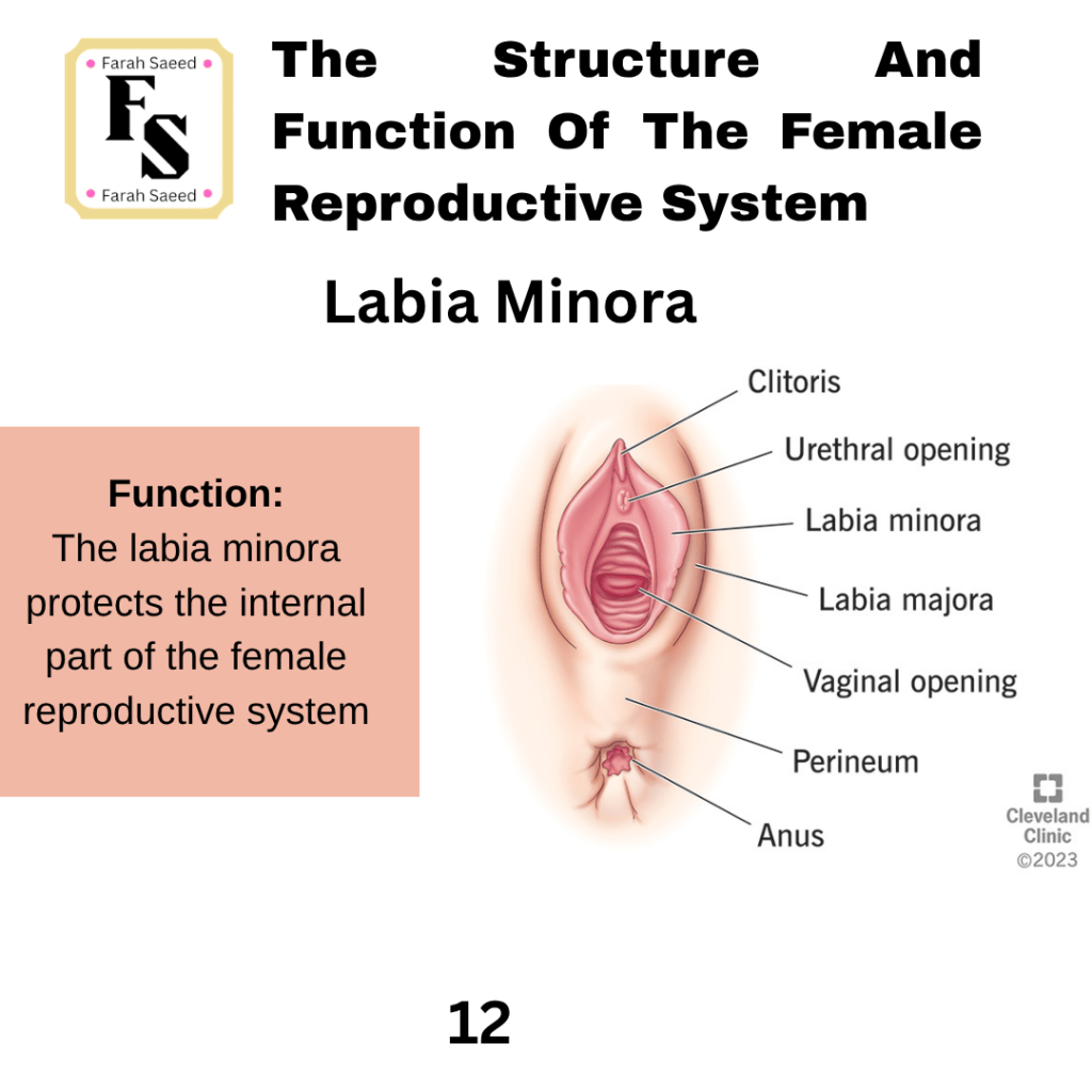
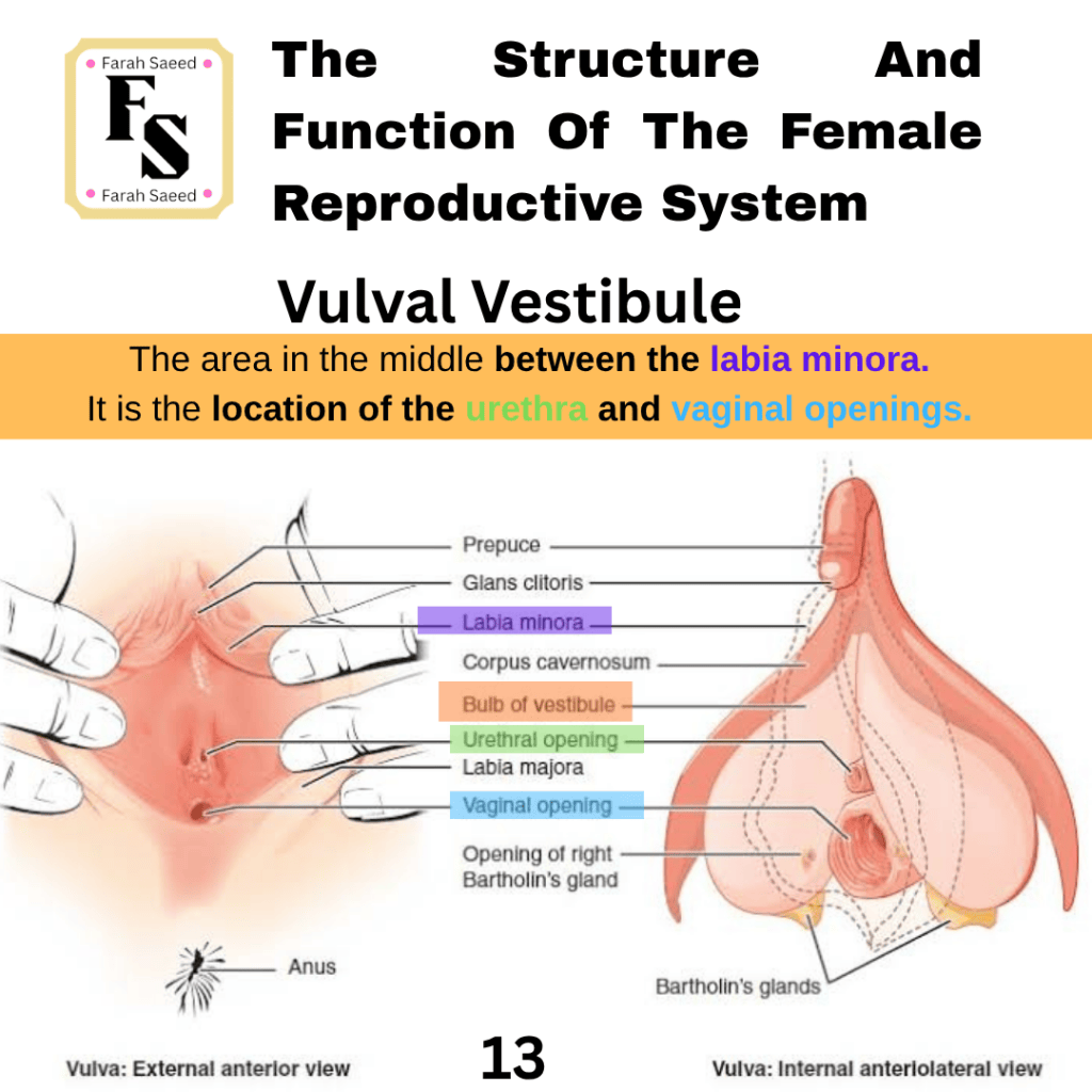
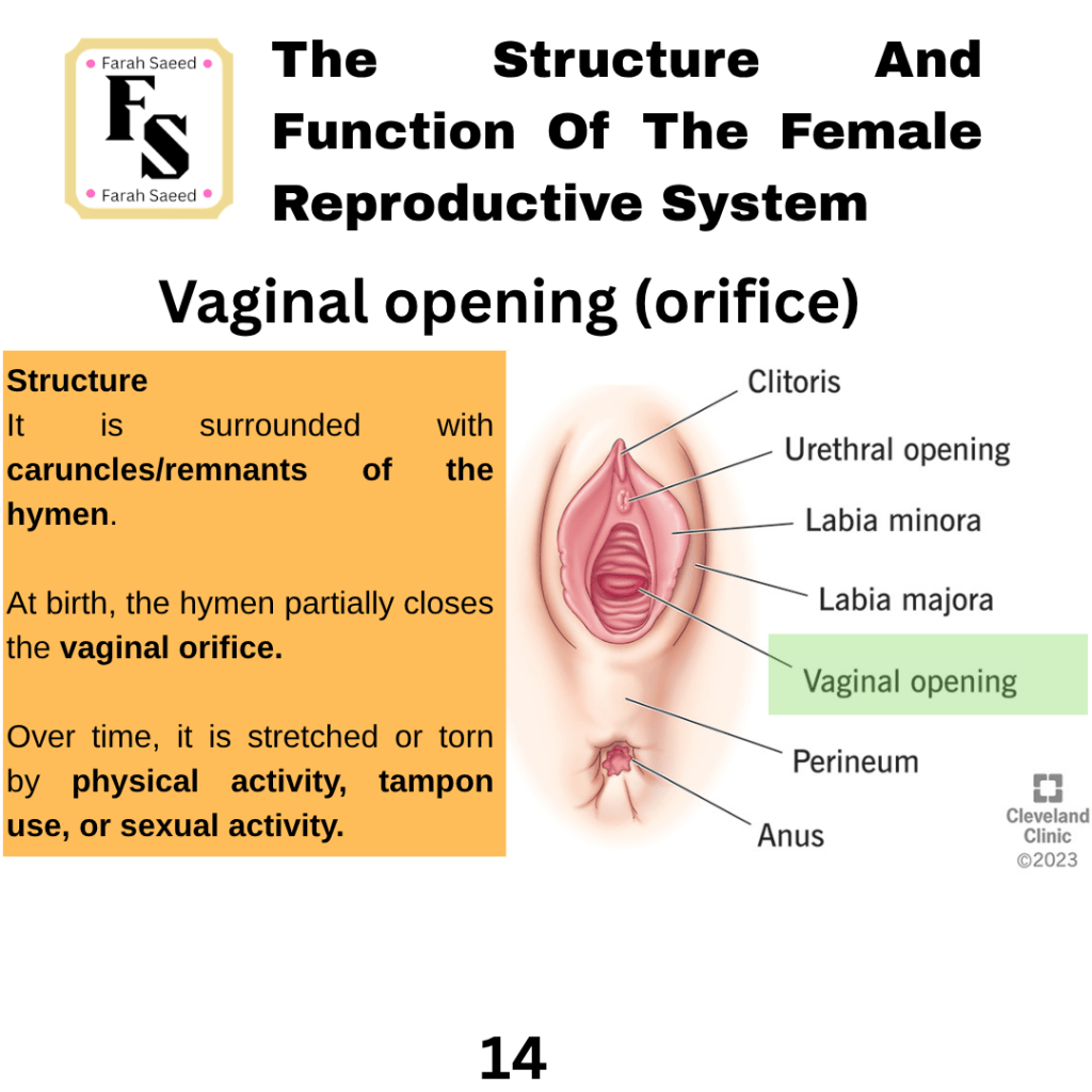
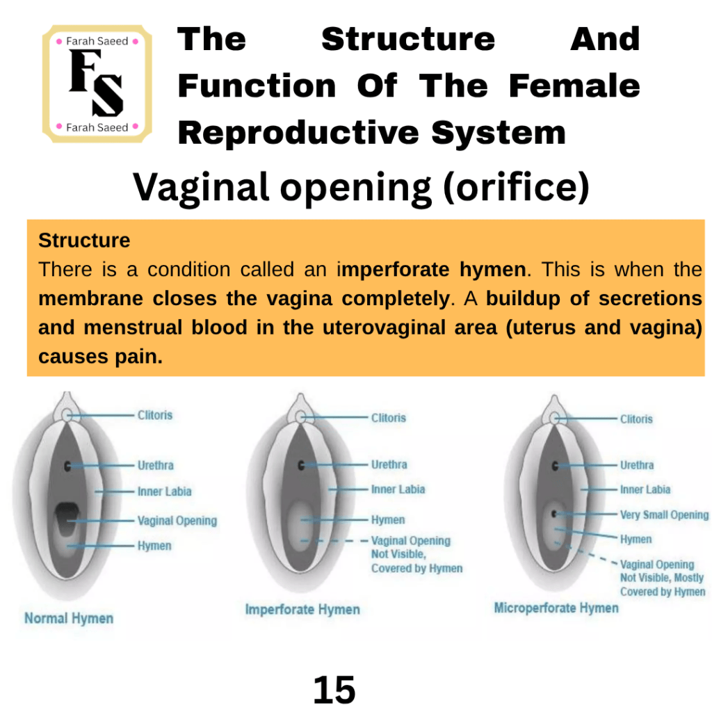
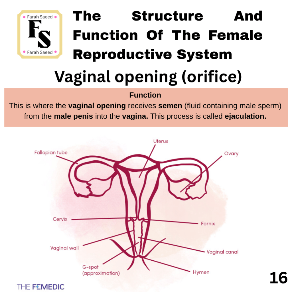
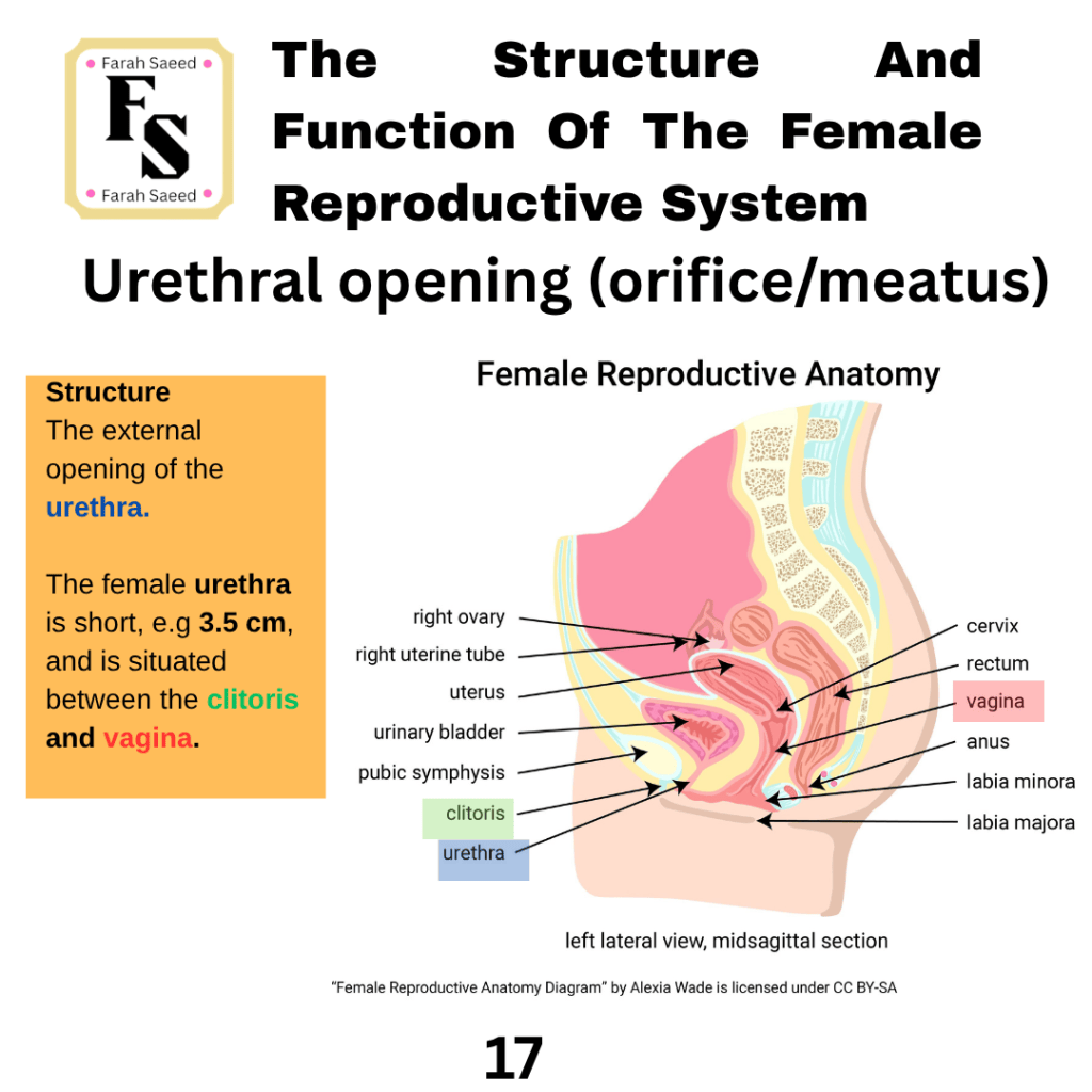
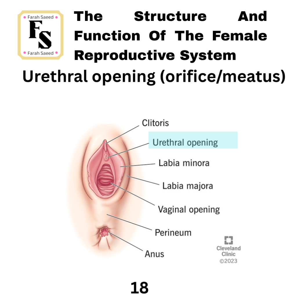
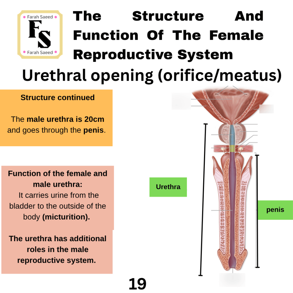
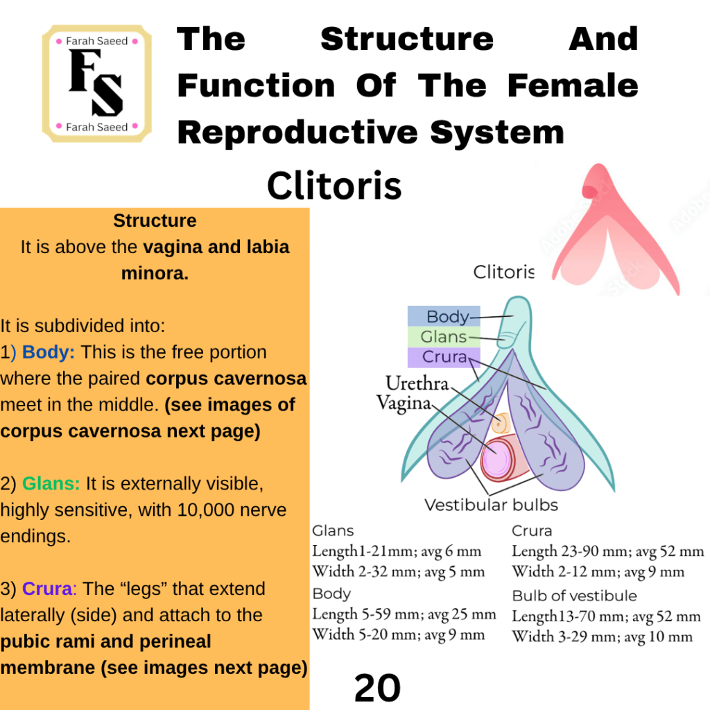
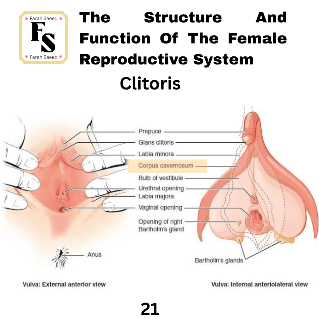
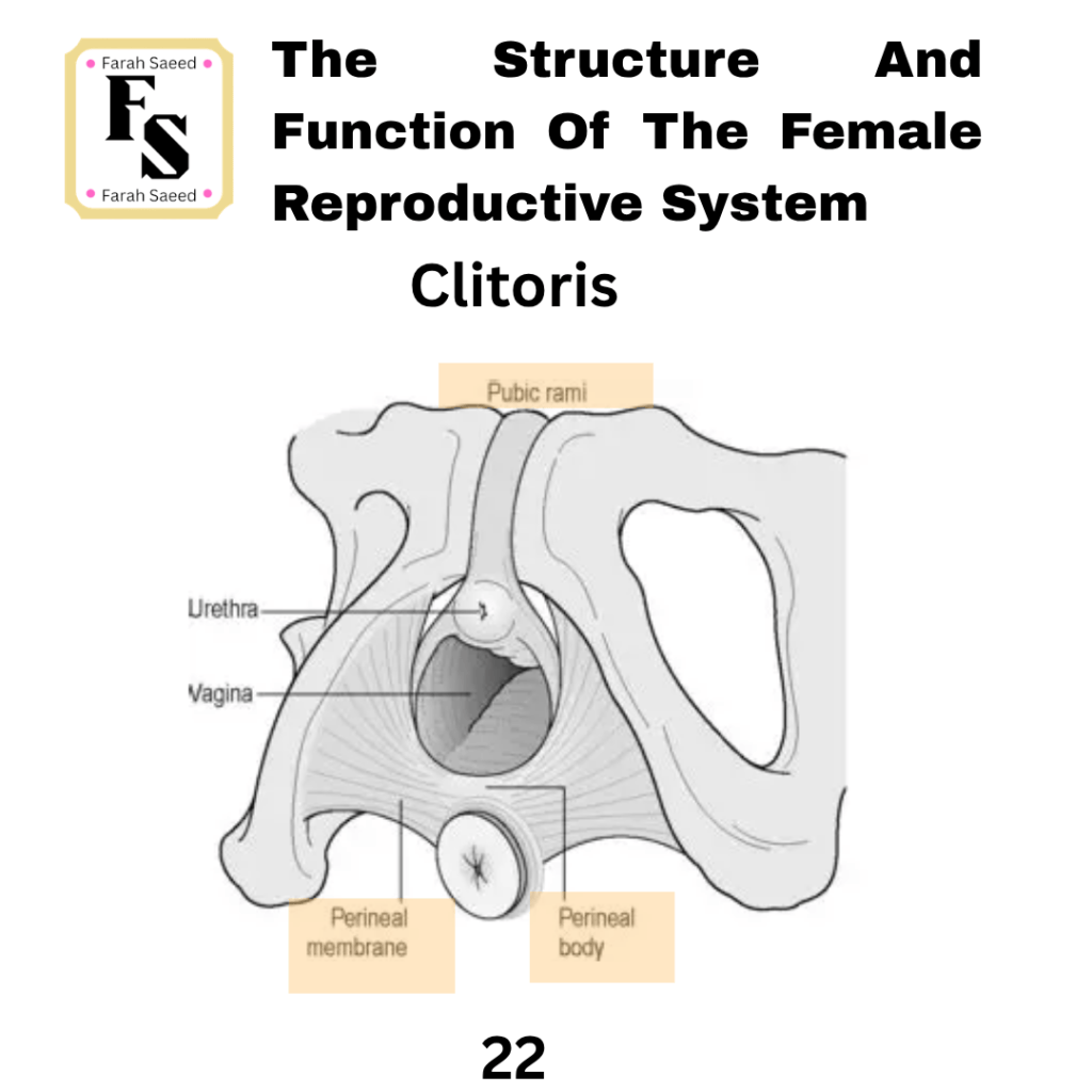
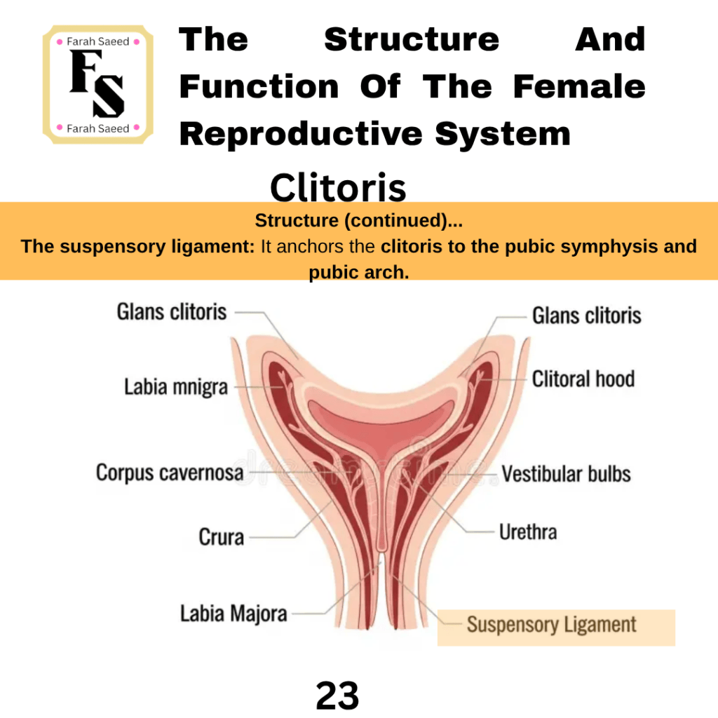
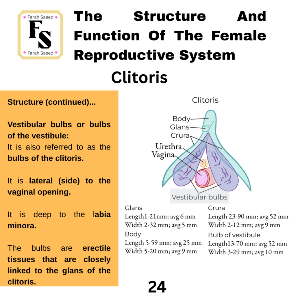
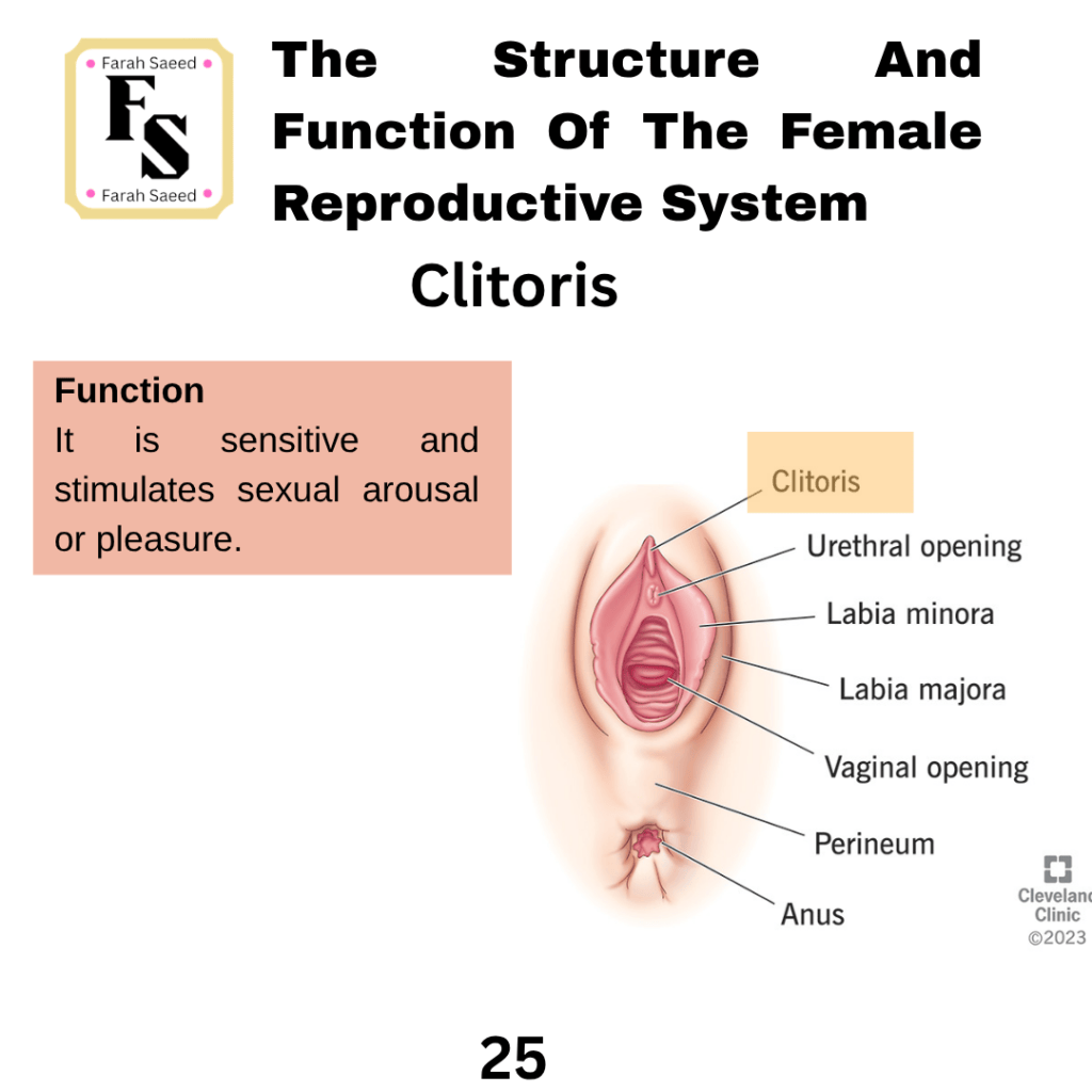
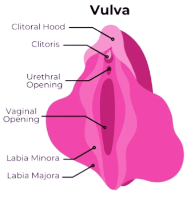
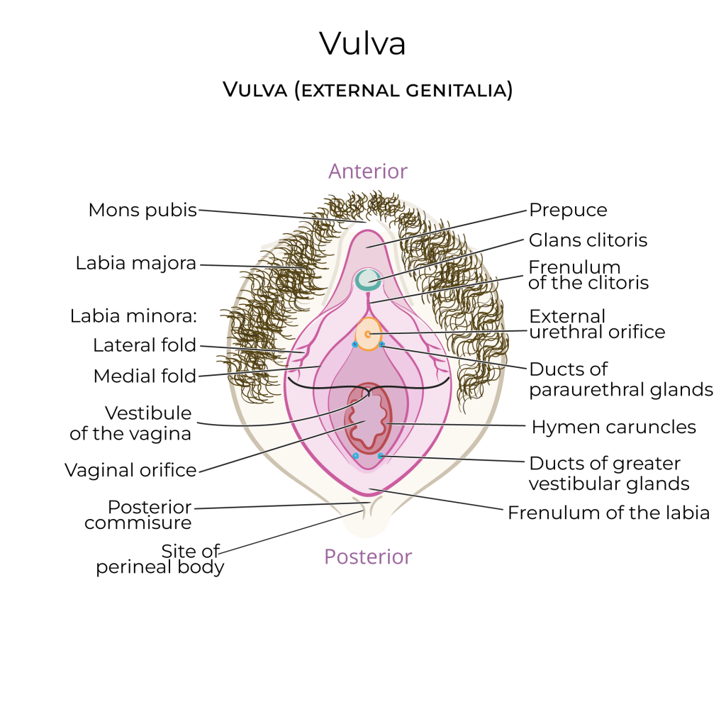
Supporting Images For External Genitalia
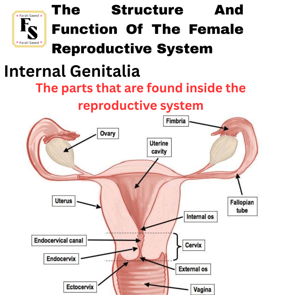
Internal Genitalia
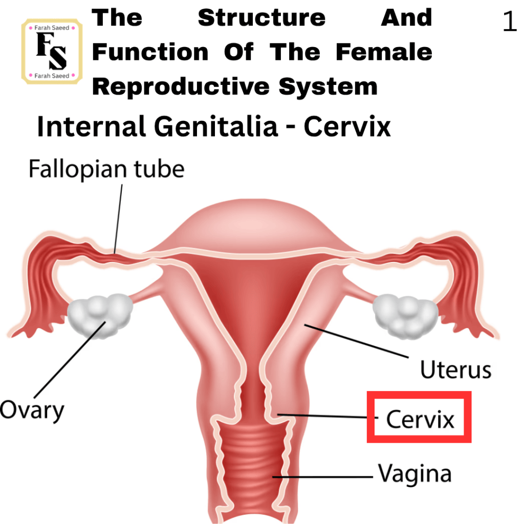
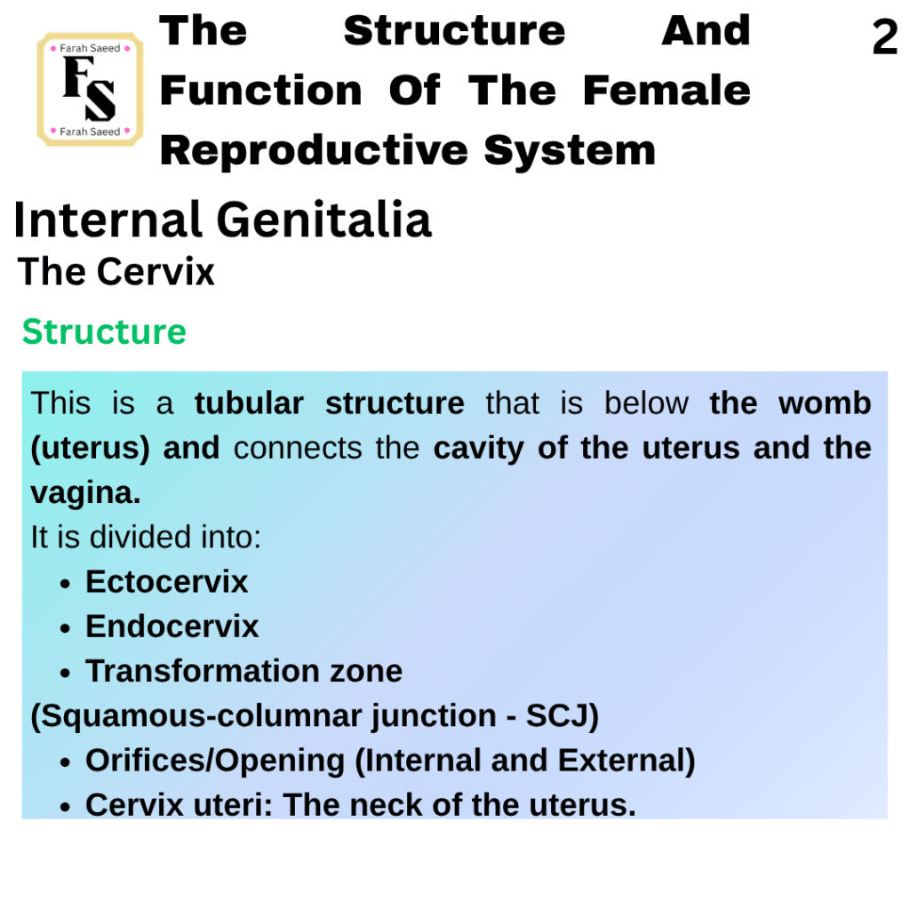
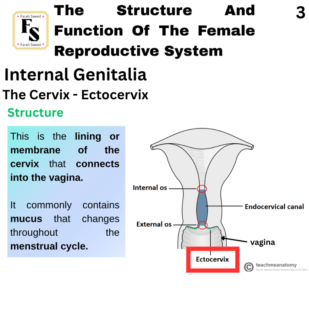
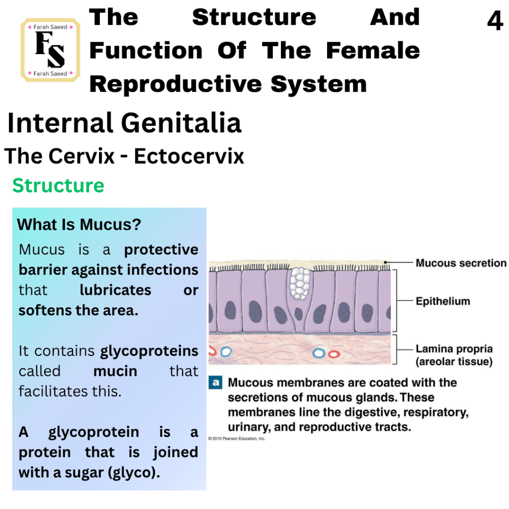
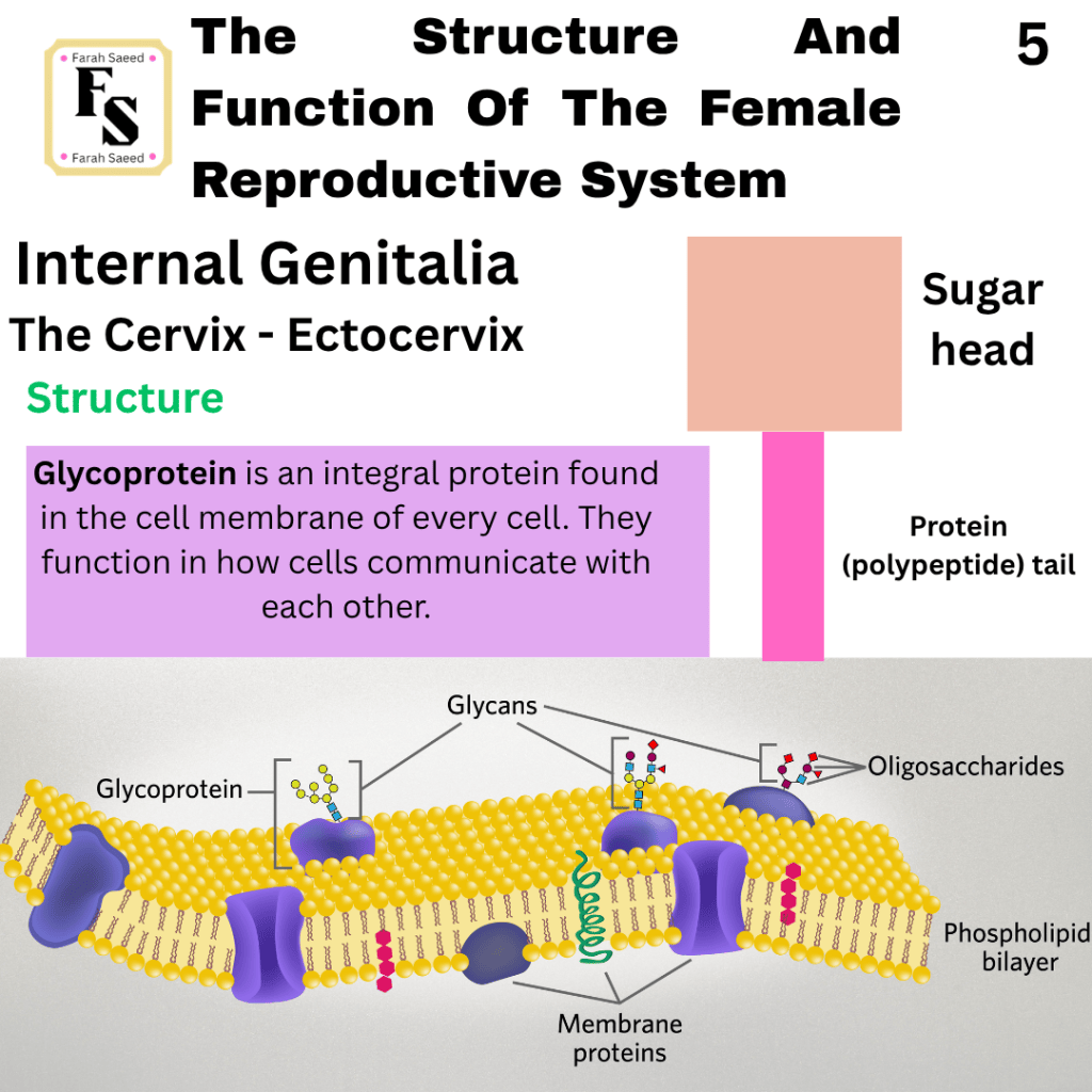
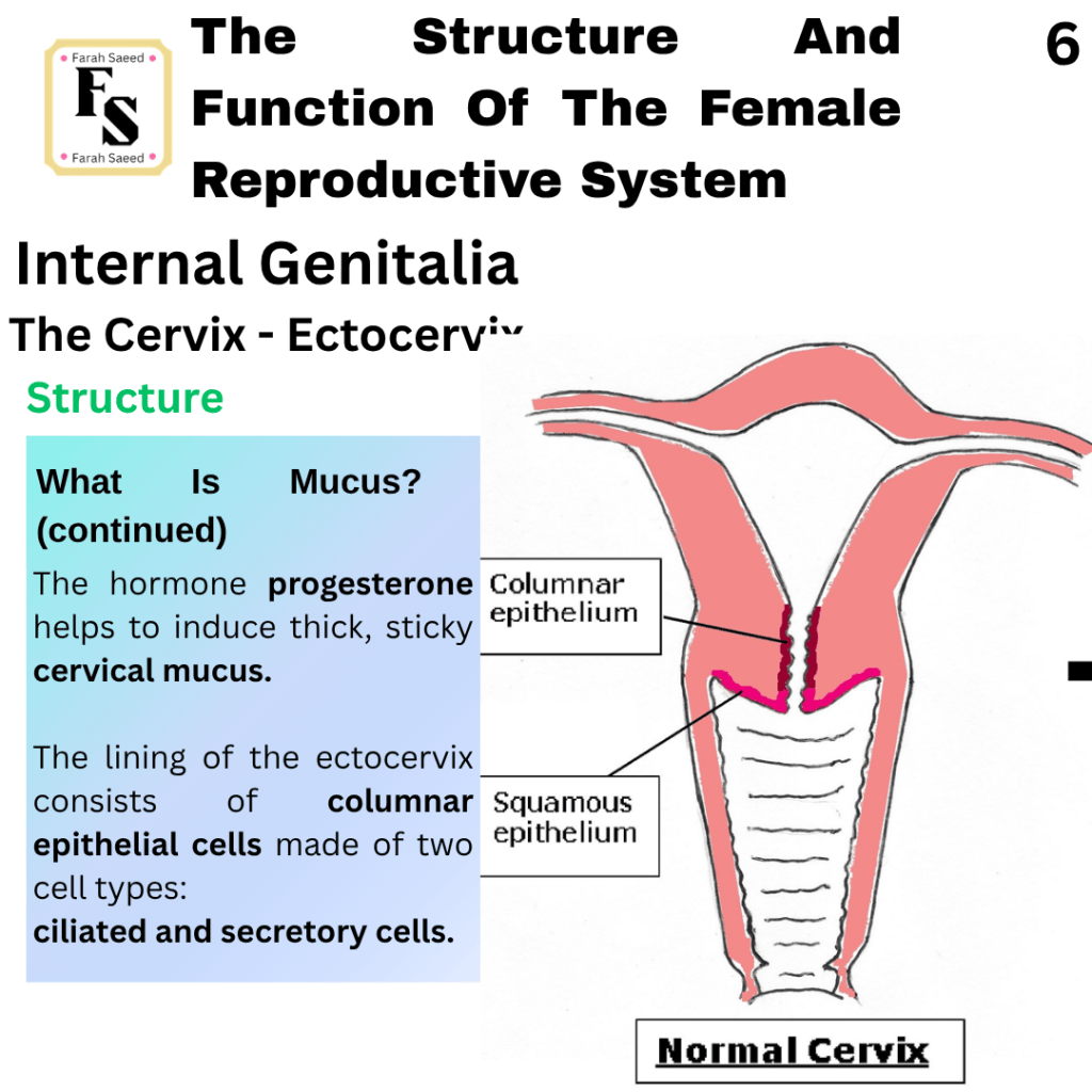
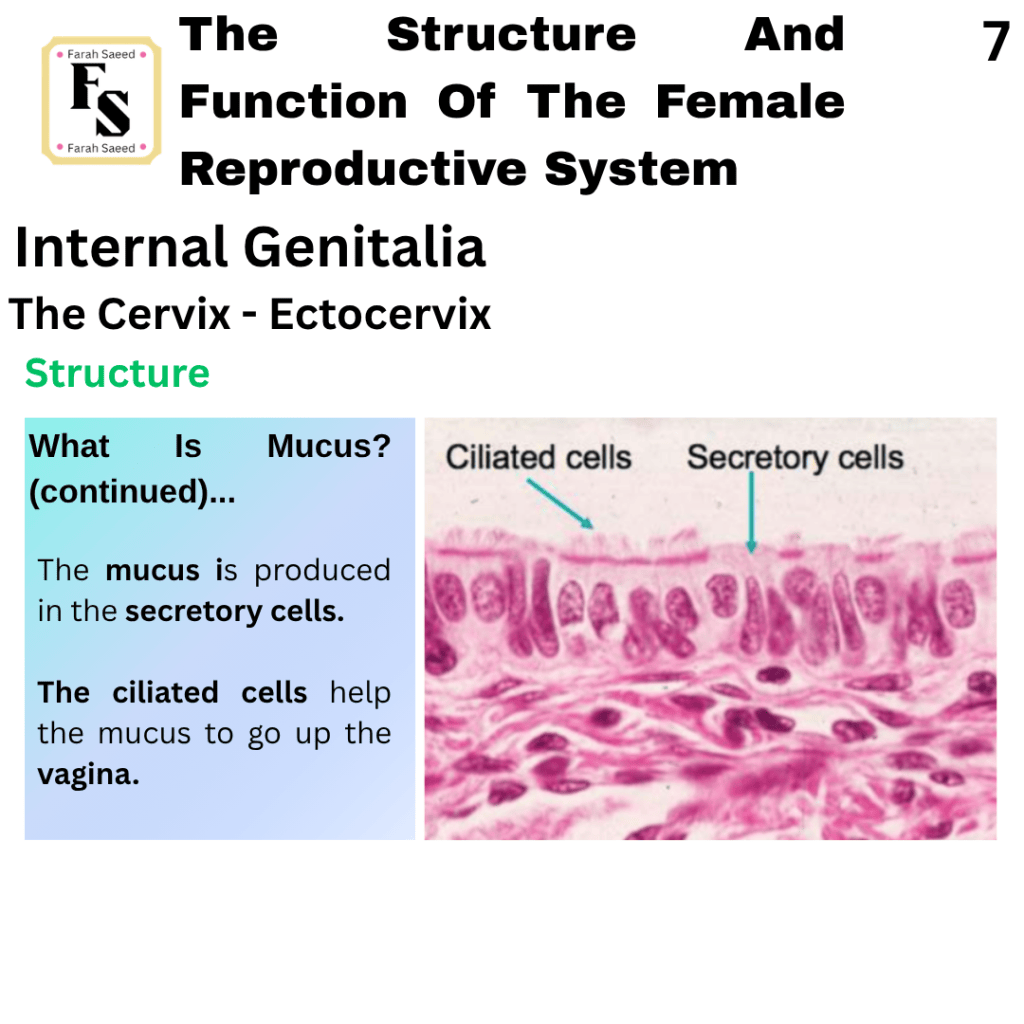
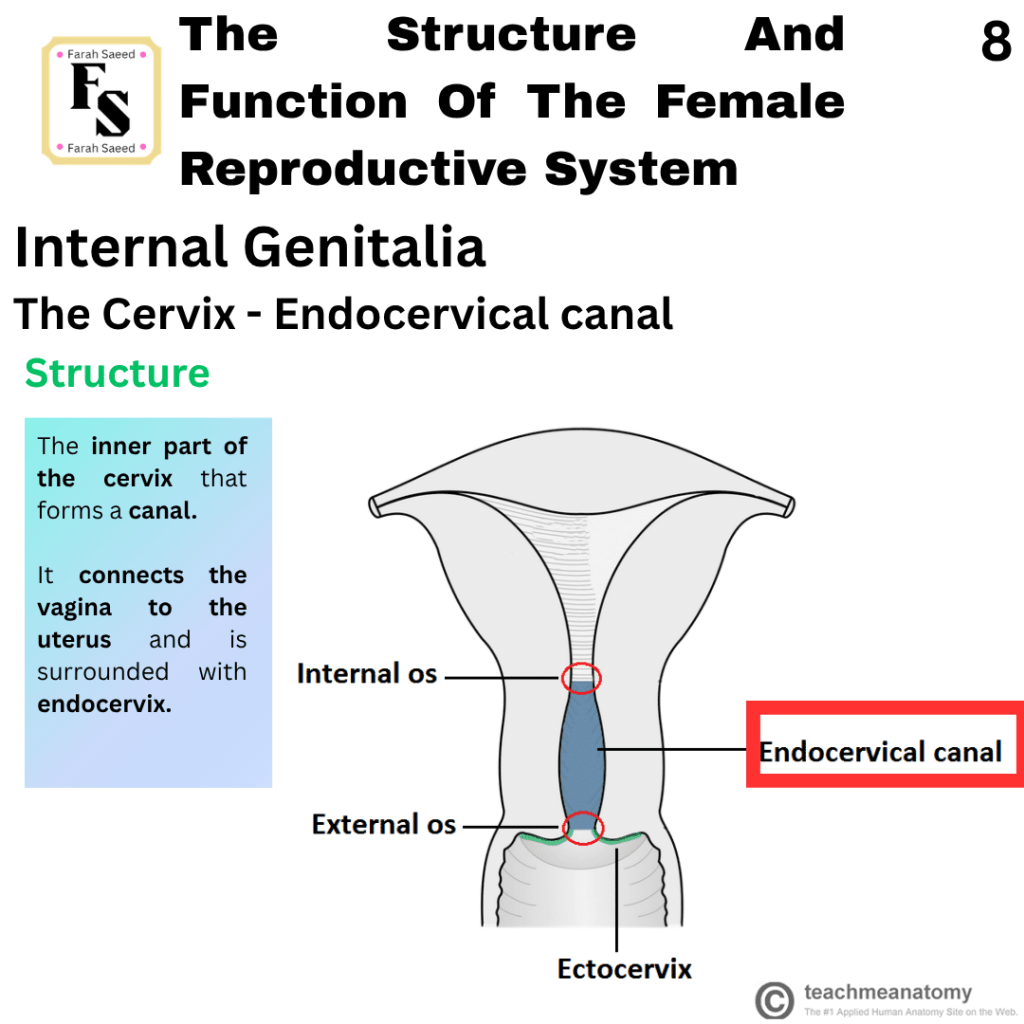
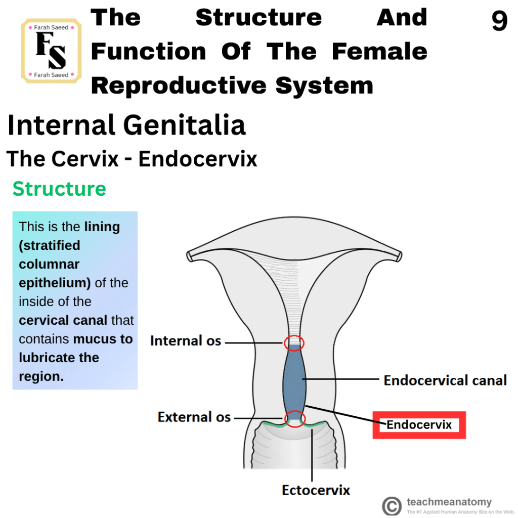
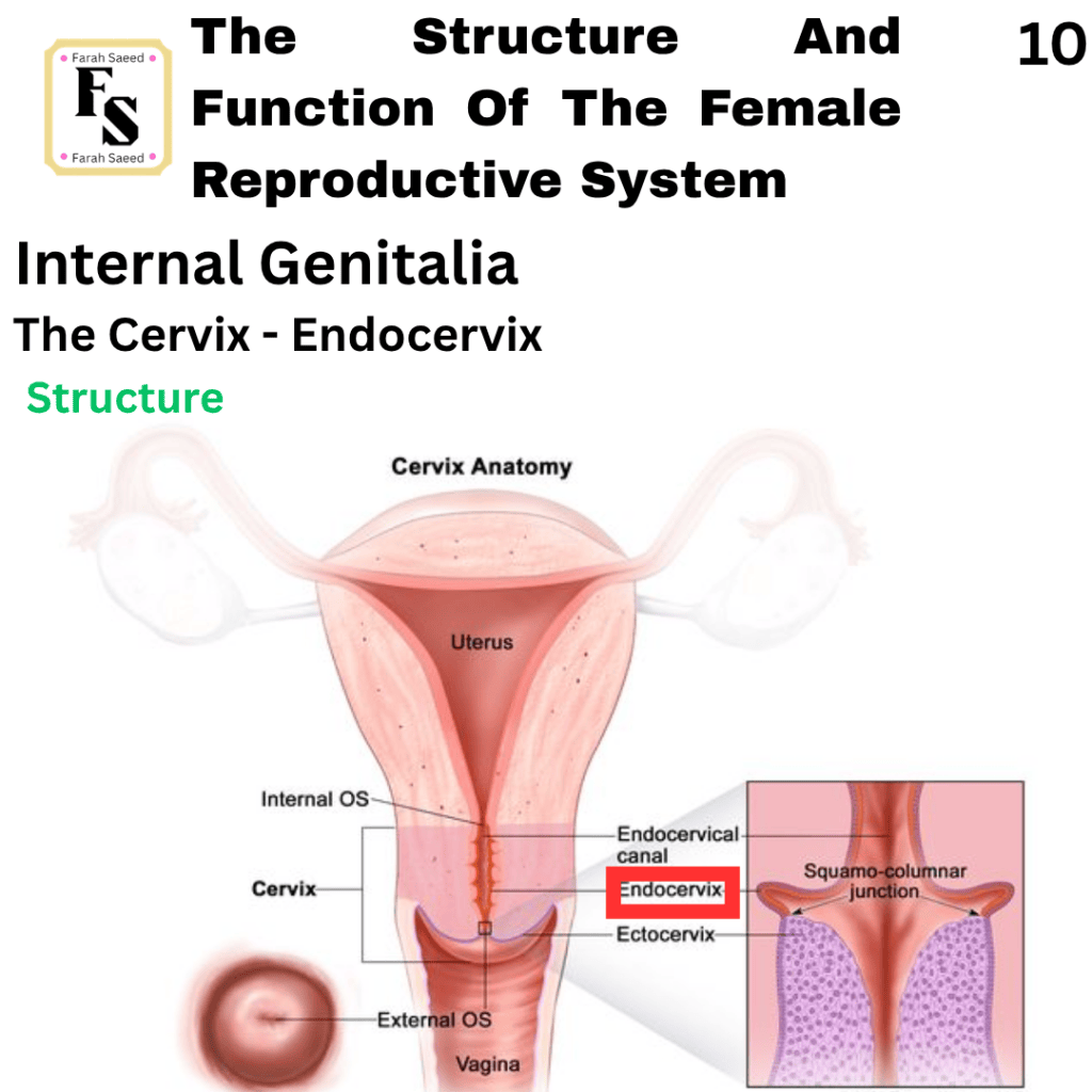
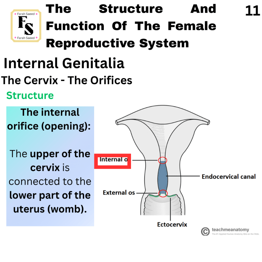
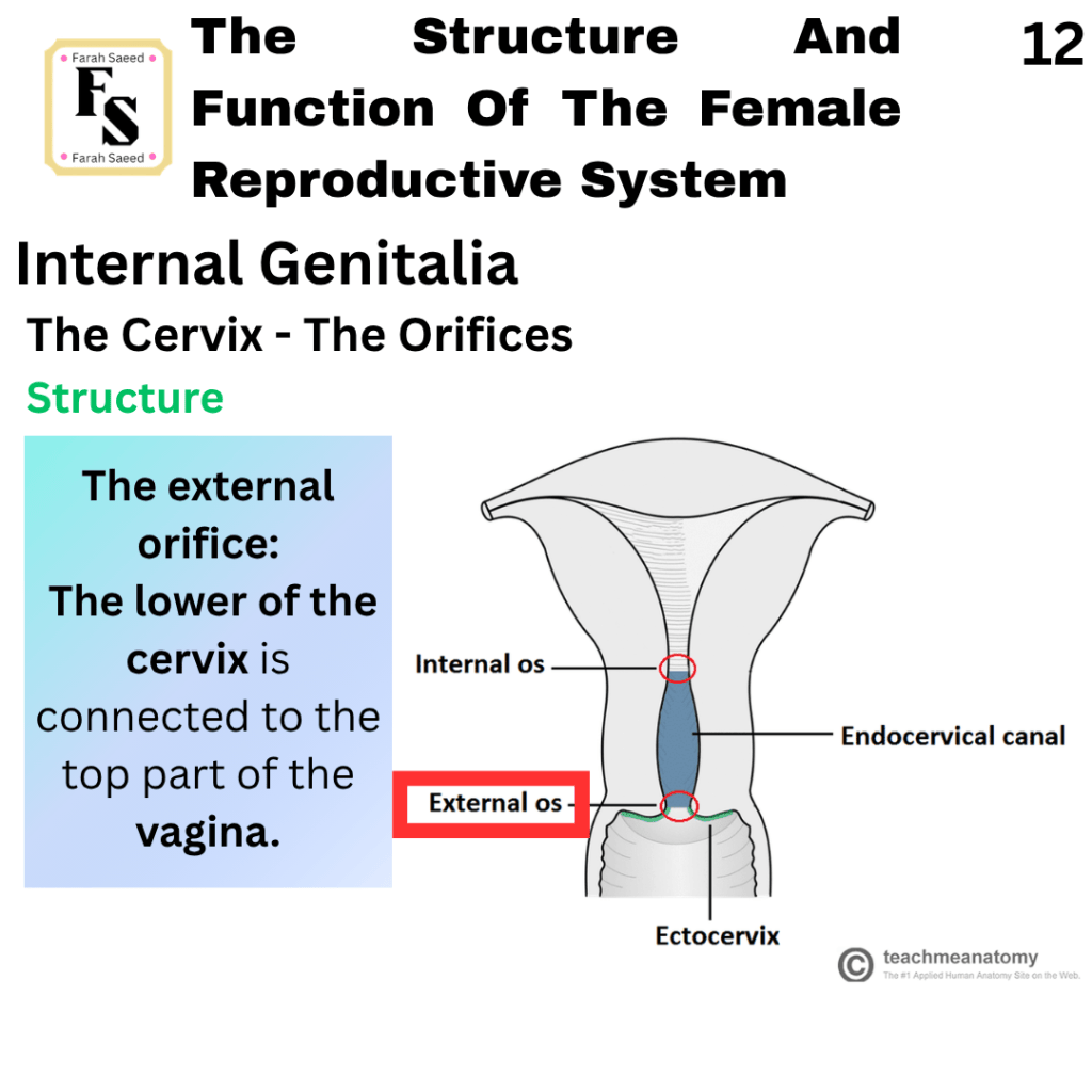
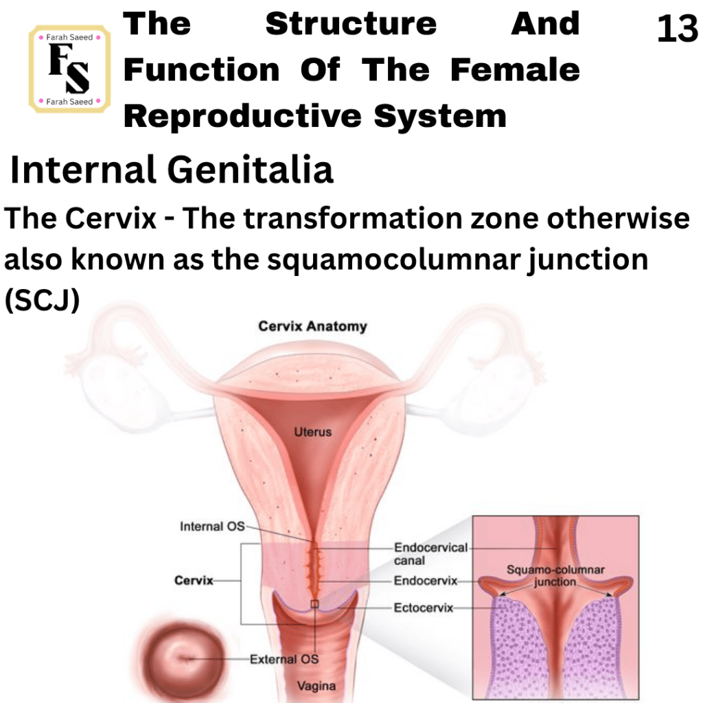
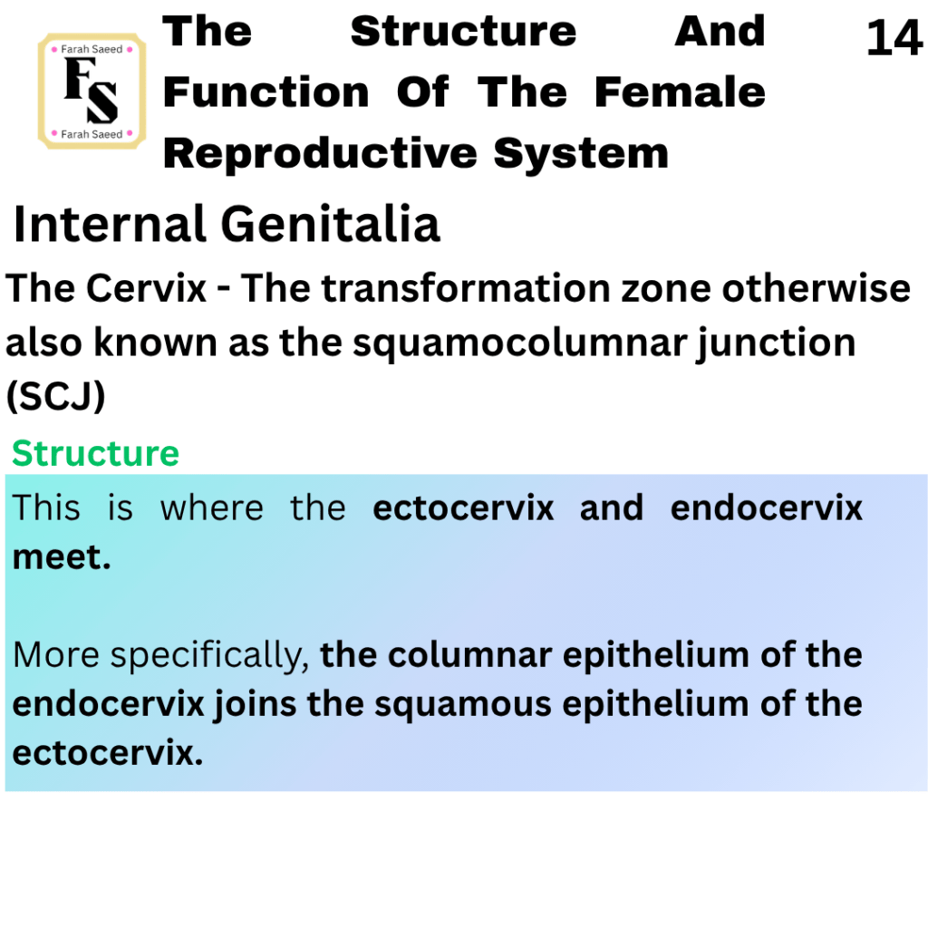
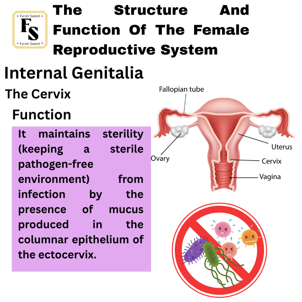
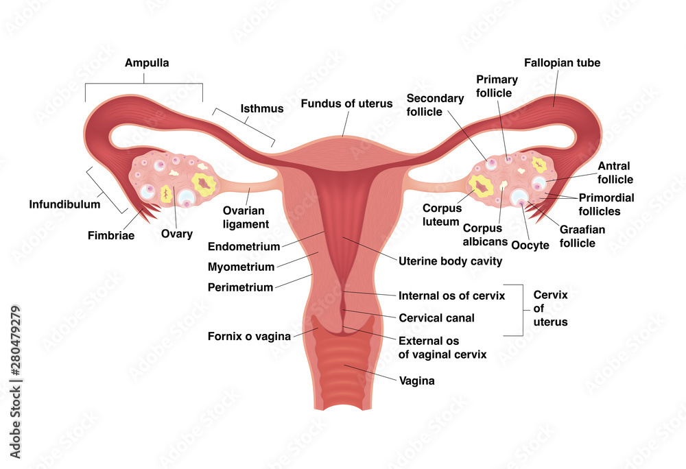
Cervix
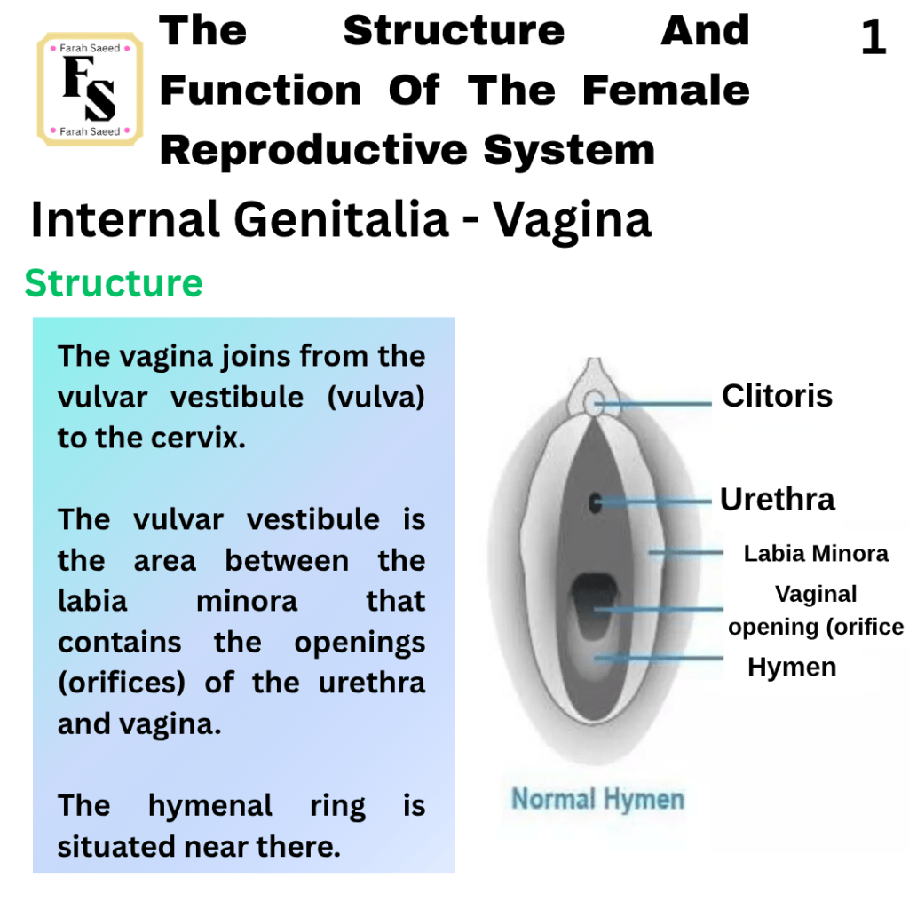
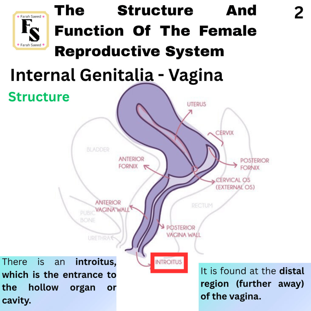
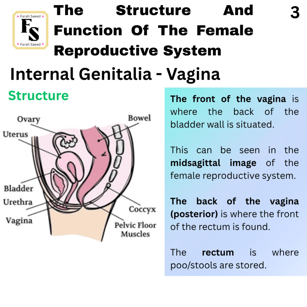
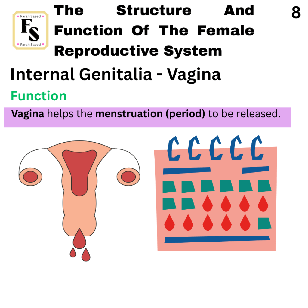
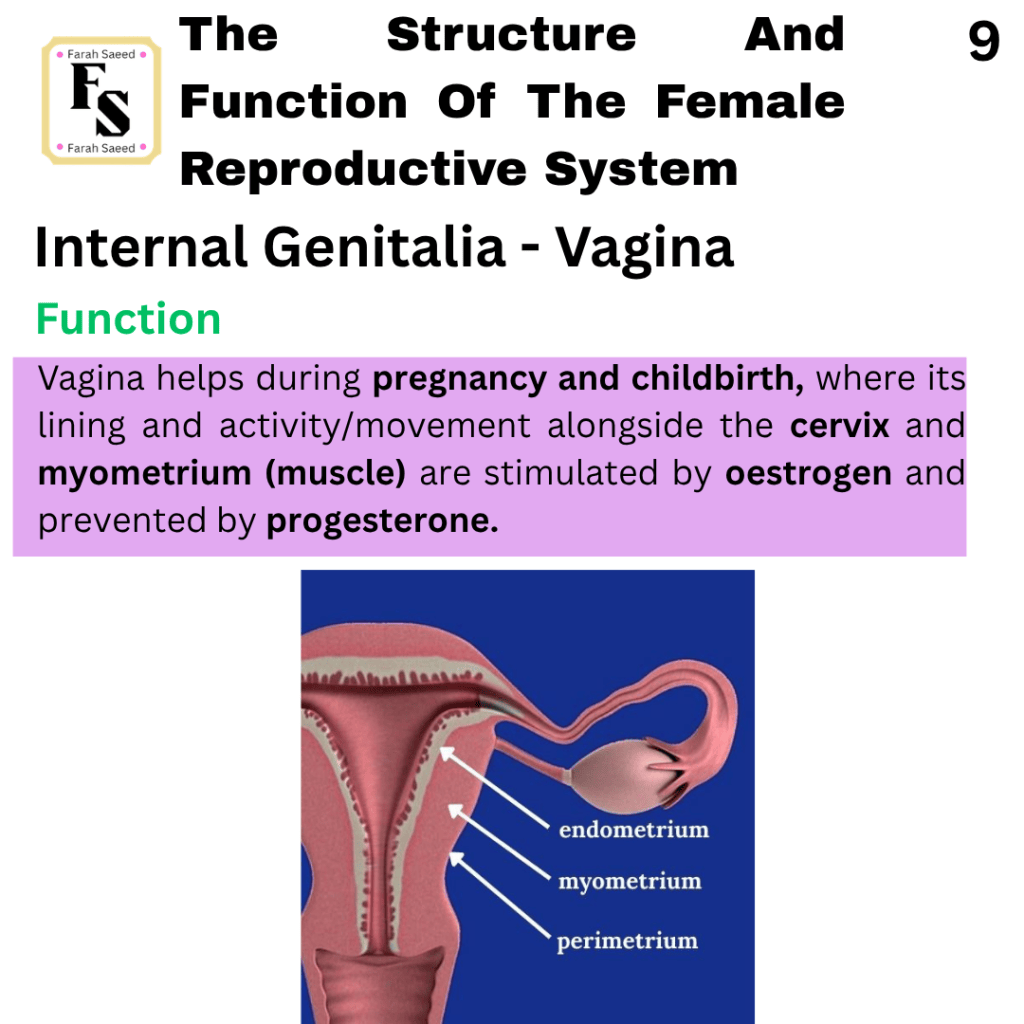
Vagina
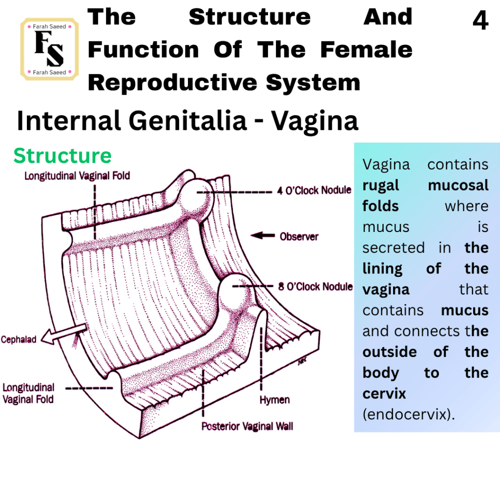
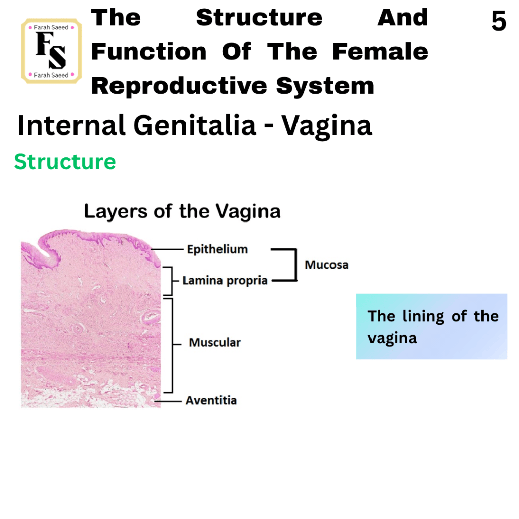
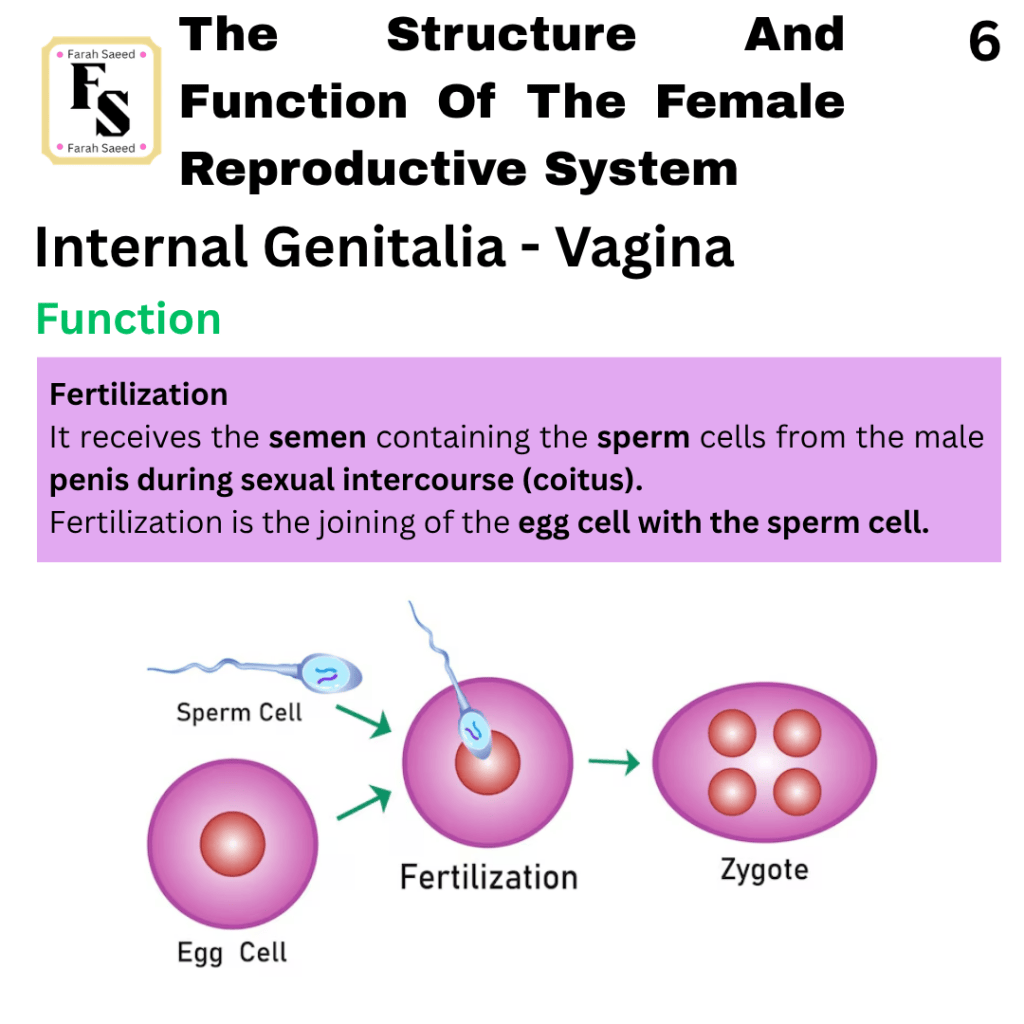
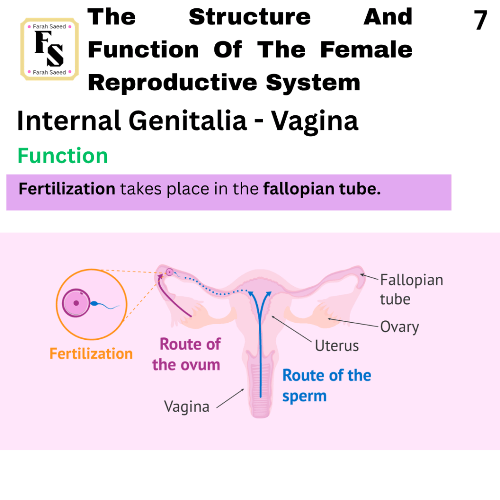
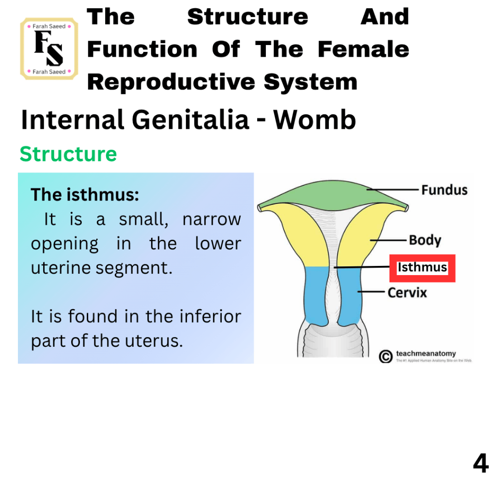
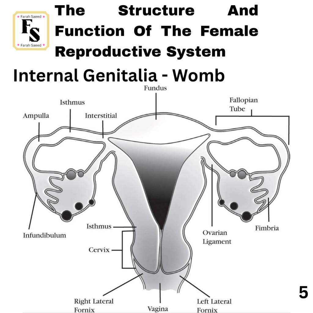
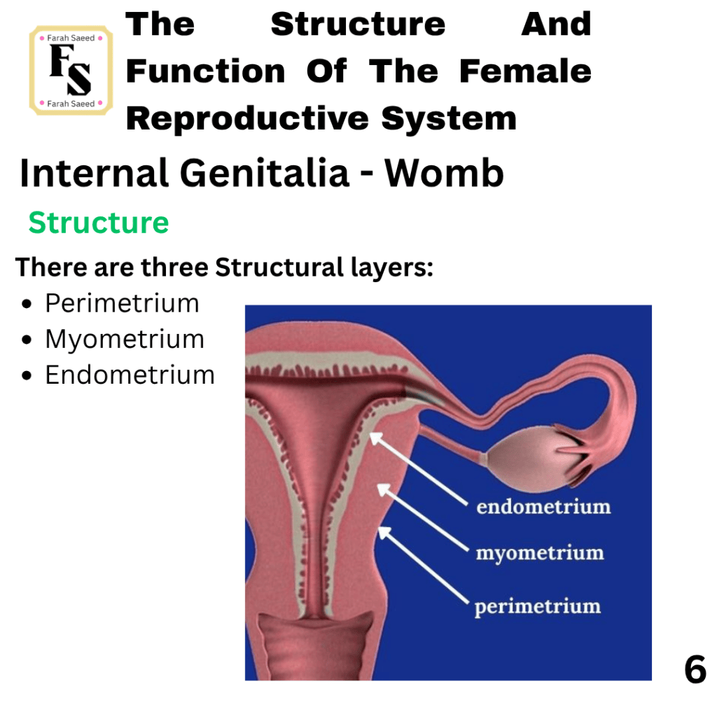
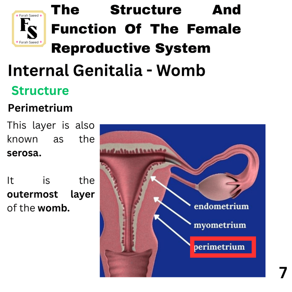
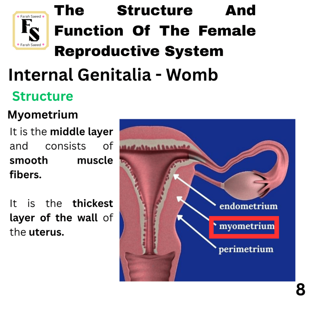
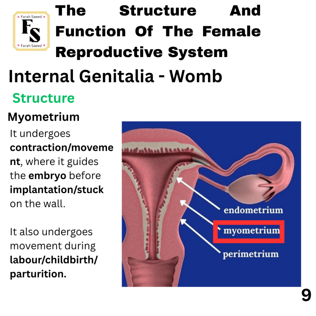
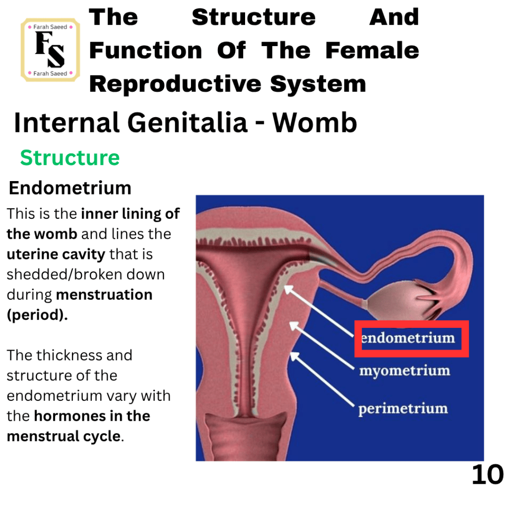
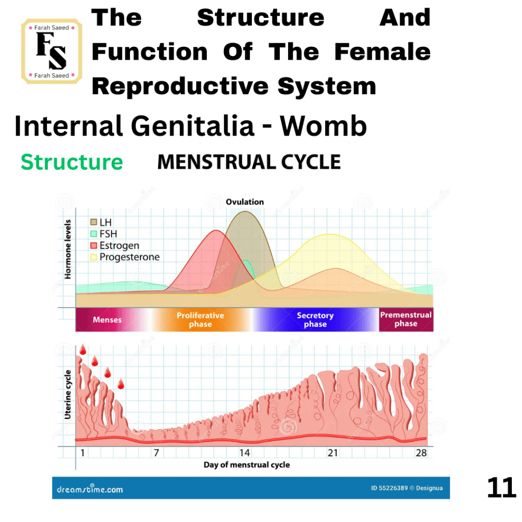
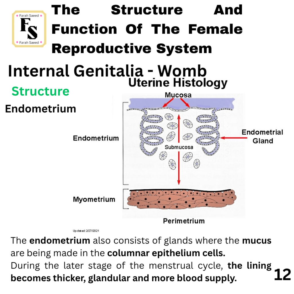
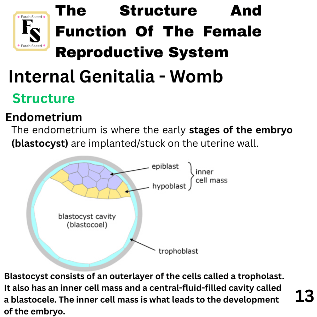
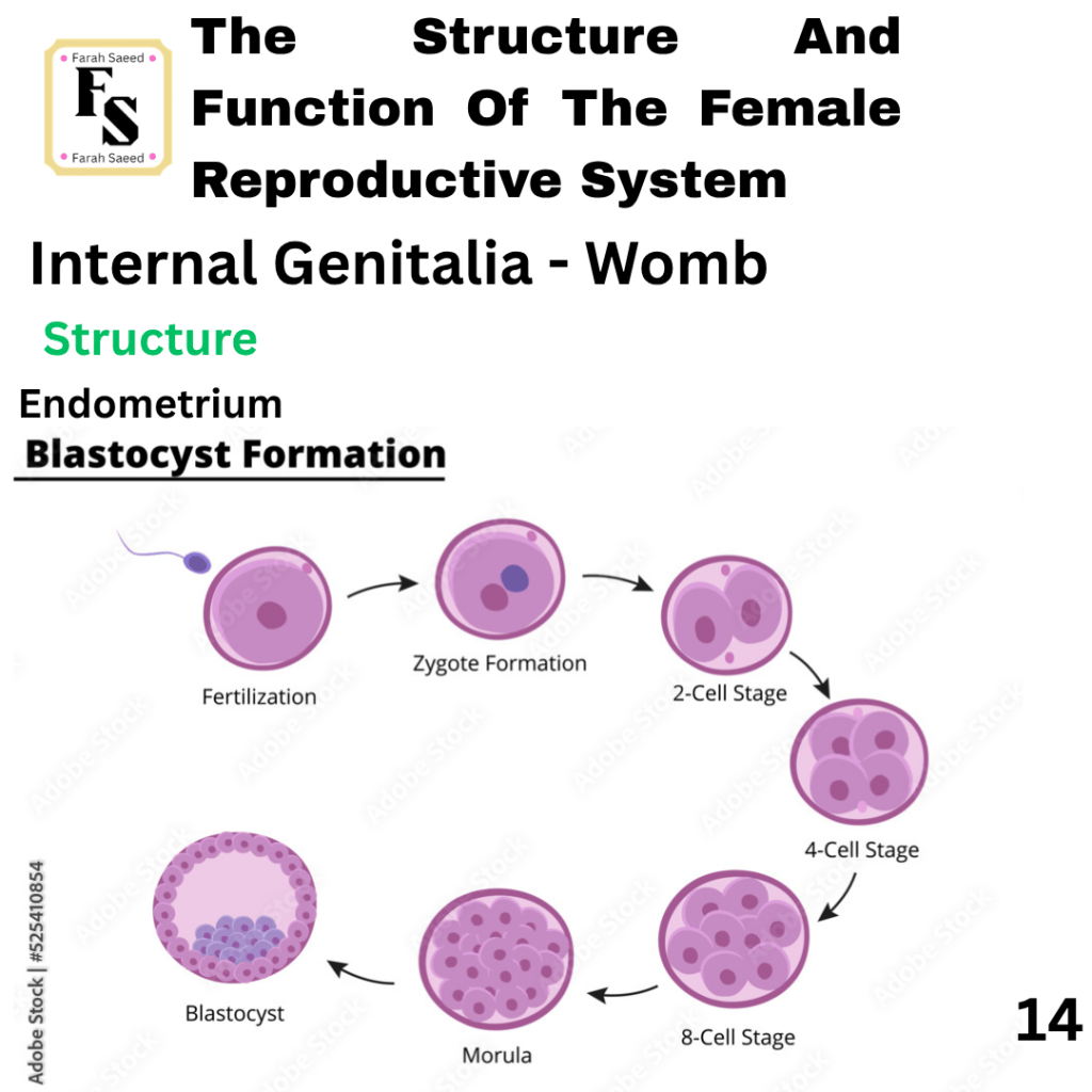
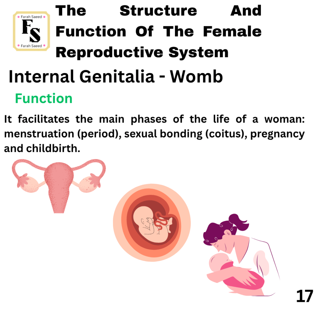
Womb
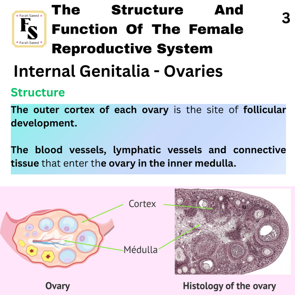
Ovaries
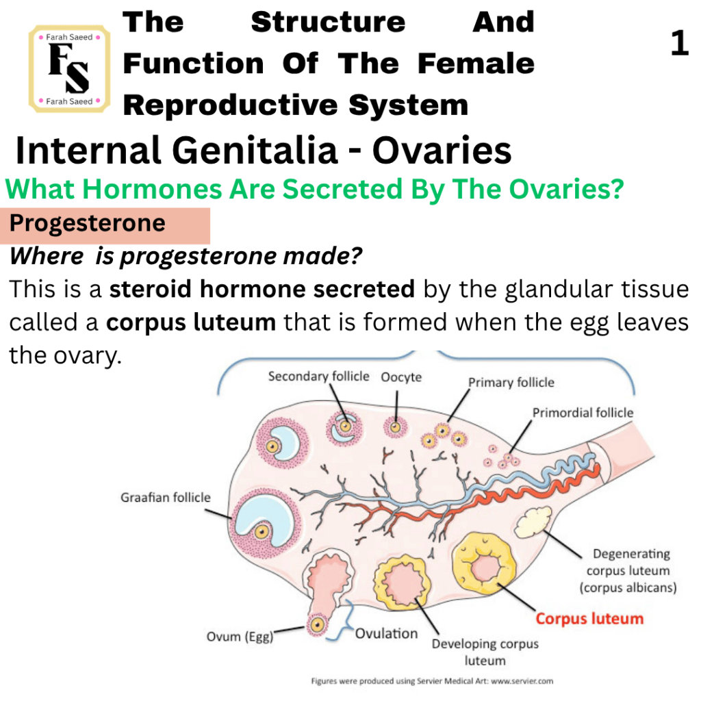
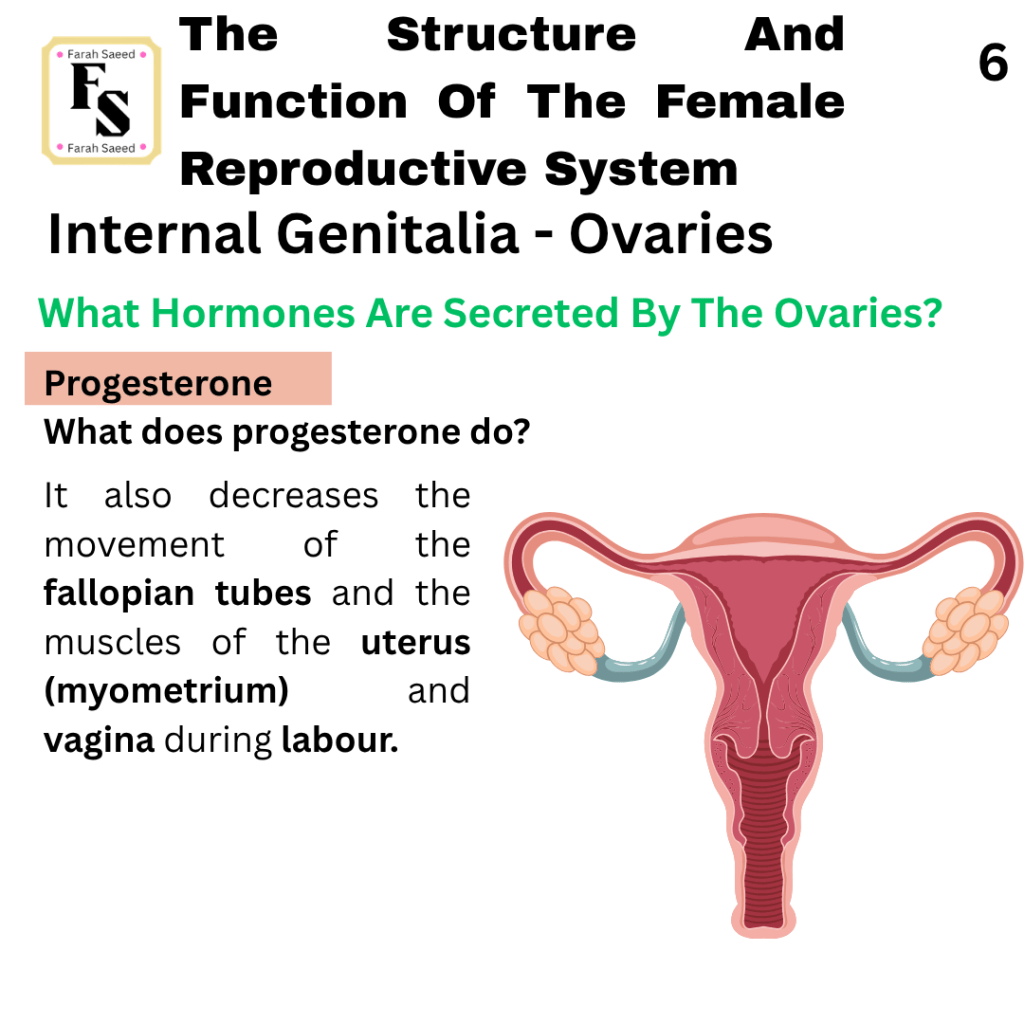
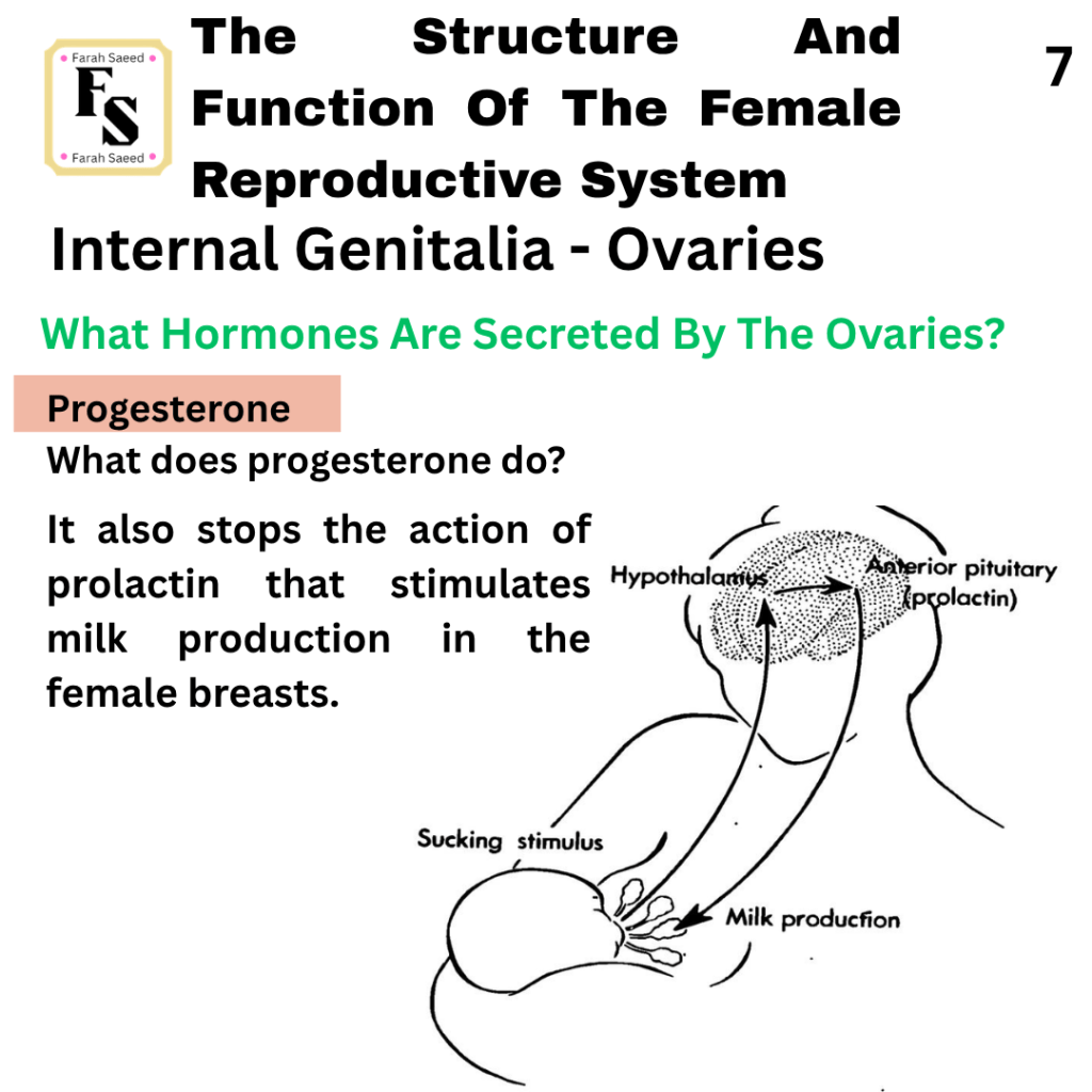
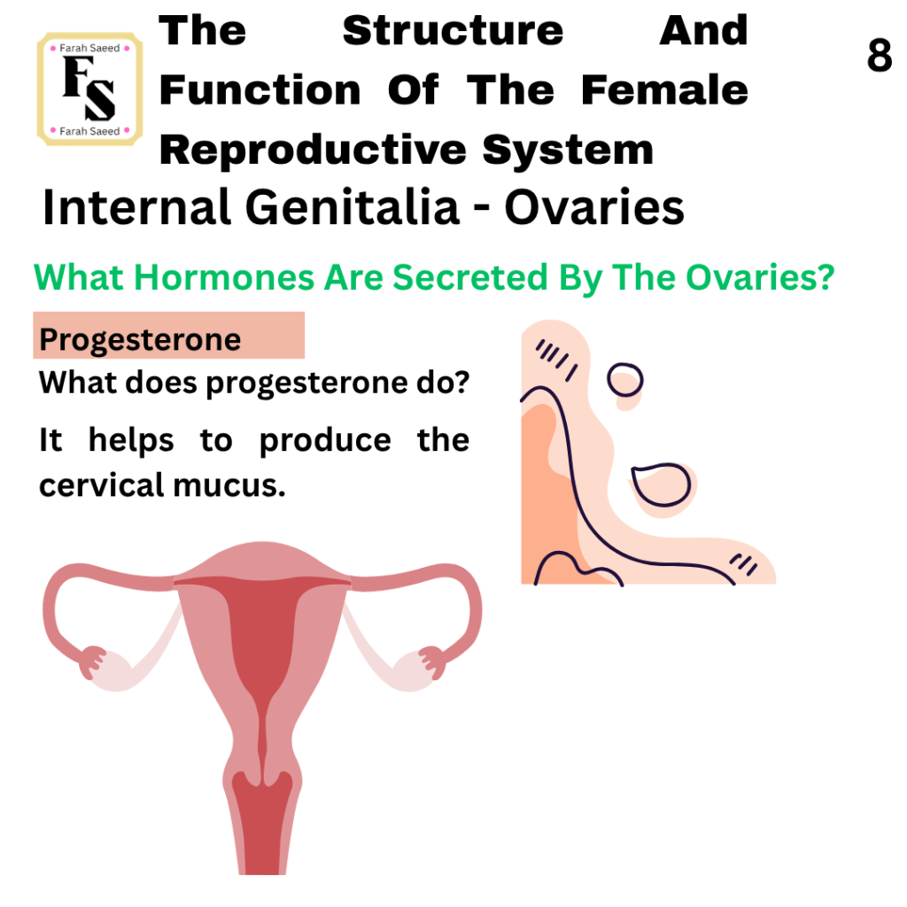
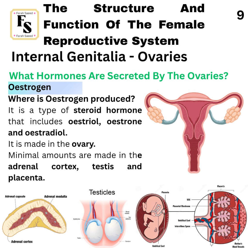
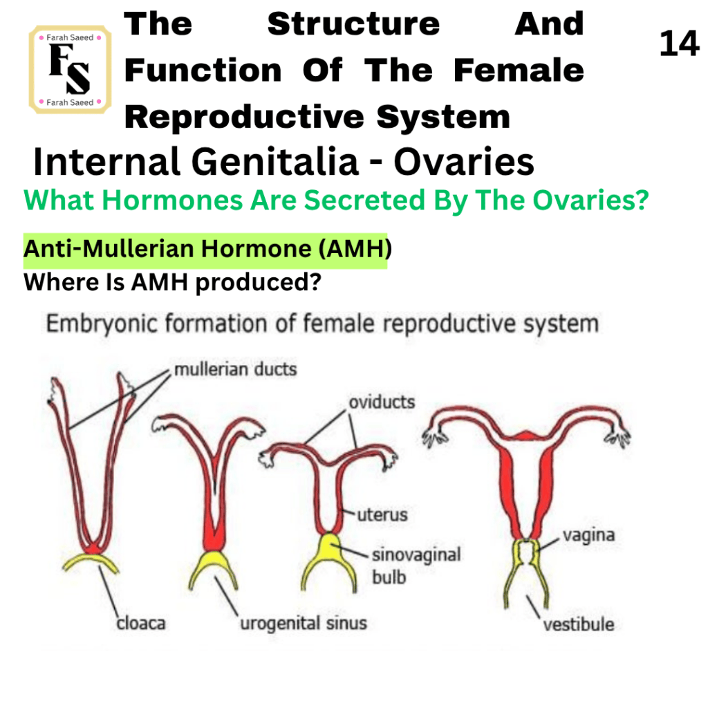
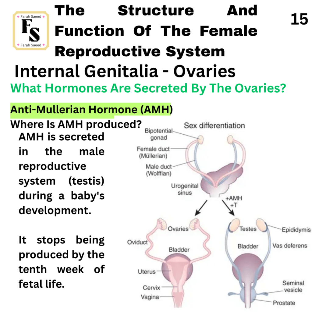
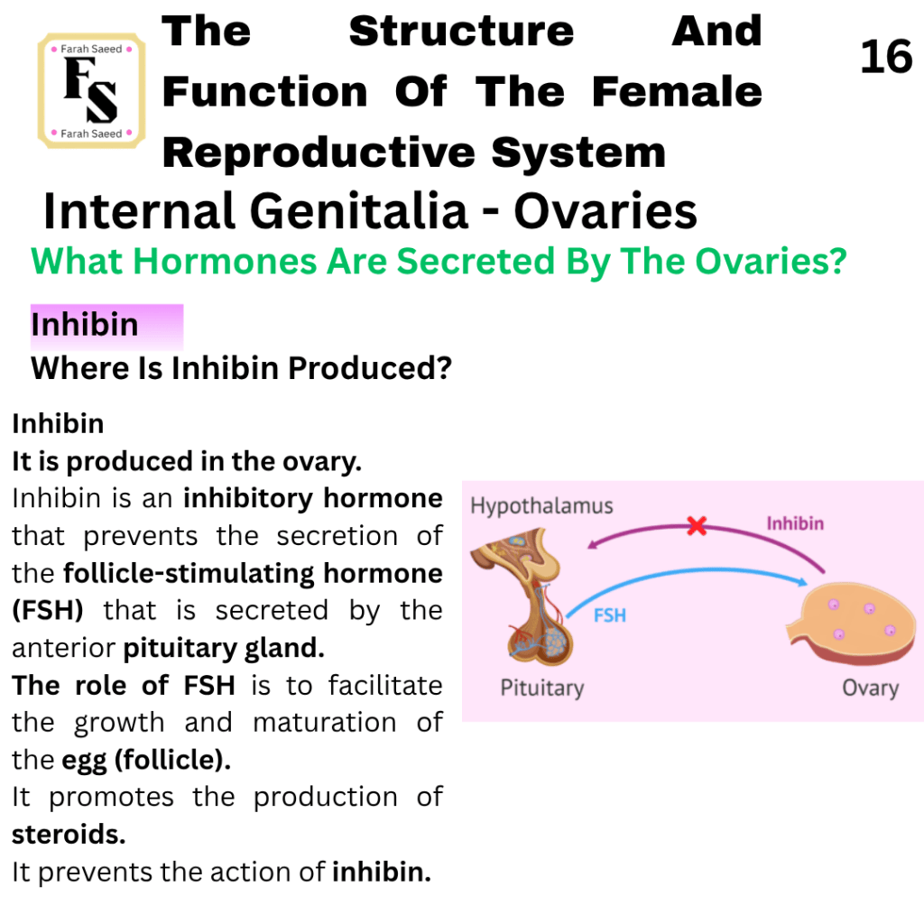
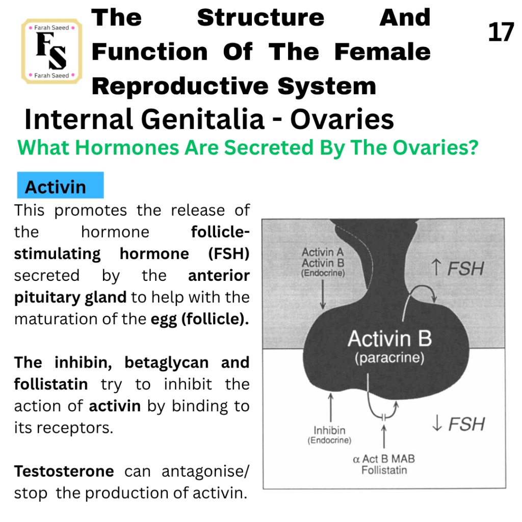
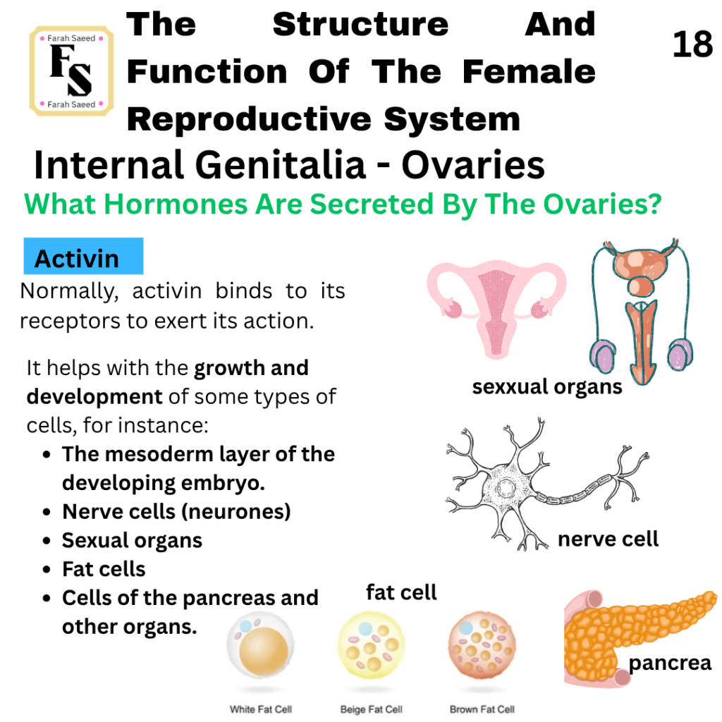
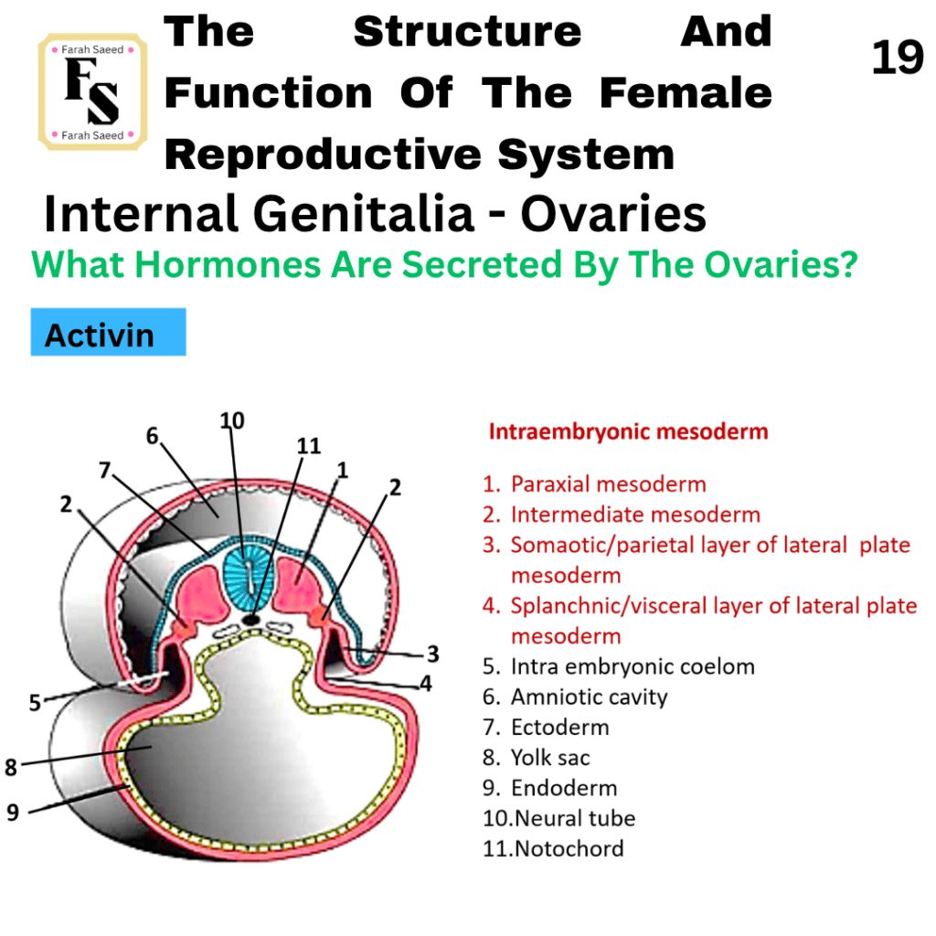
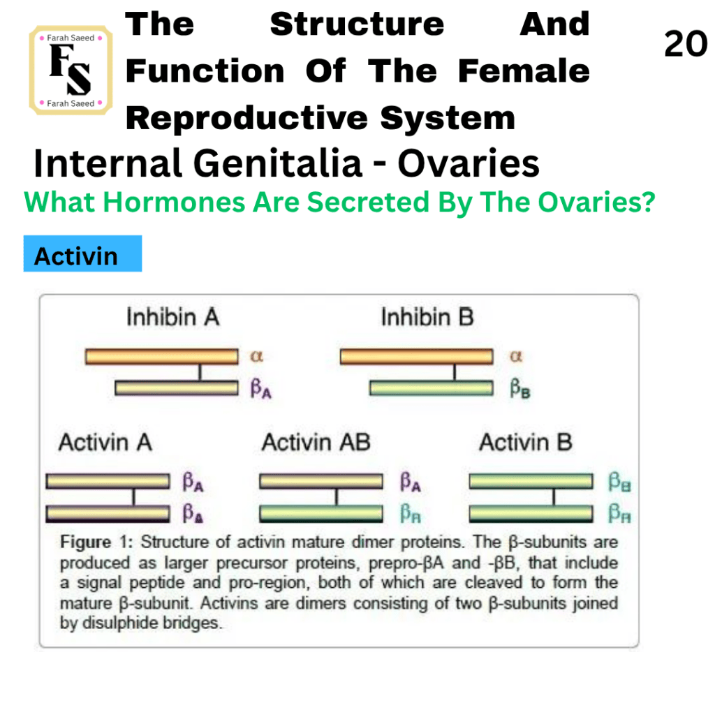
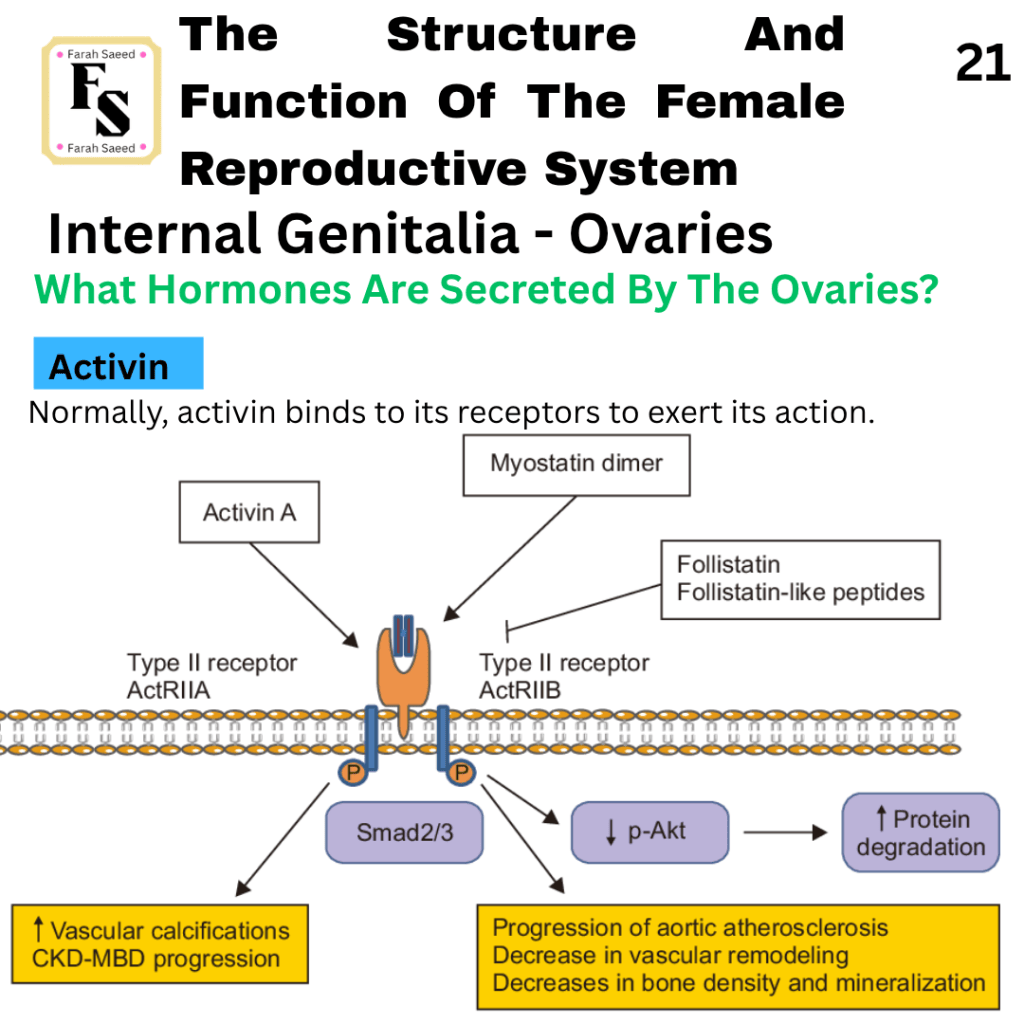
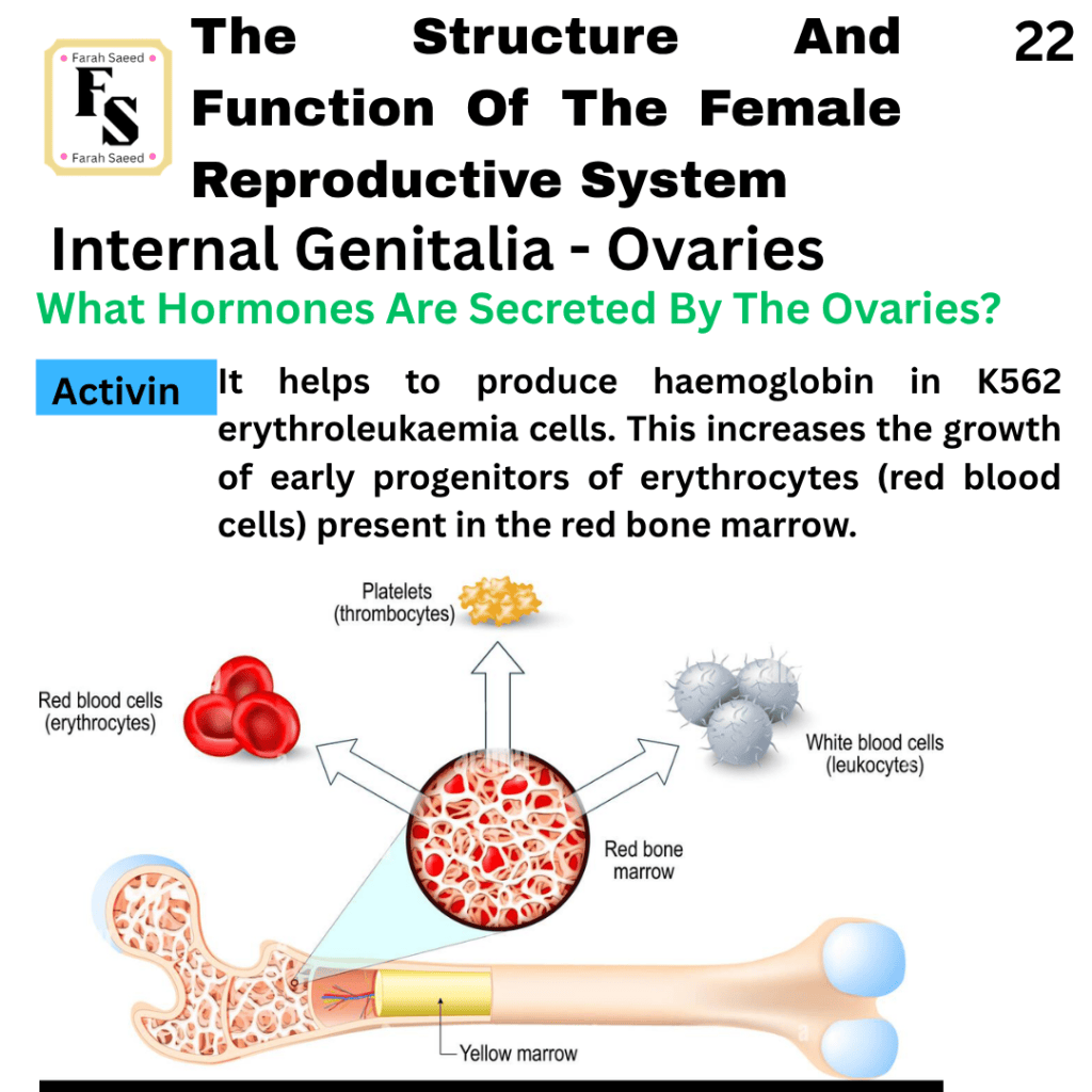
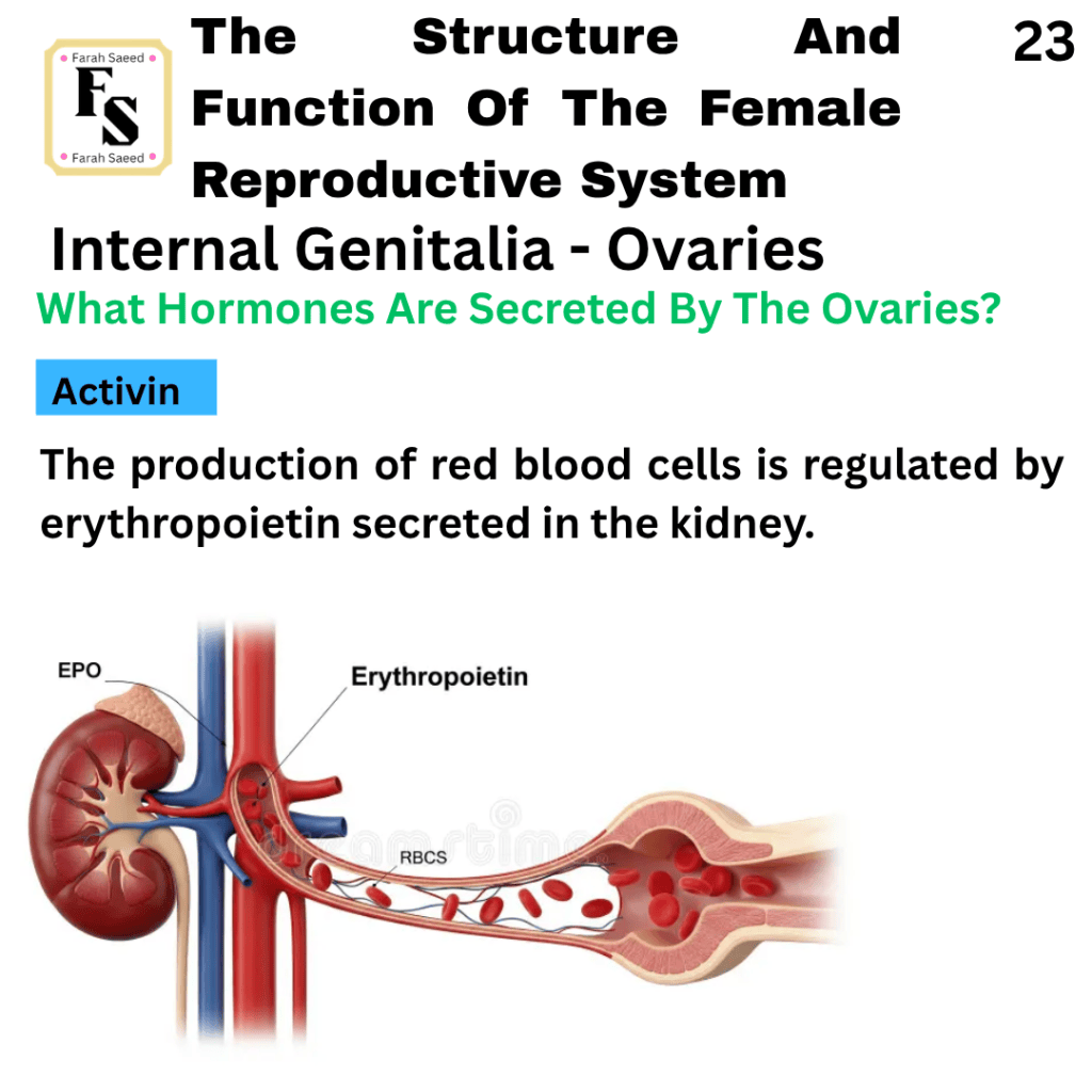
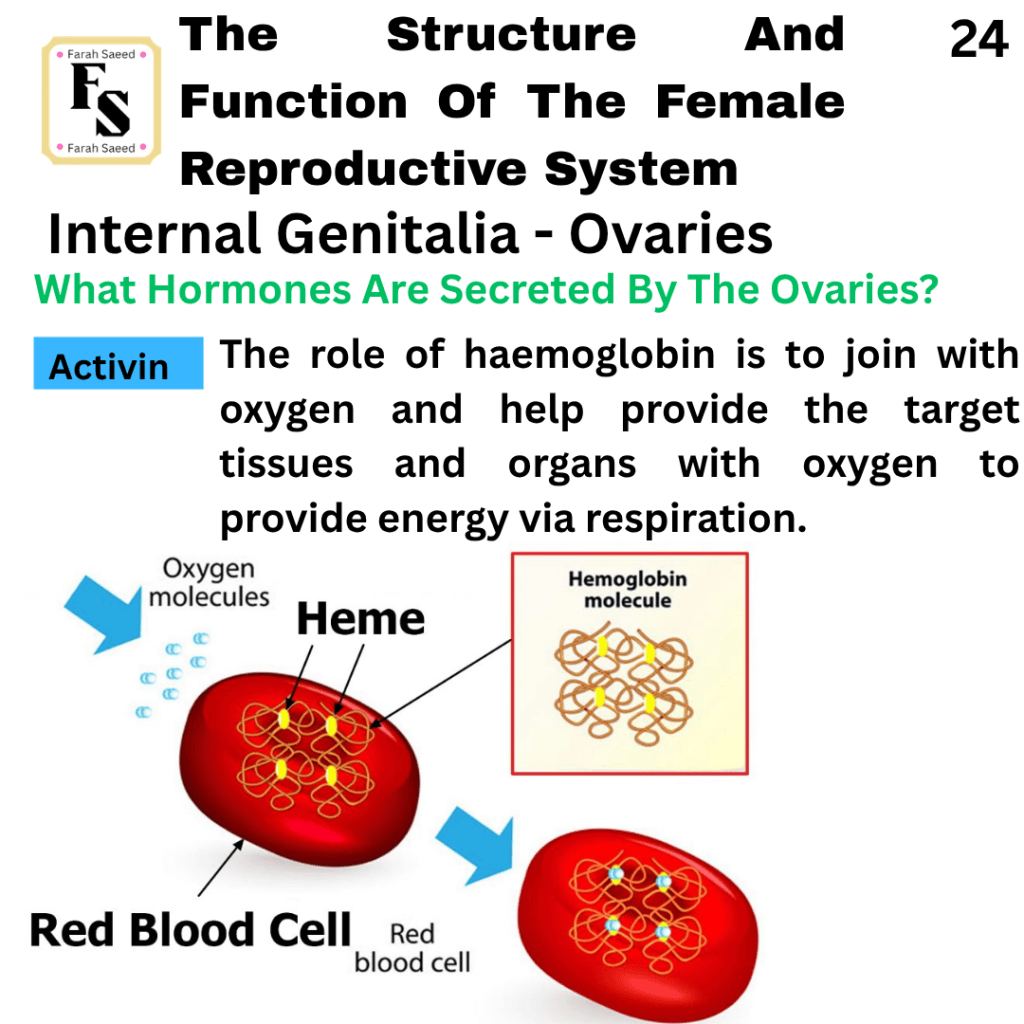
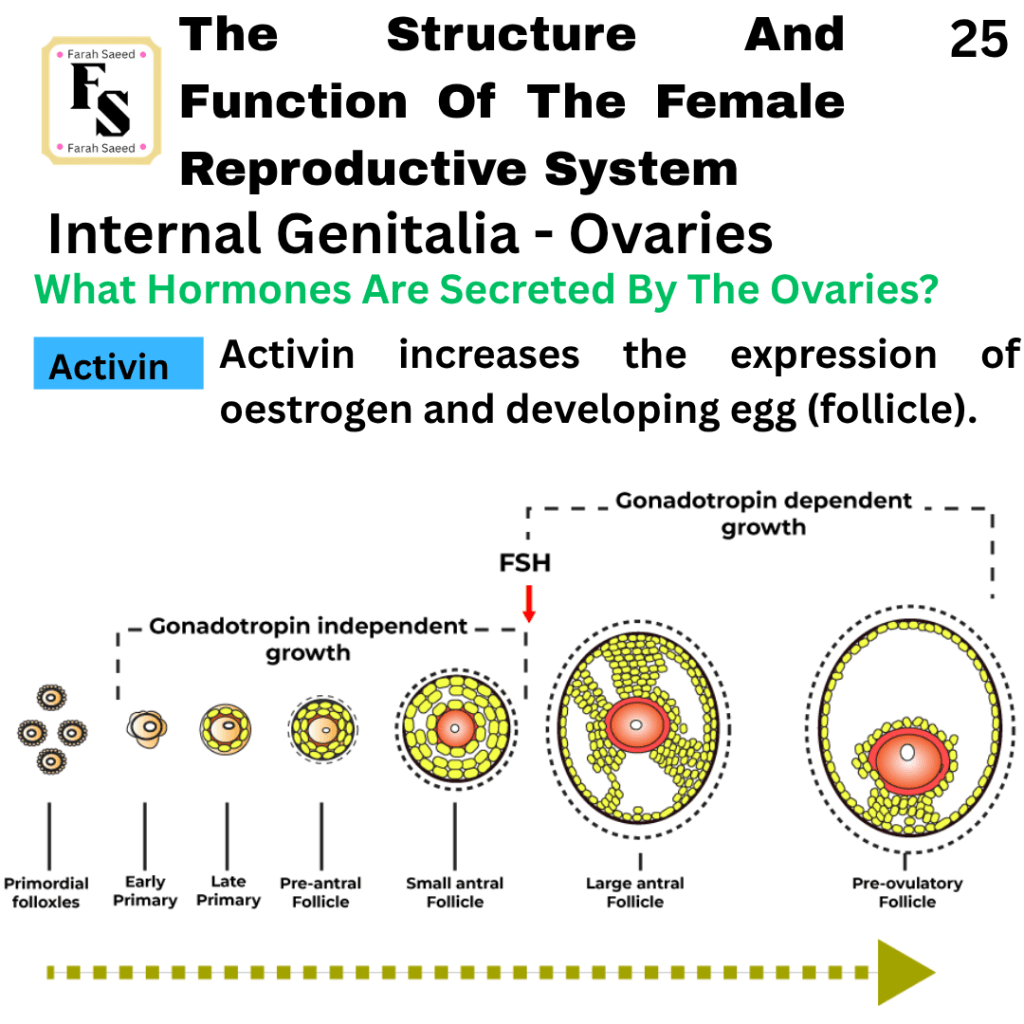
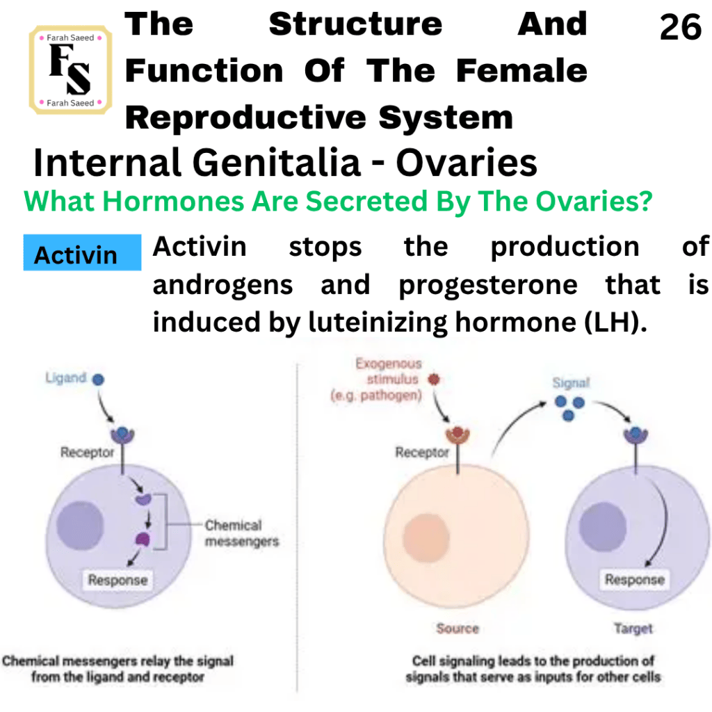
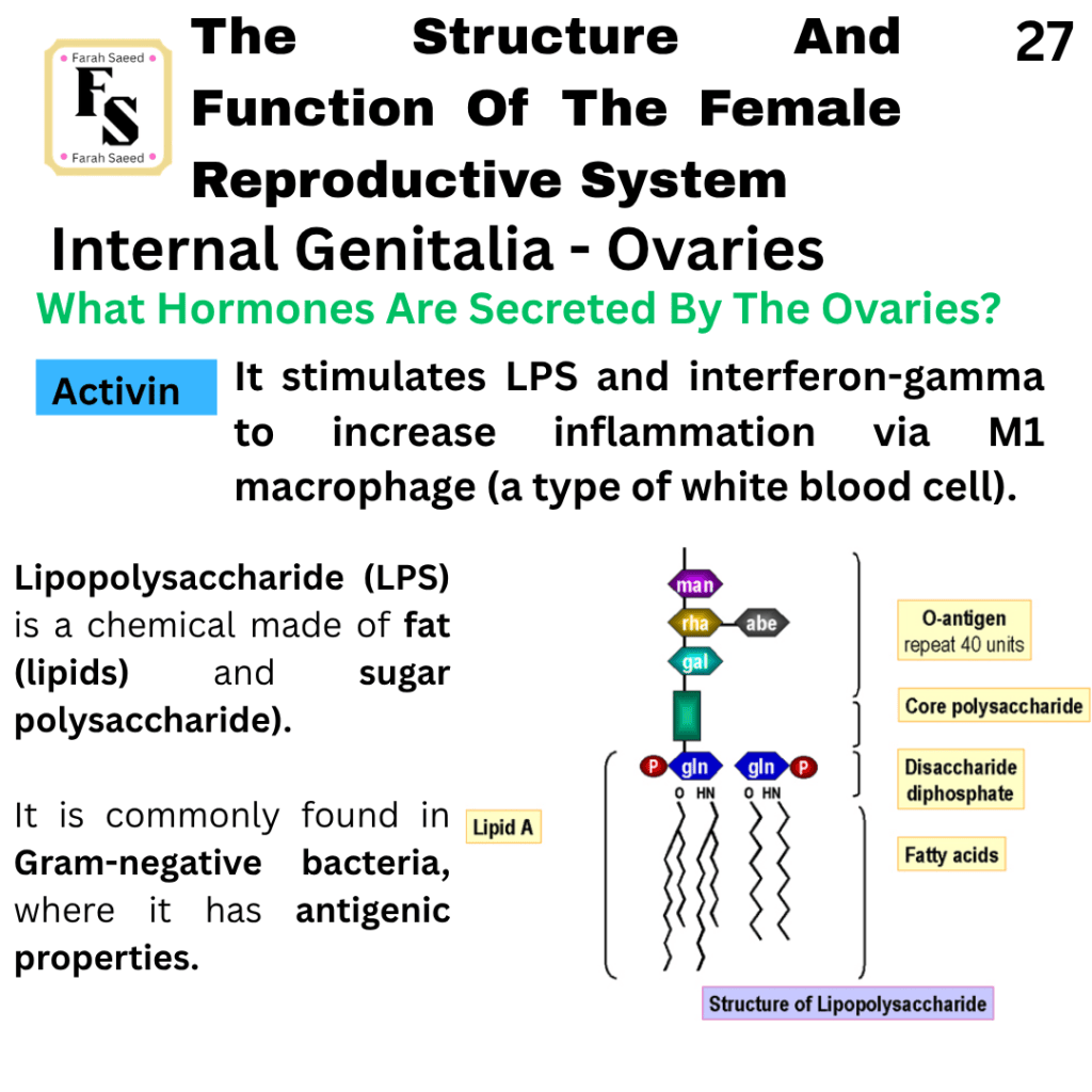
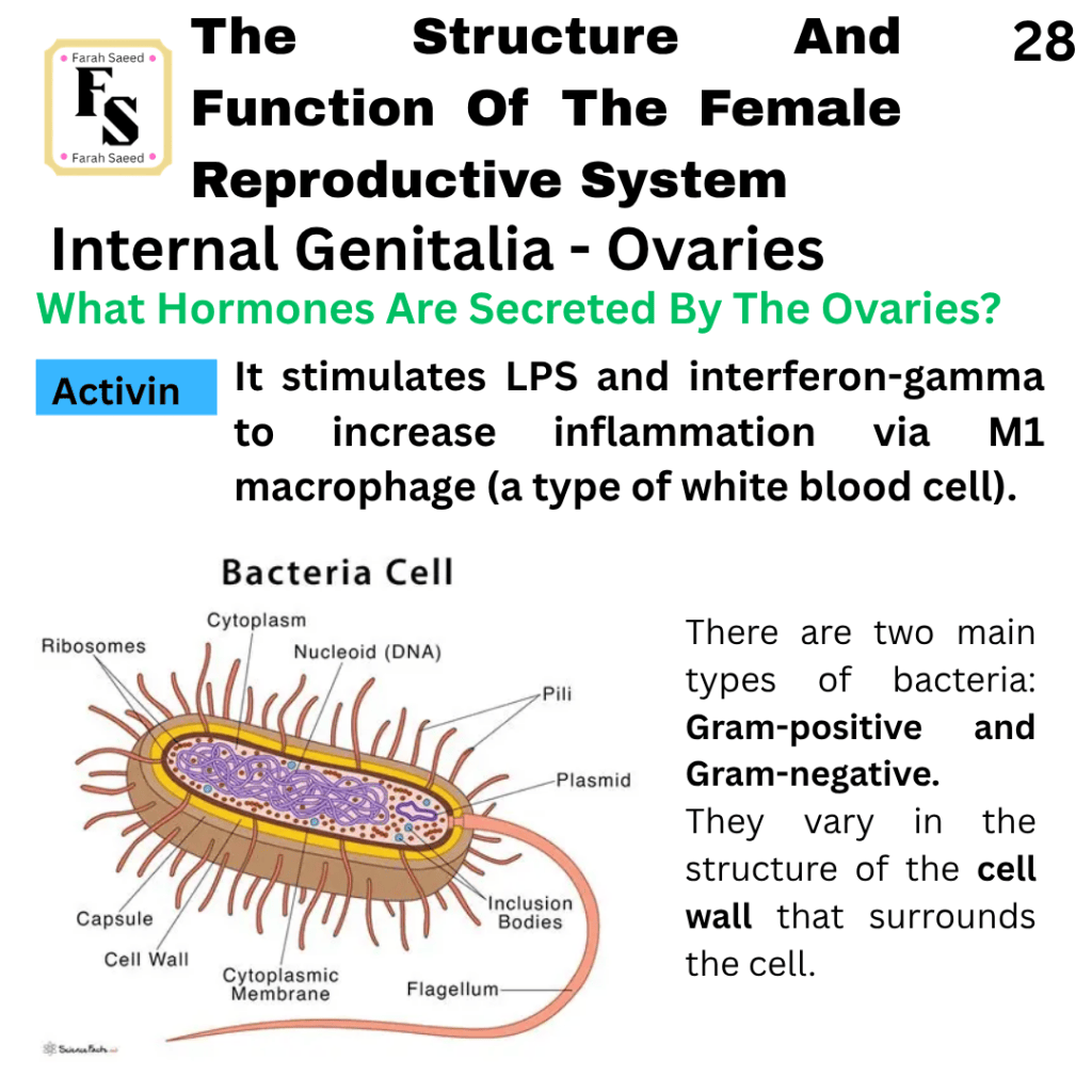
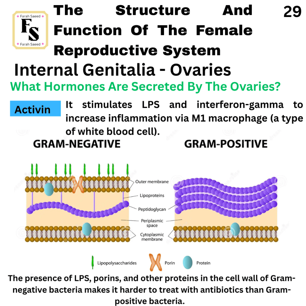
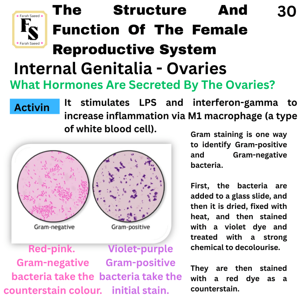
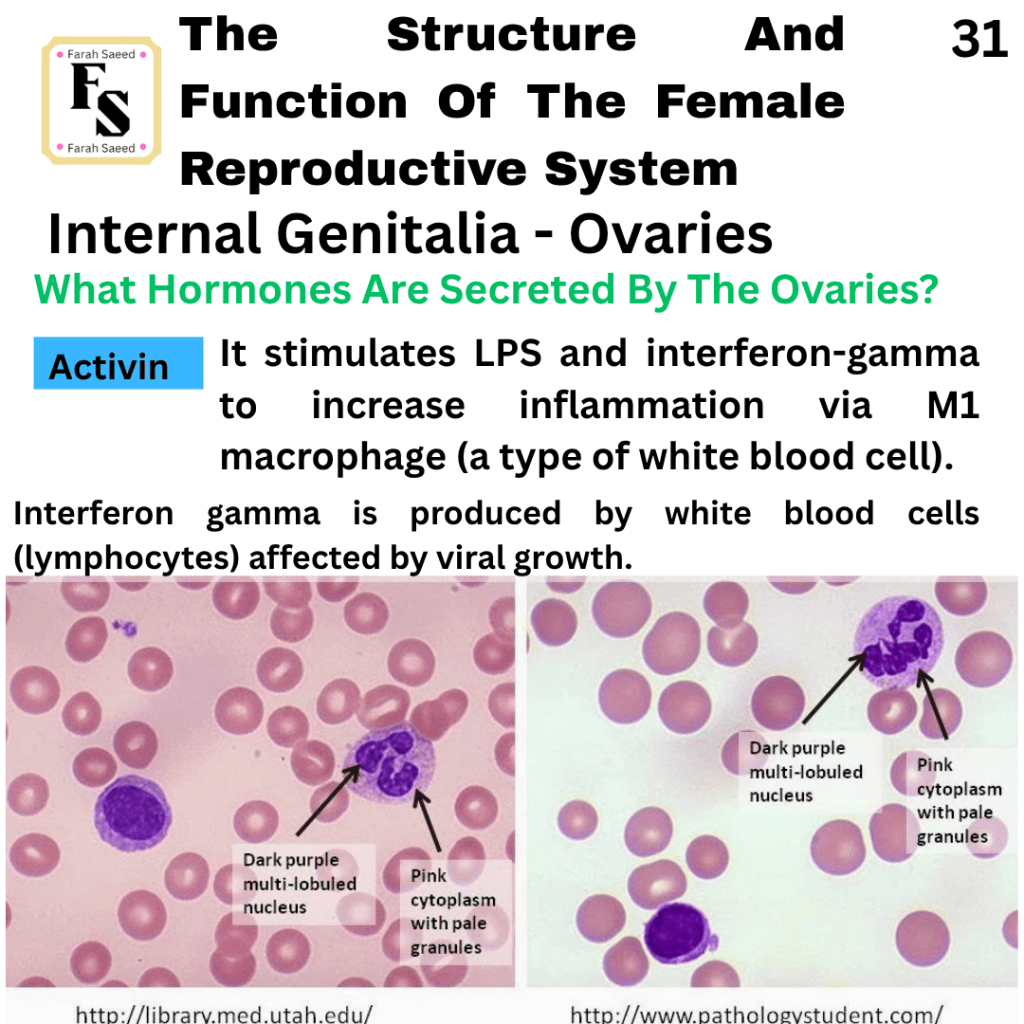
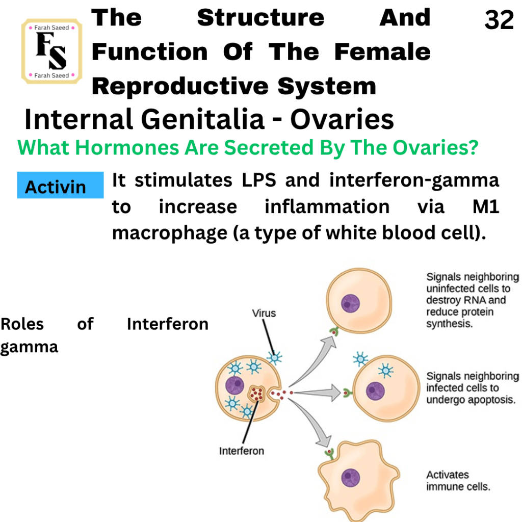
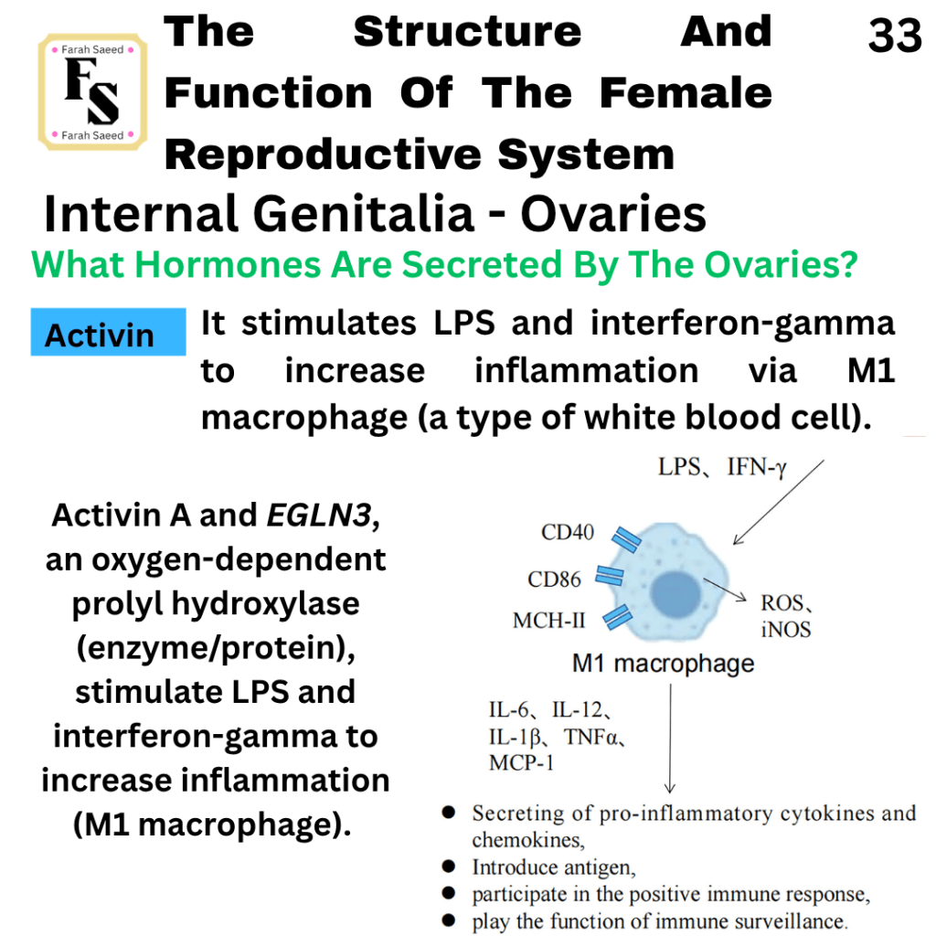
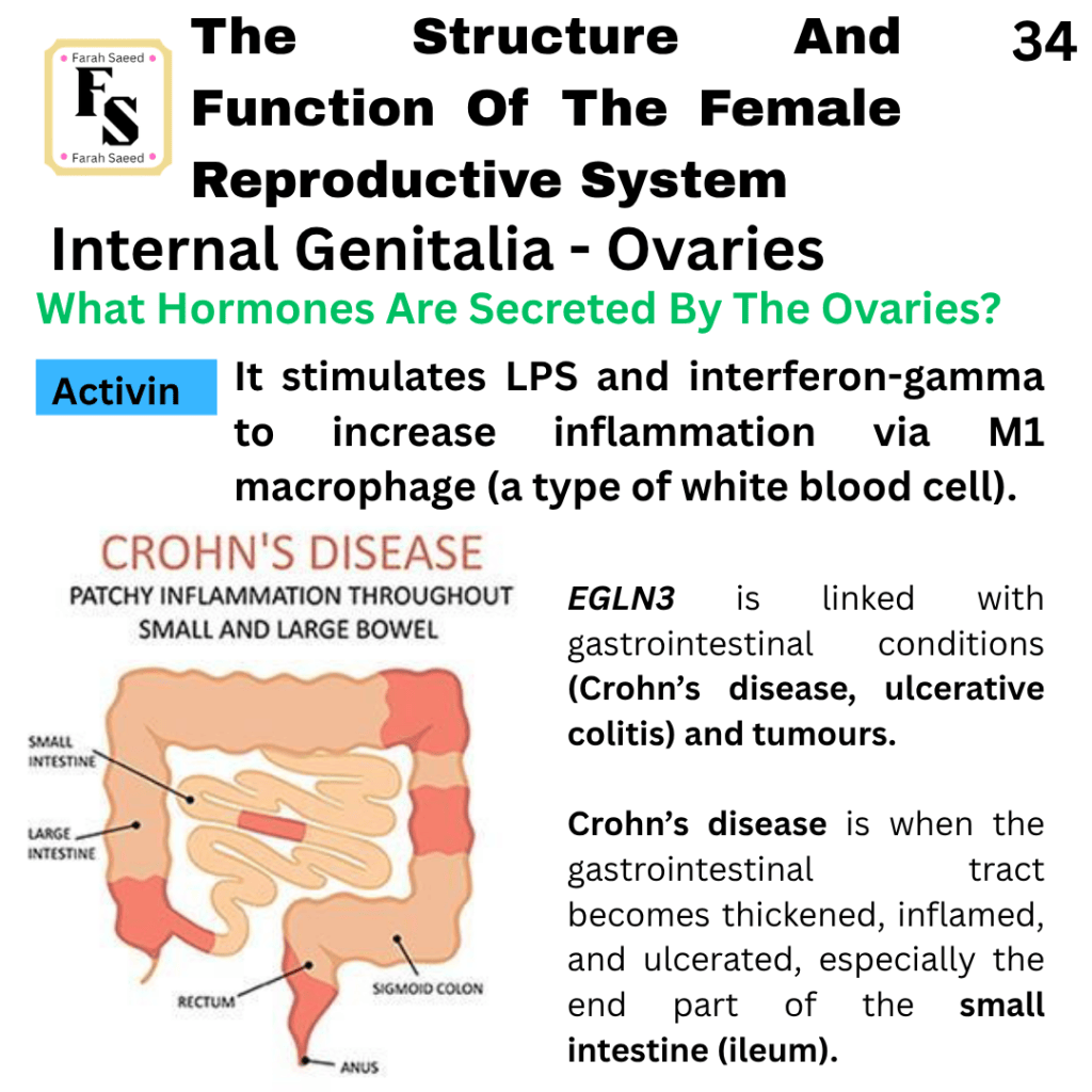
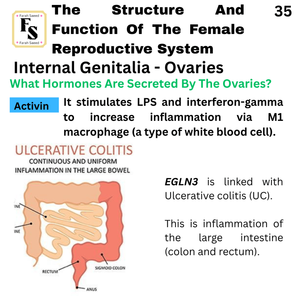
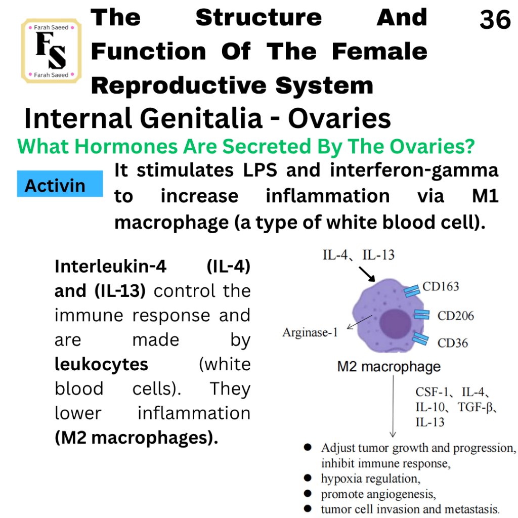
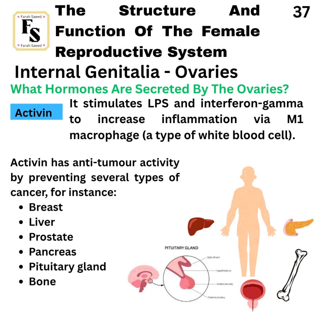
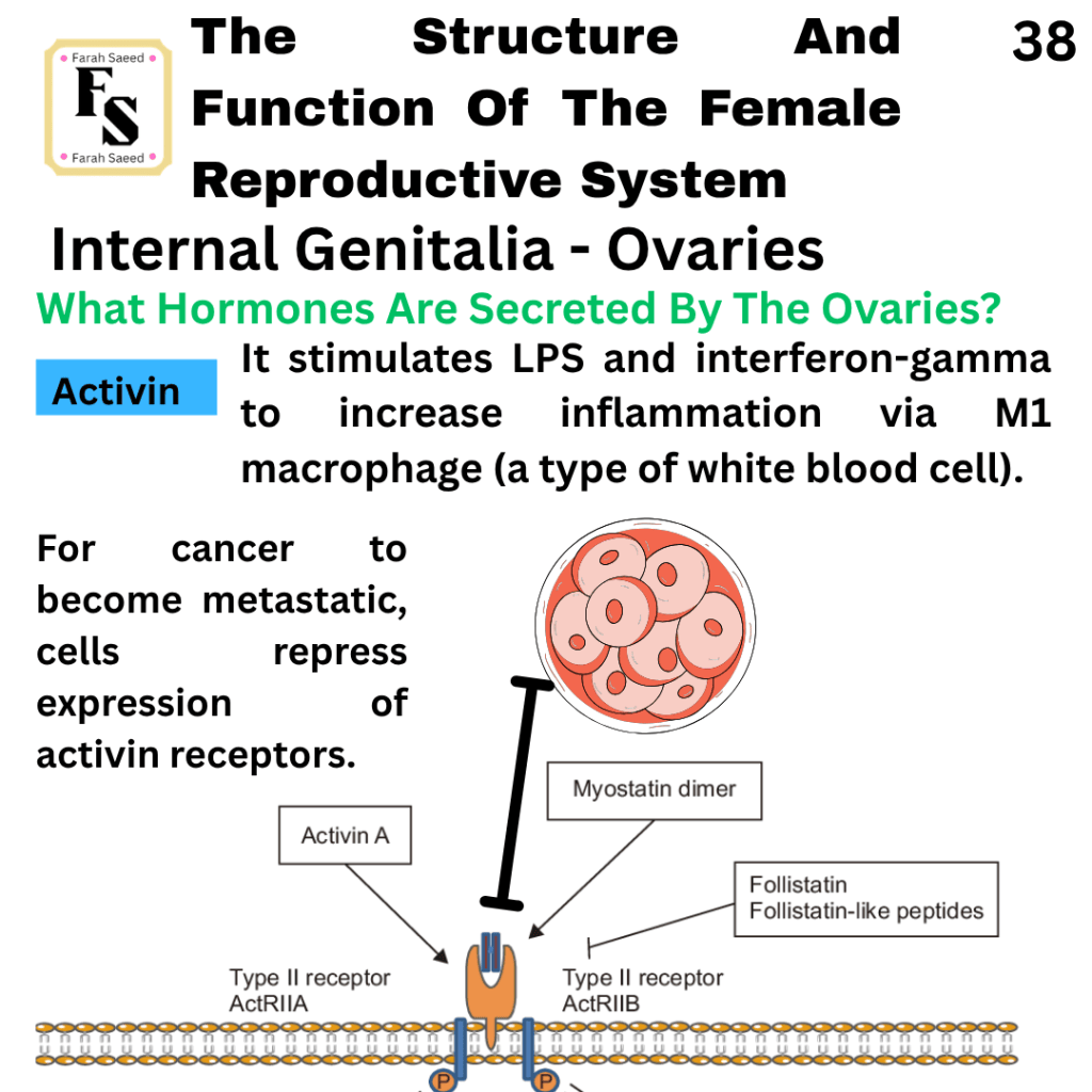
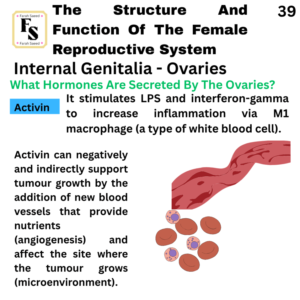
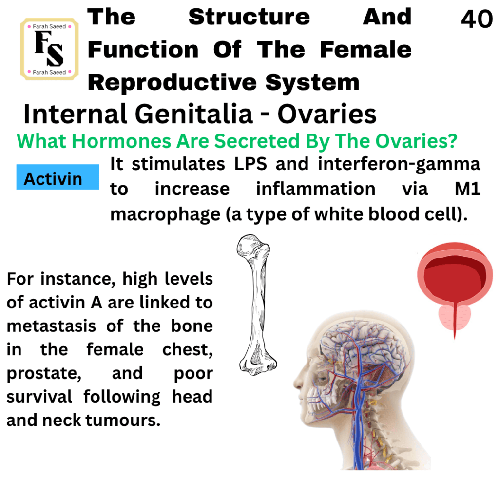
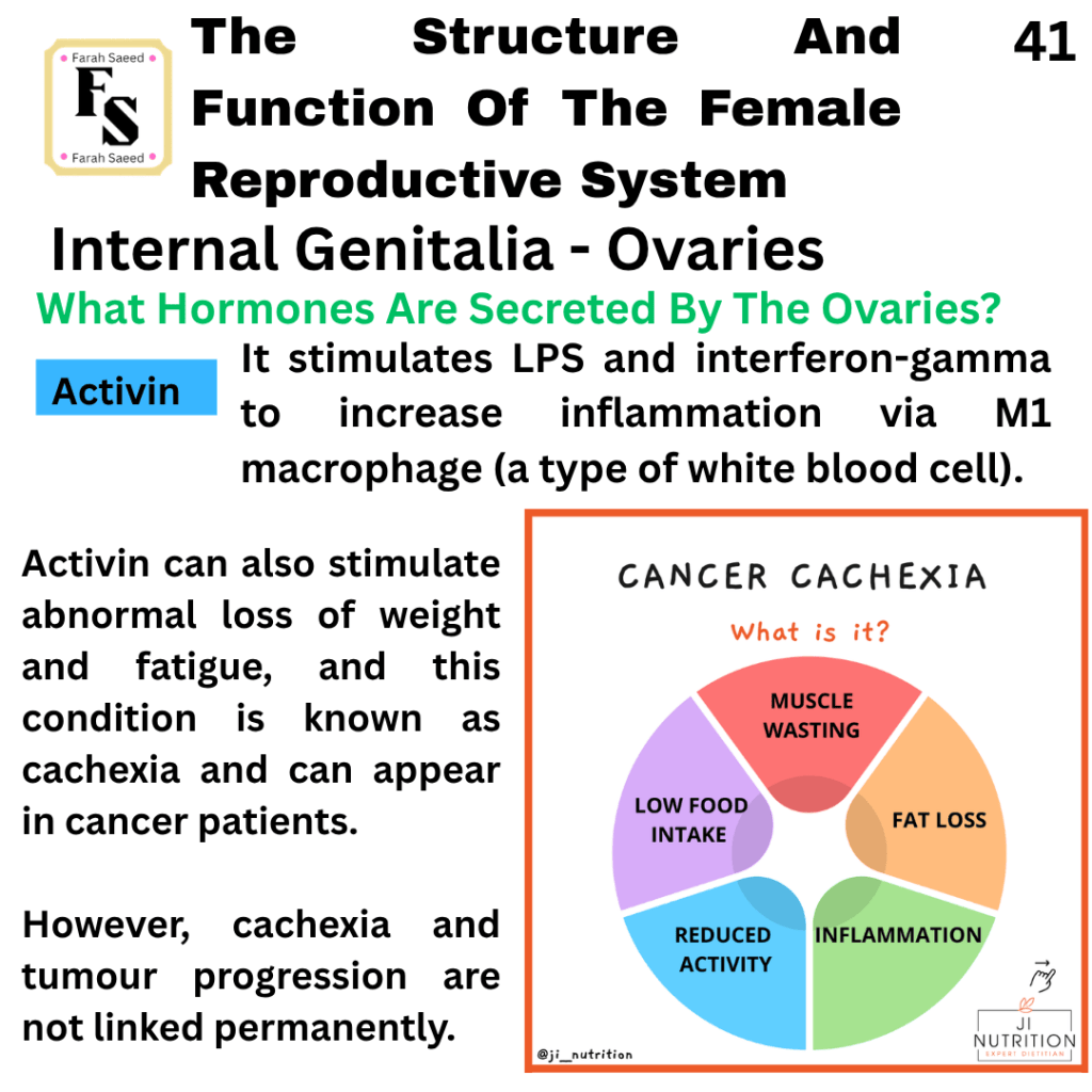
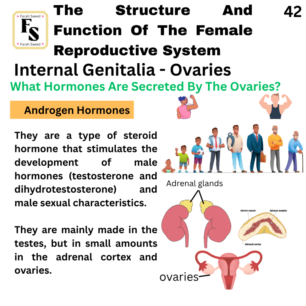
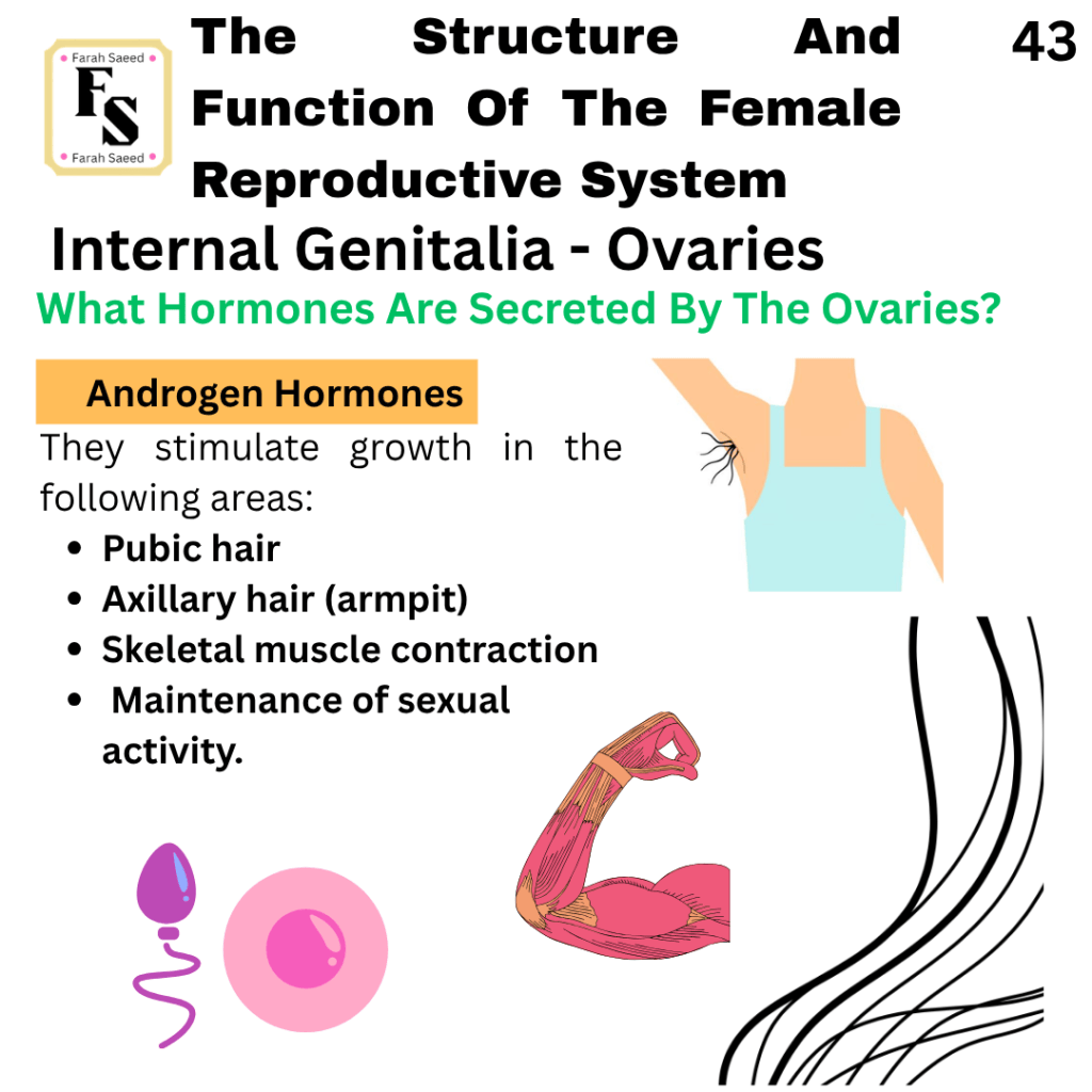
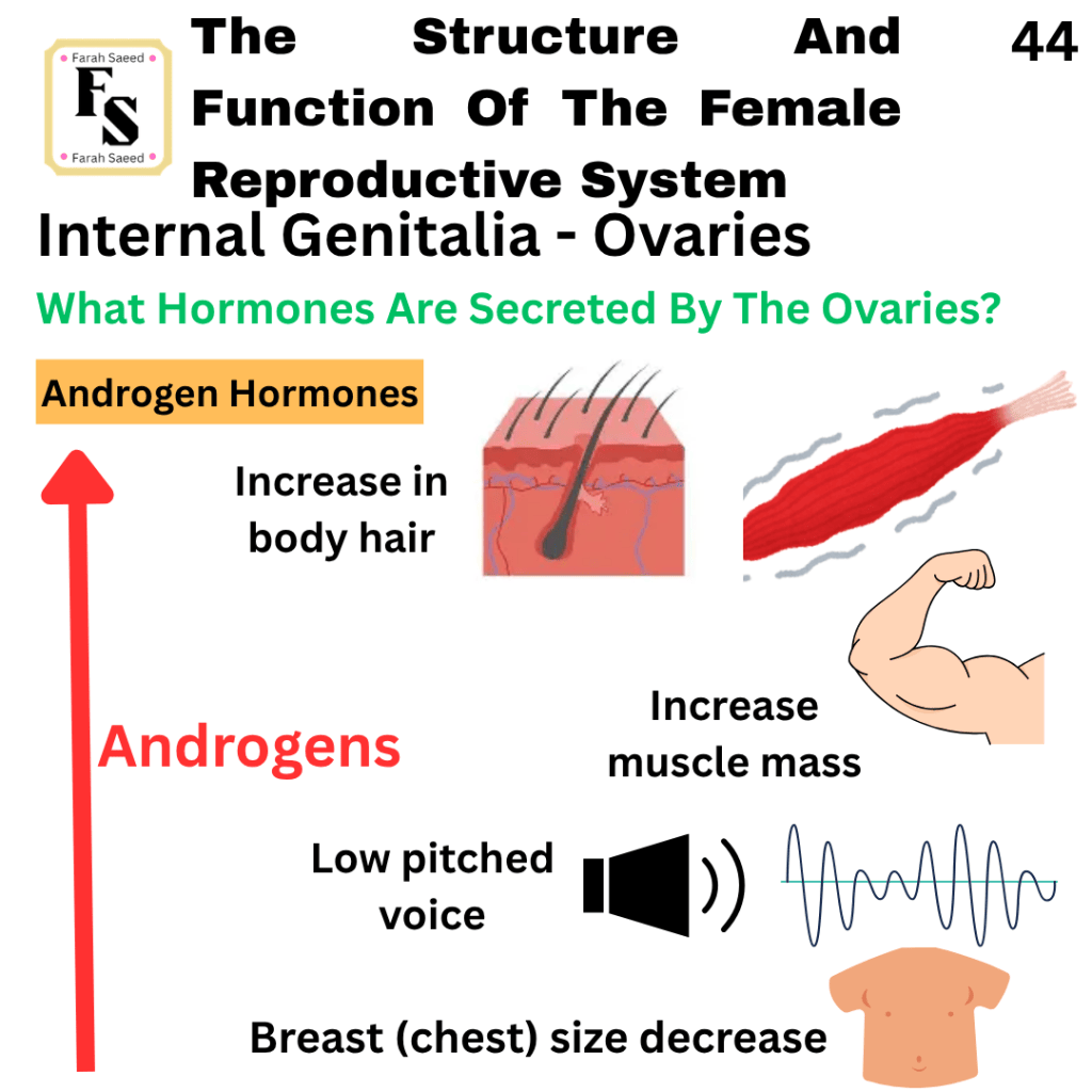
What Hormones Are Secreted By The Ovaries?
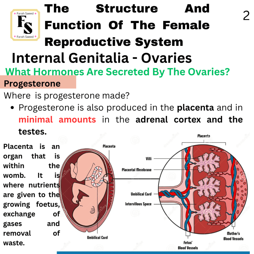
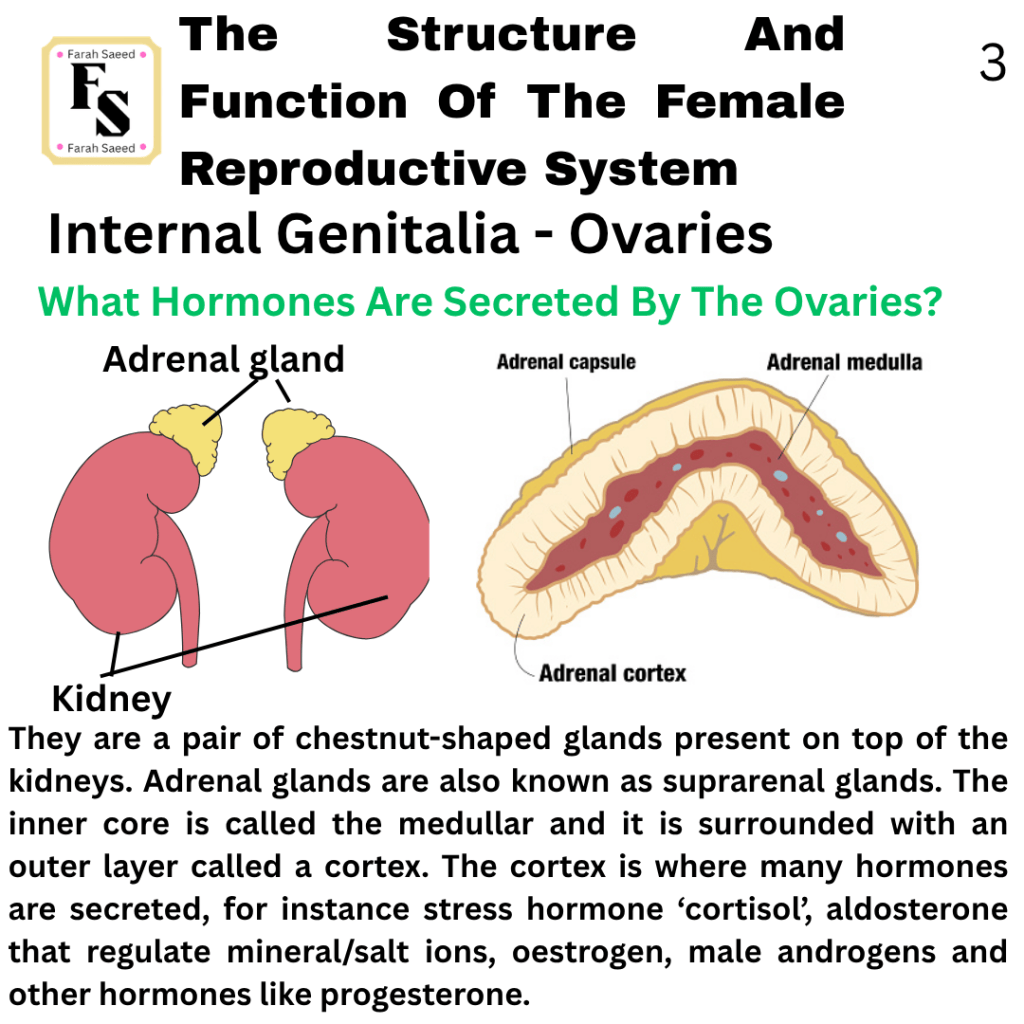
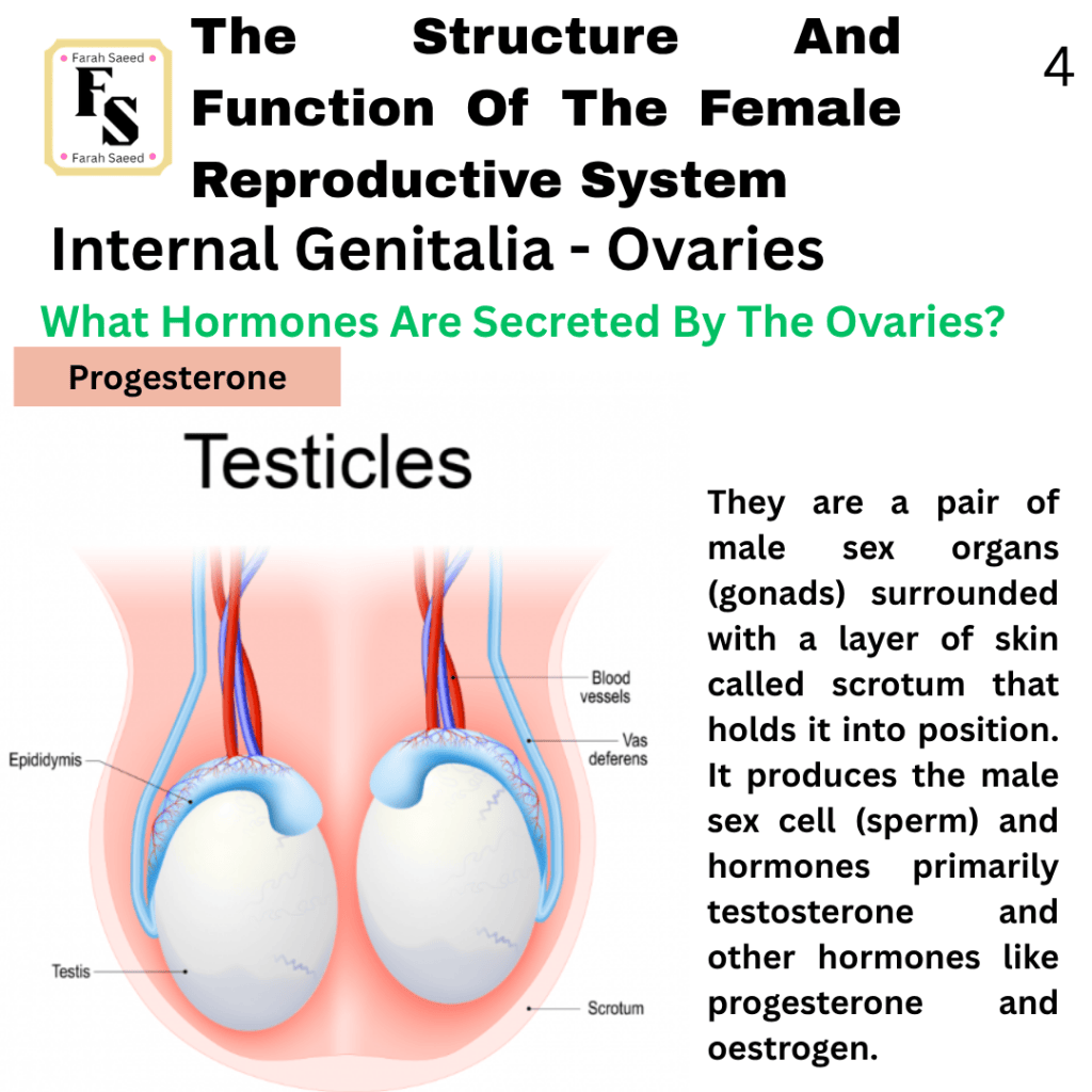
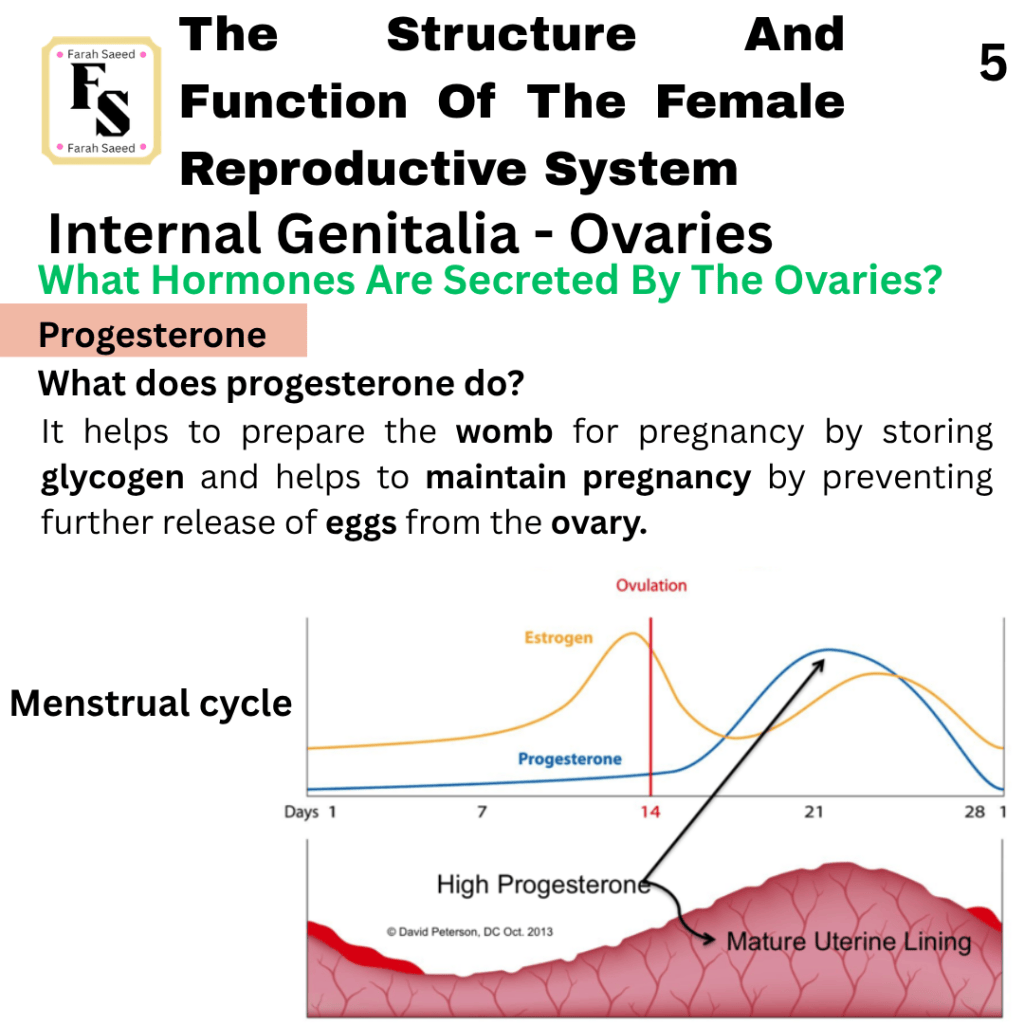
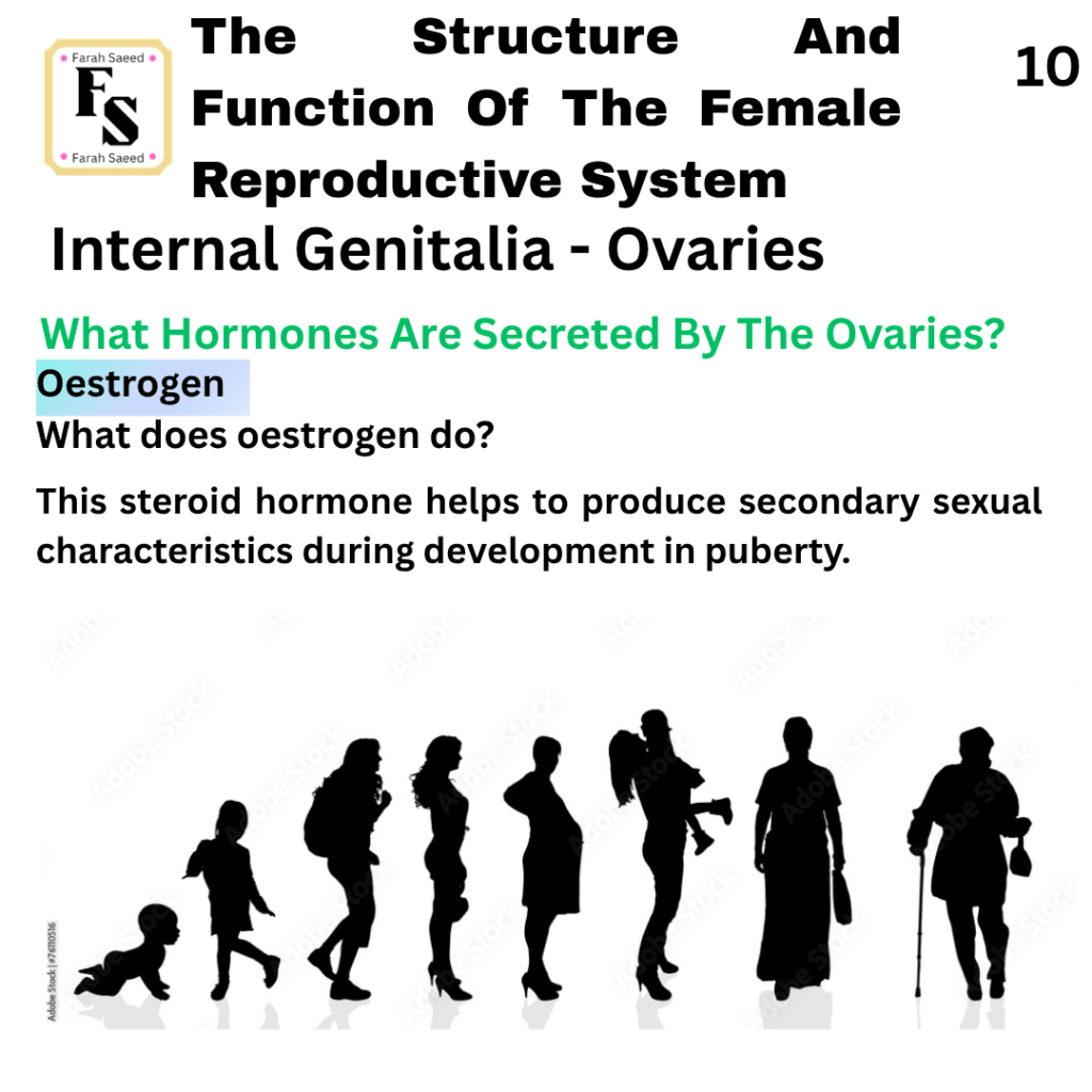
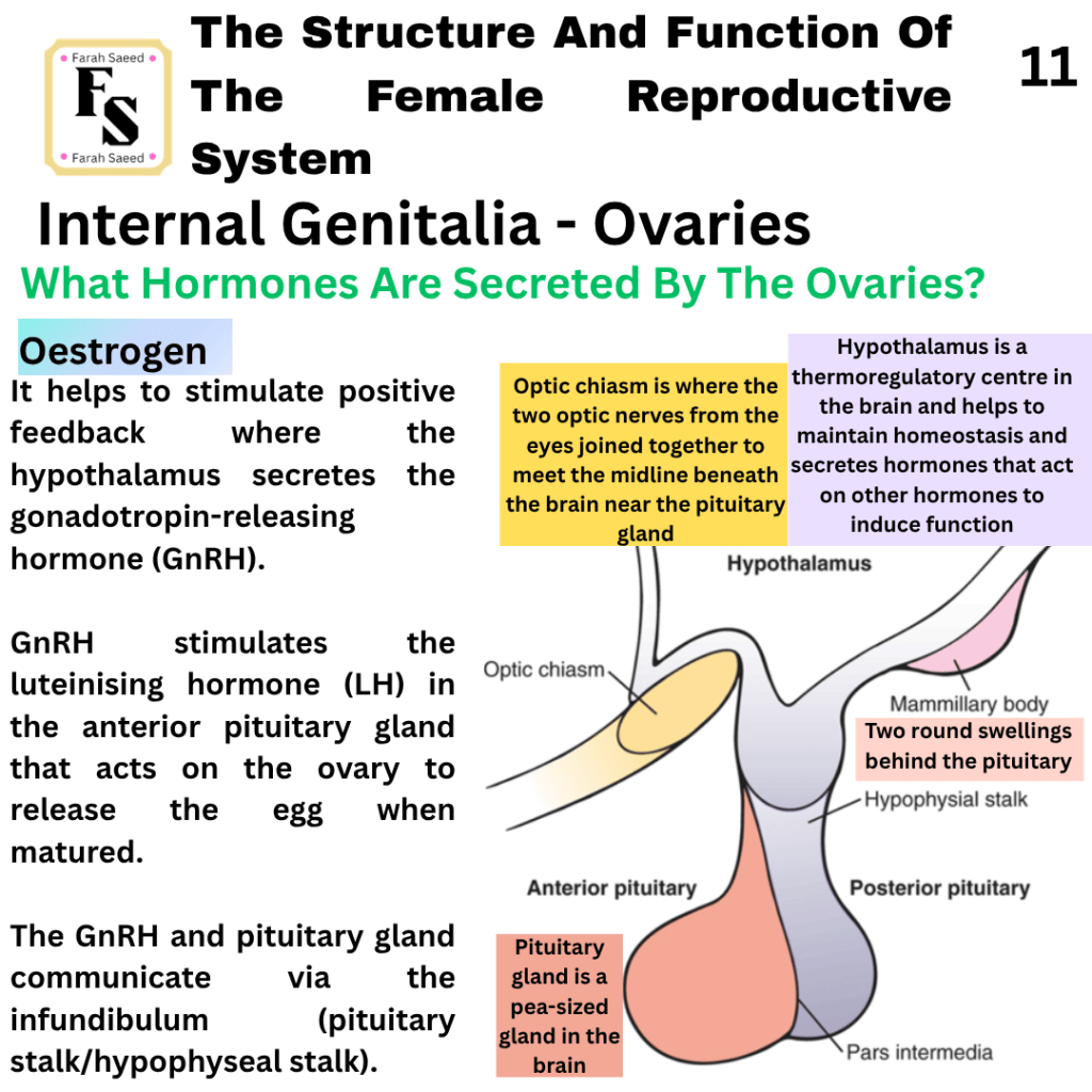
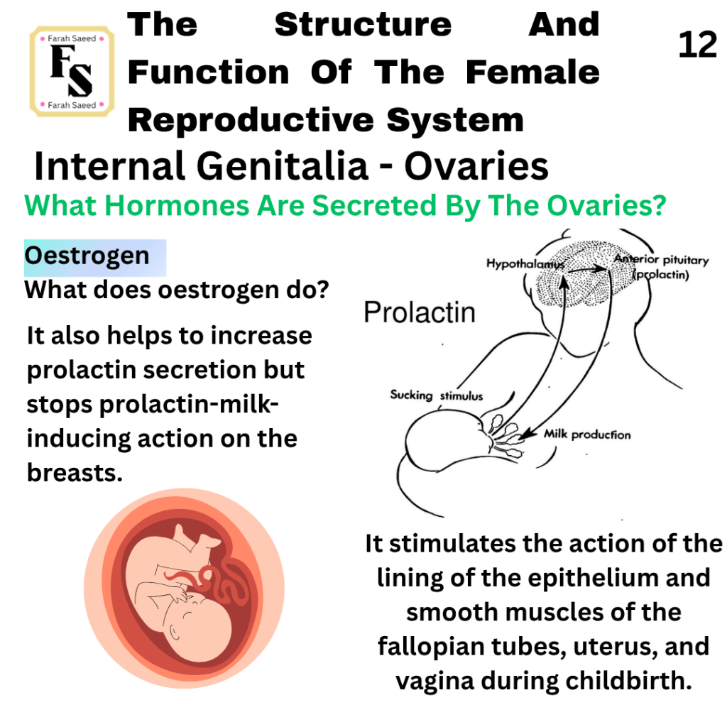
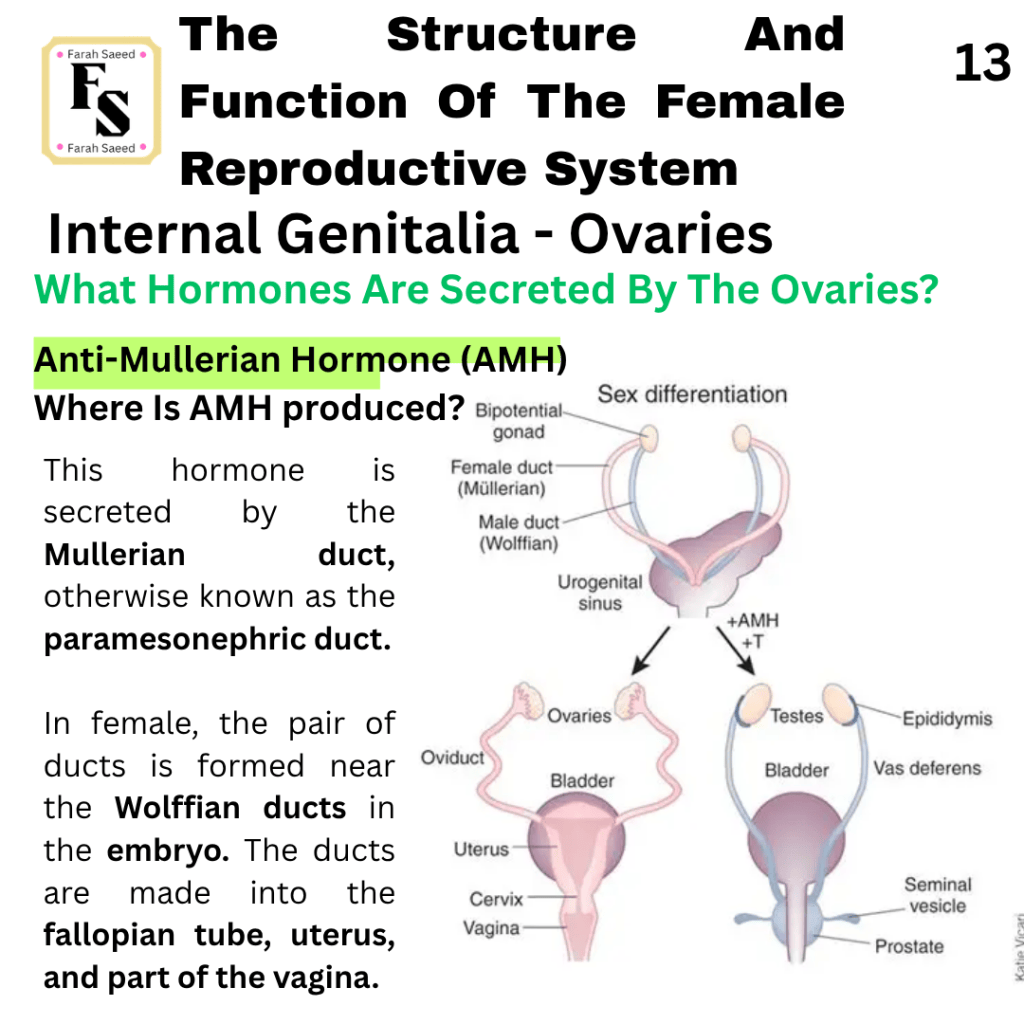
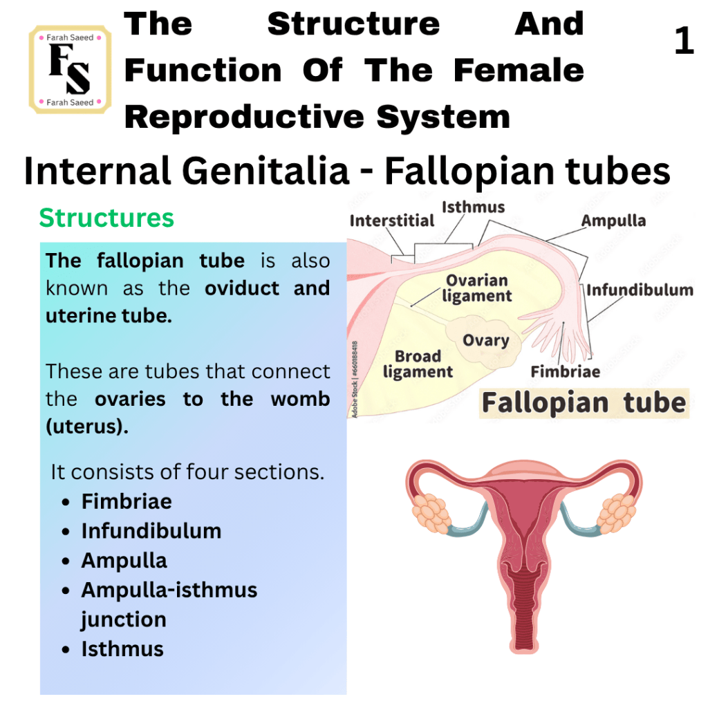
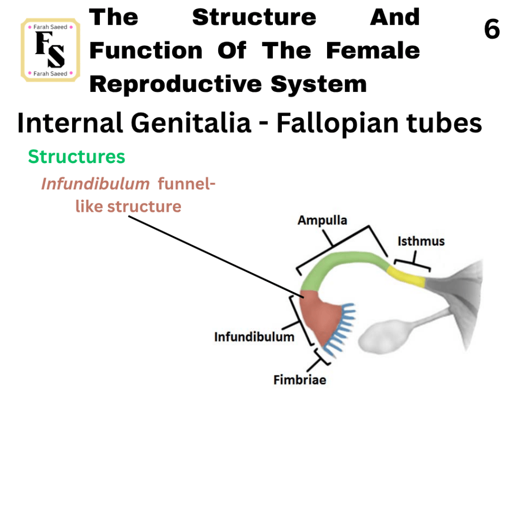
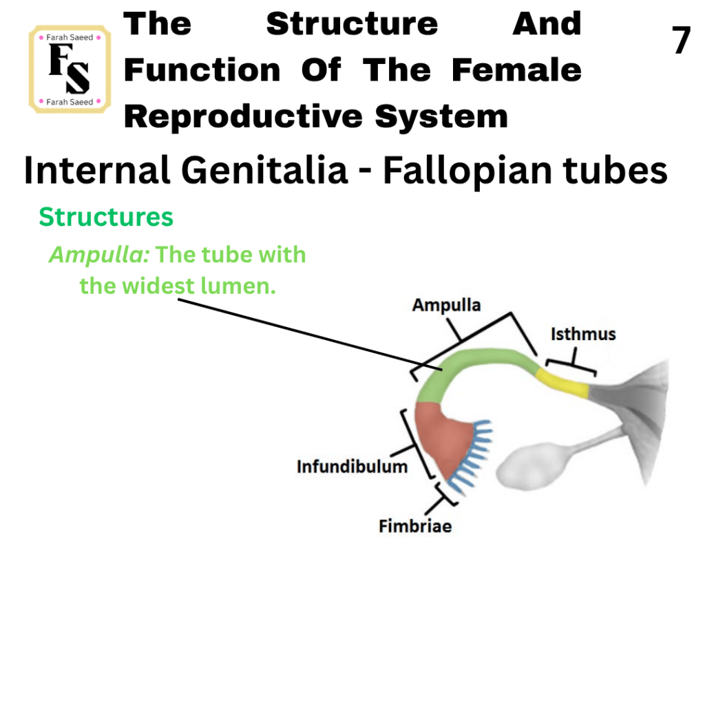
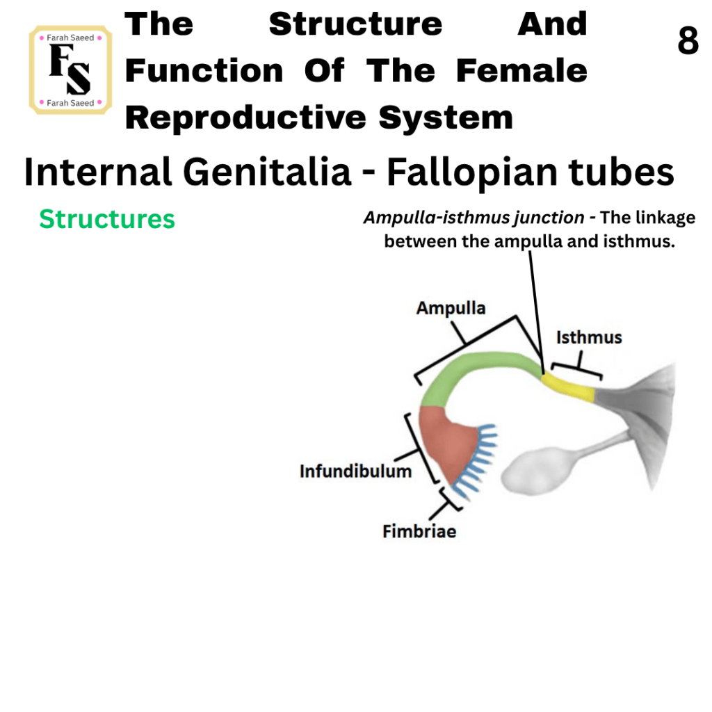
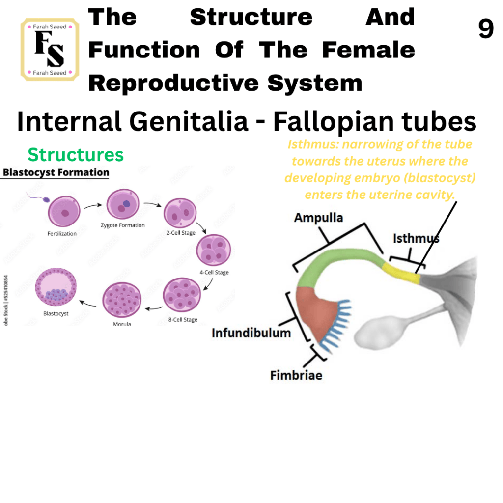
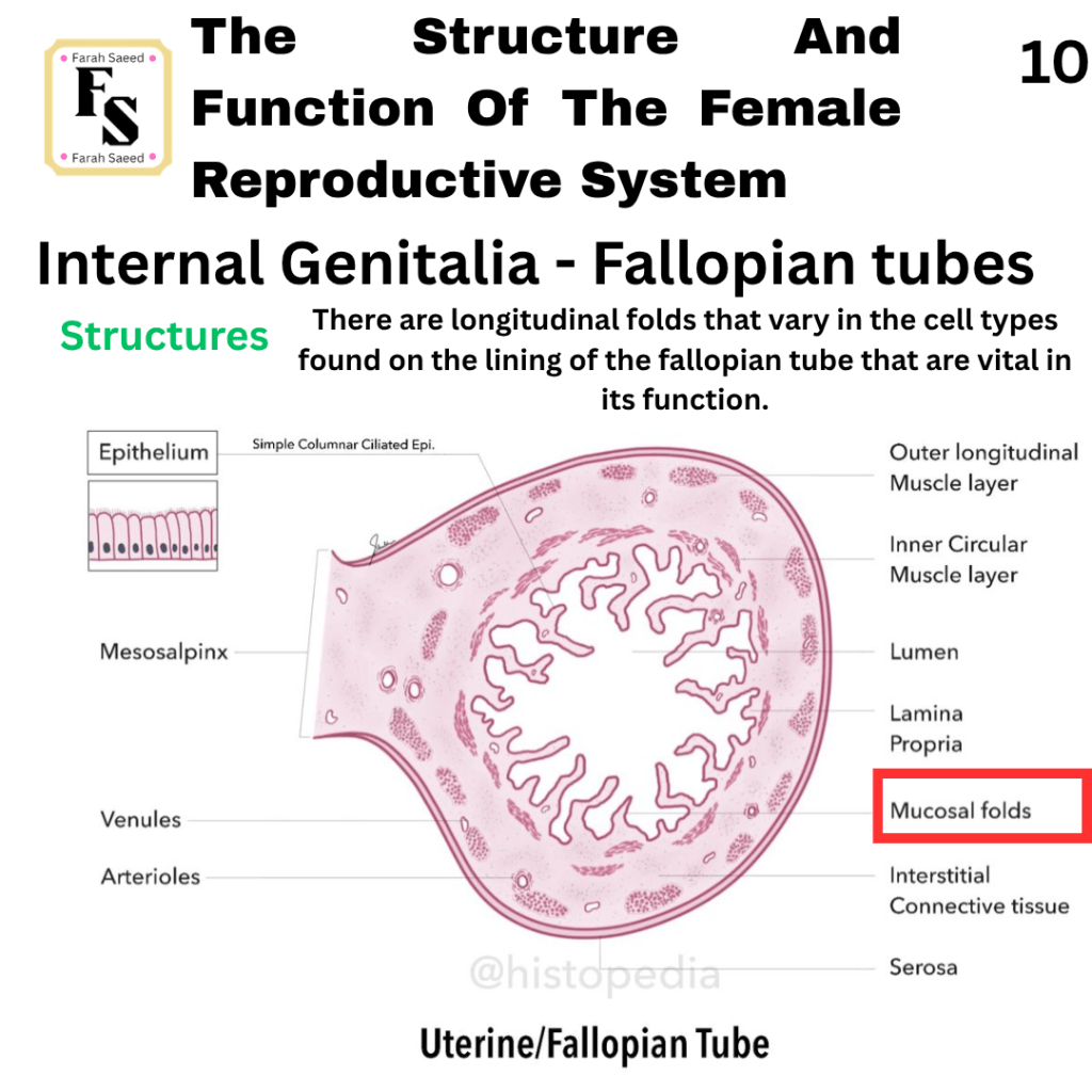
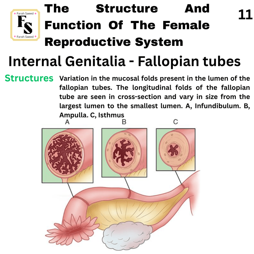
The Fallopian Tubes
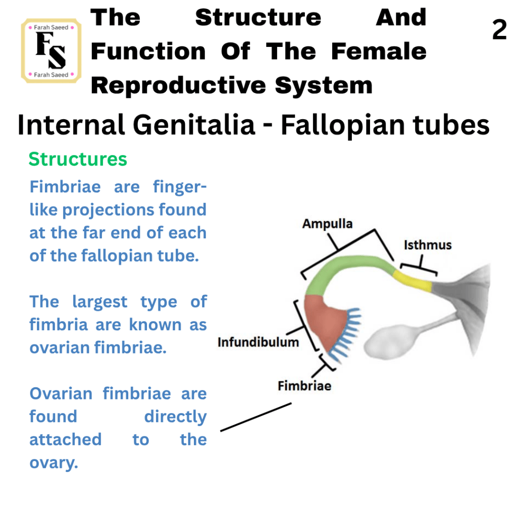
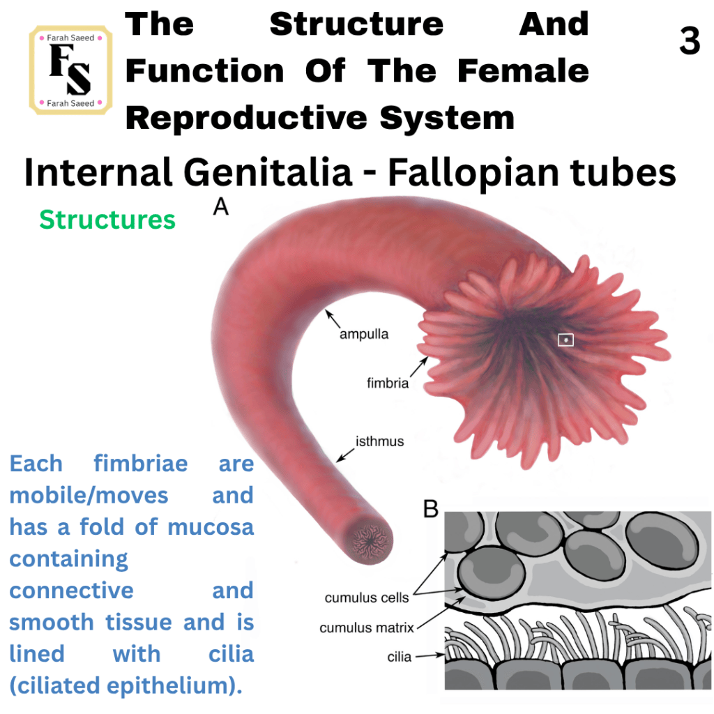
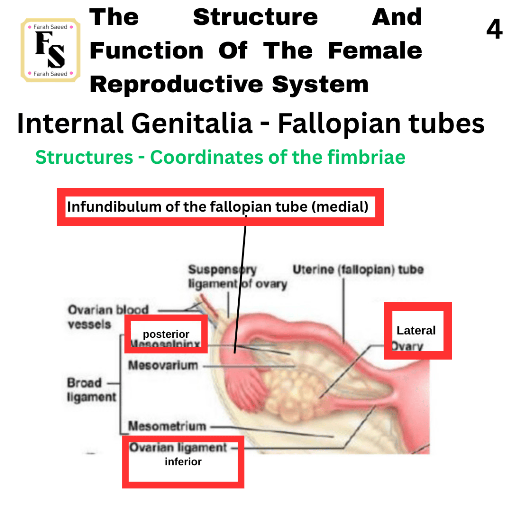
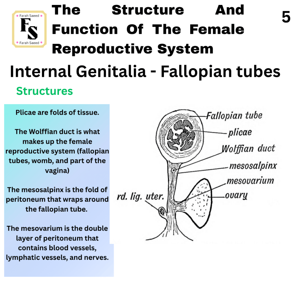
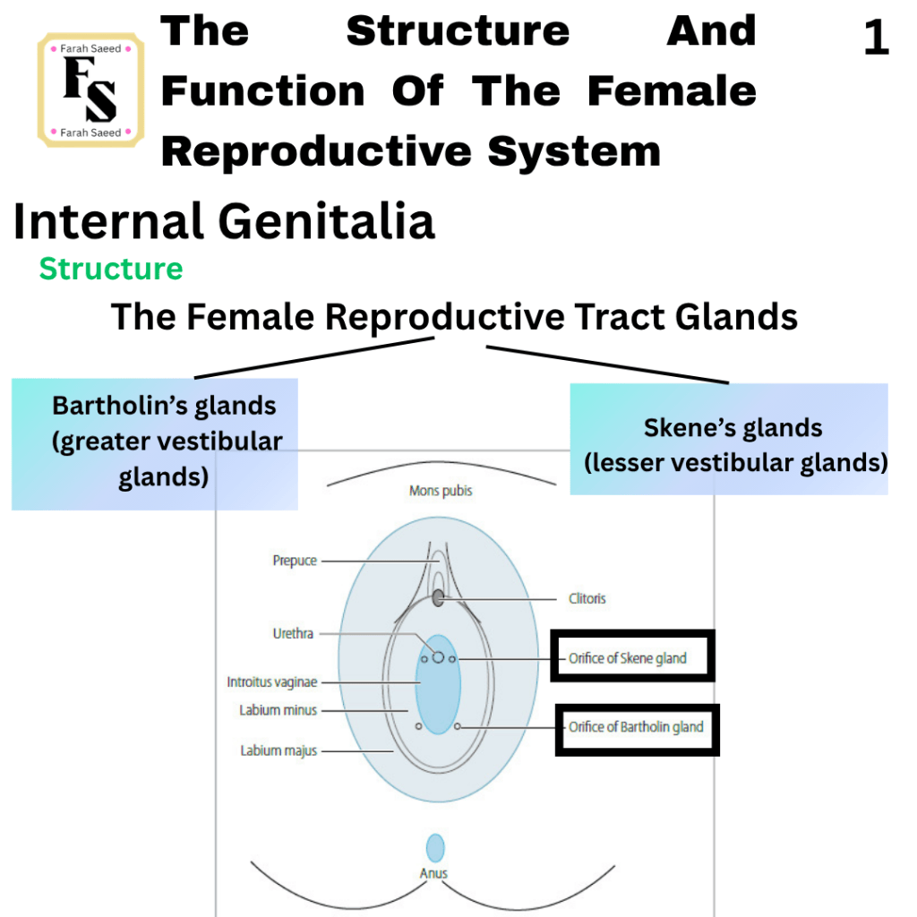
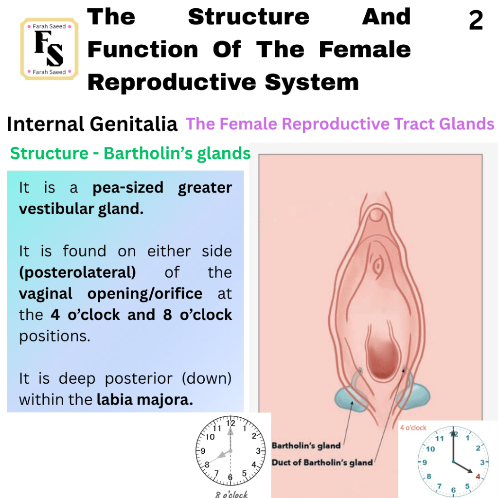
The Female Reproductive Tract Glands
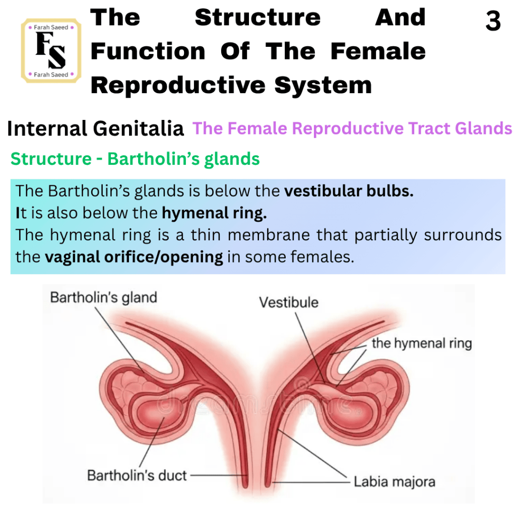
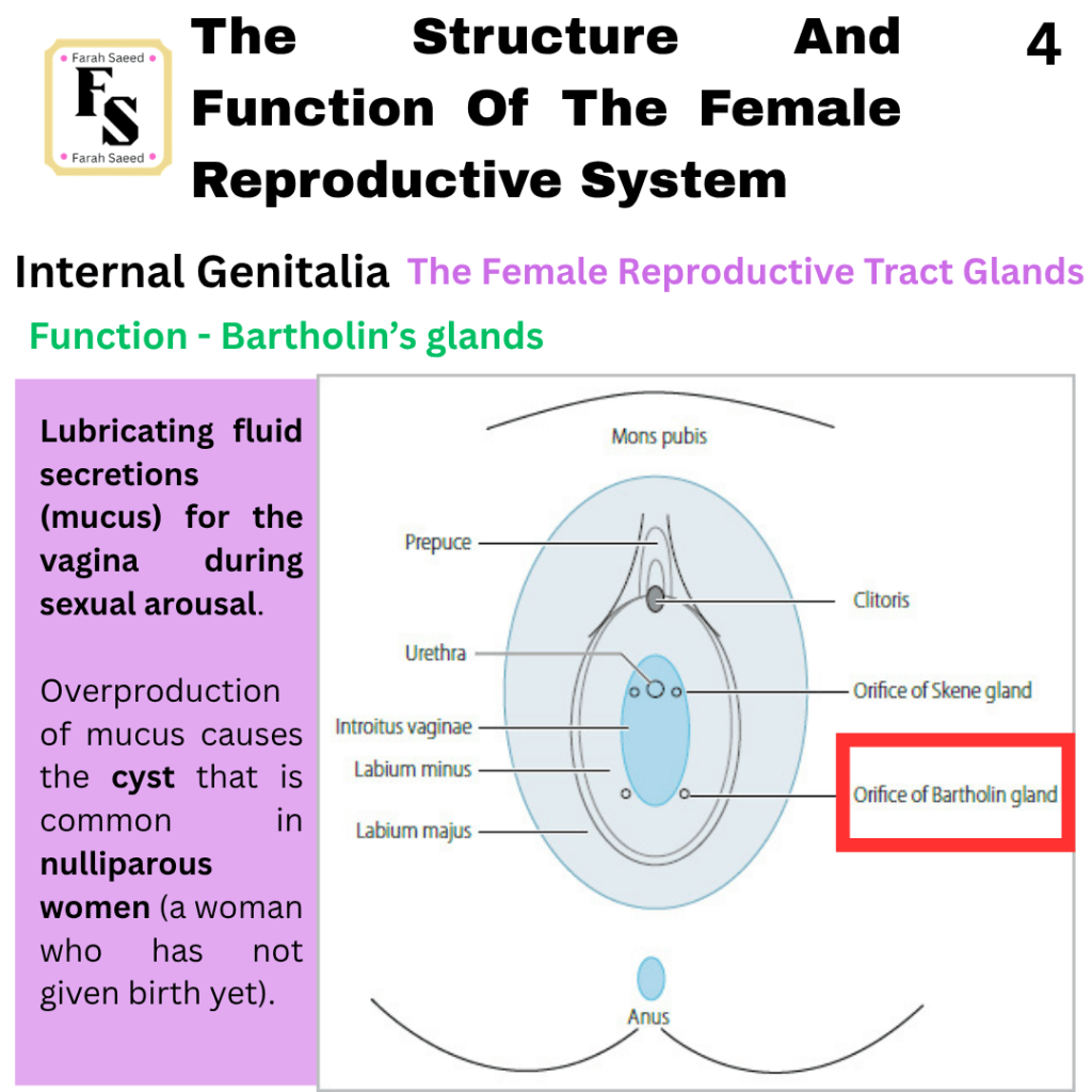
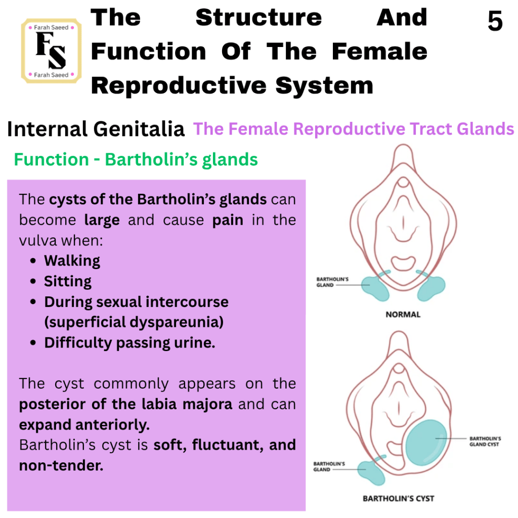
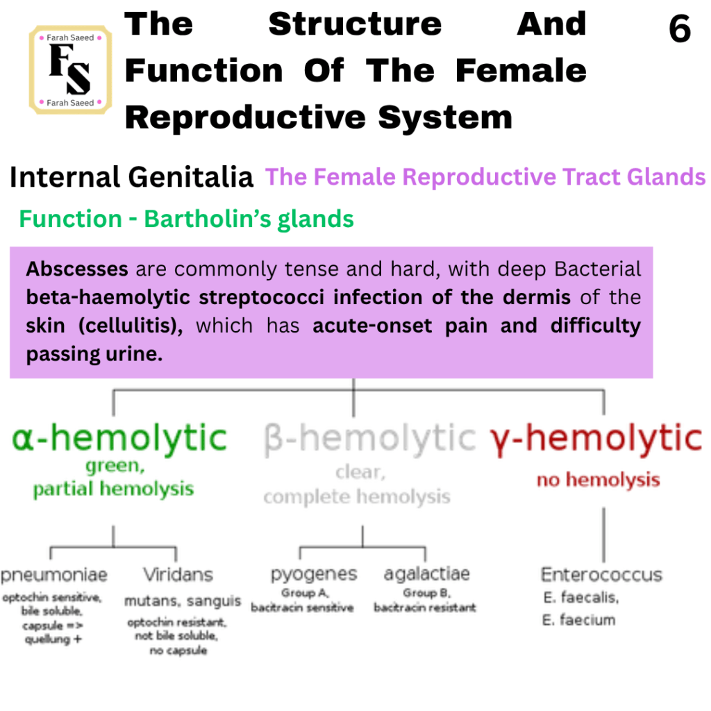
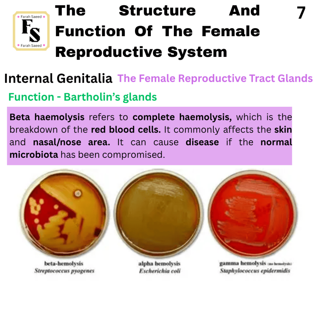
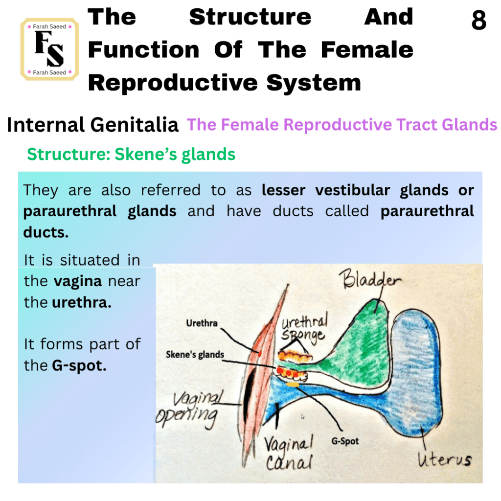
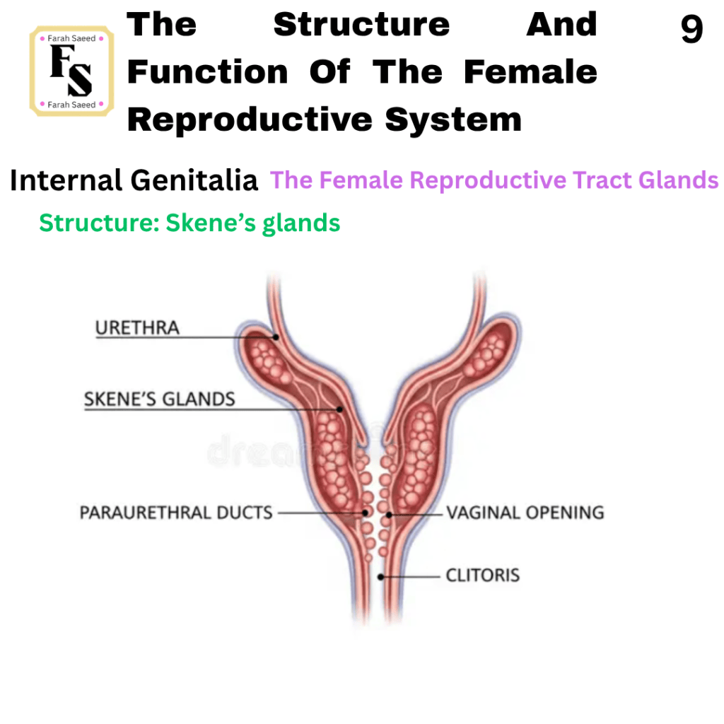
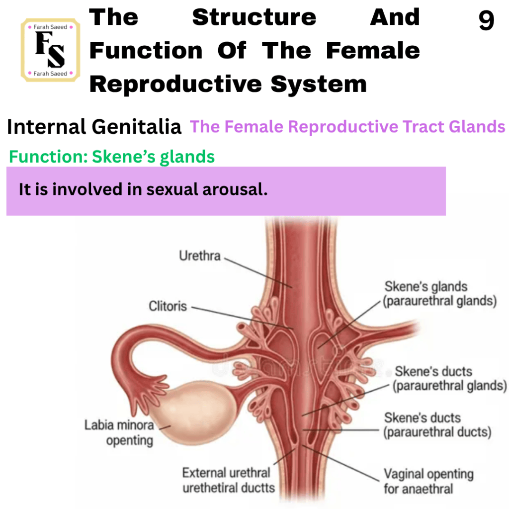
Supporting Images And Video Resources For Internal Genitalia
3D Anatomy – Female Reproductive System
Anatomy Zone: The Anatomy Of The Female Reproductive System
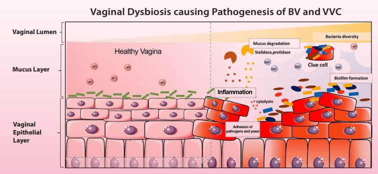
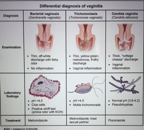
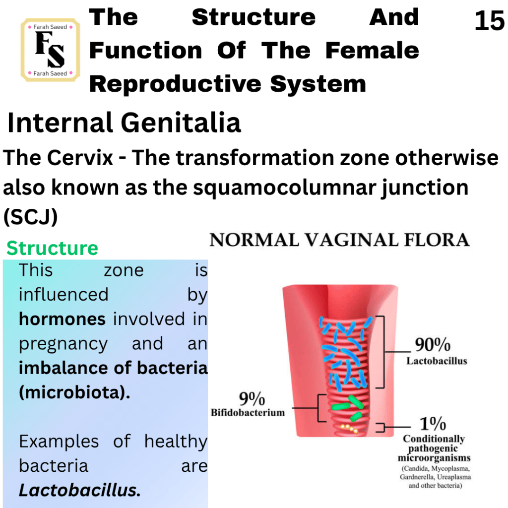
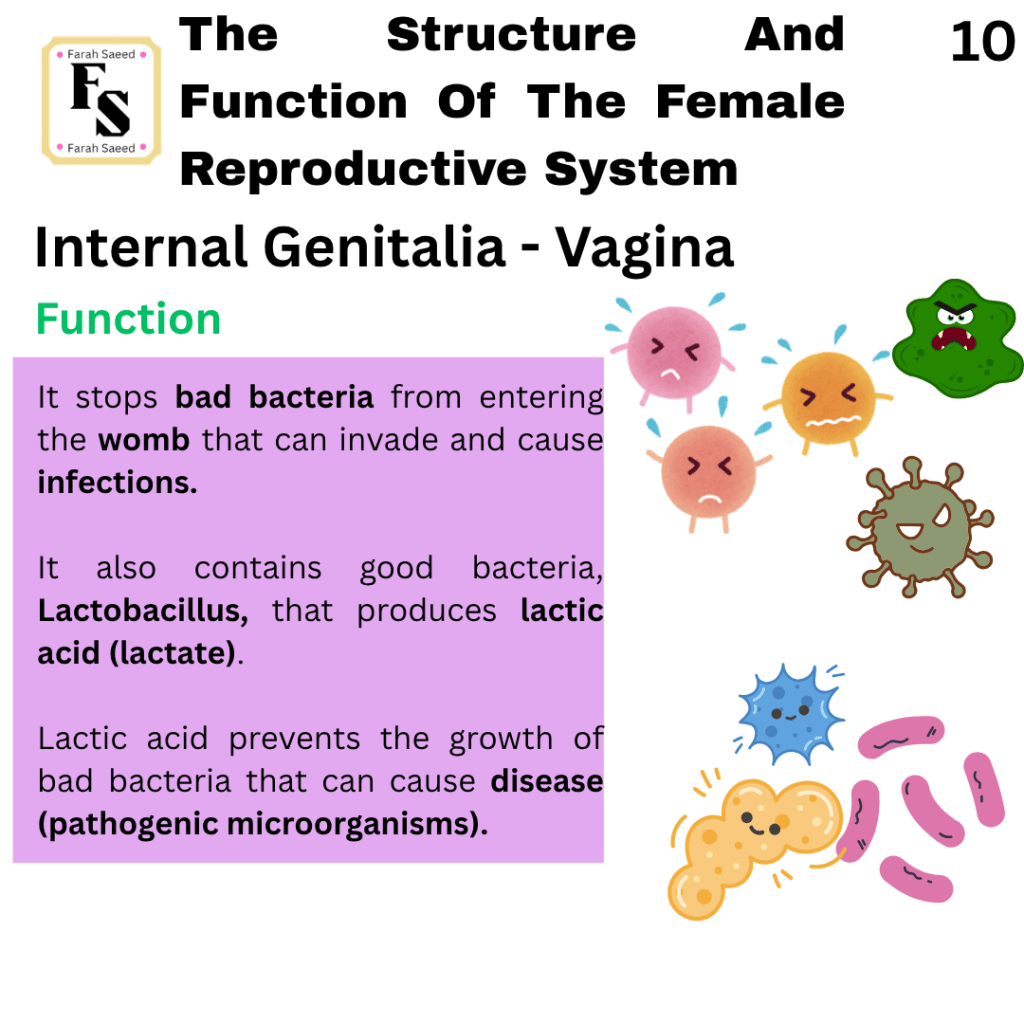
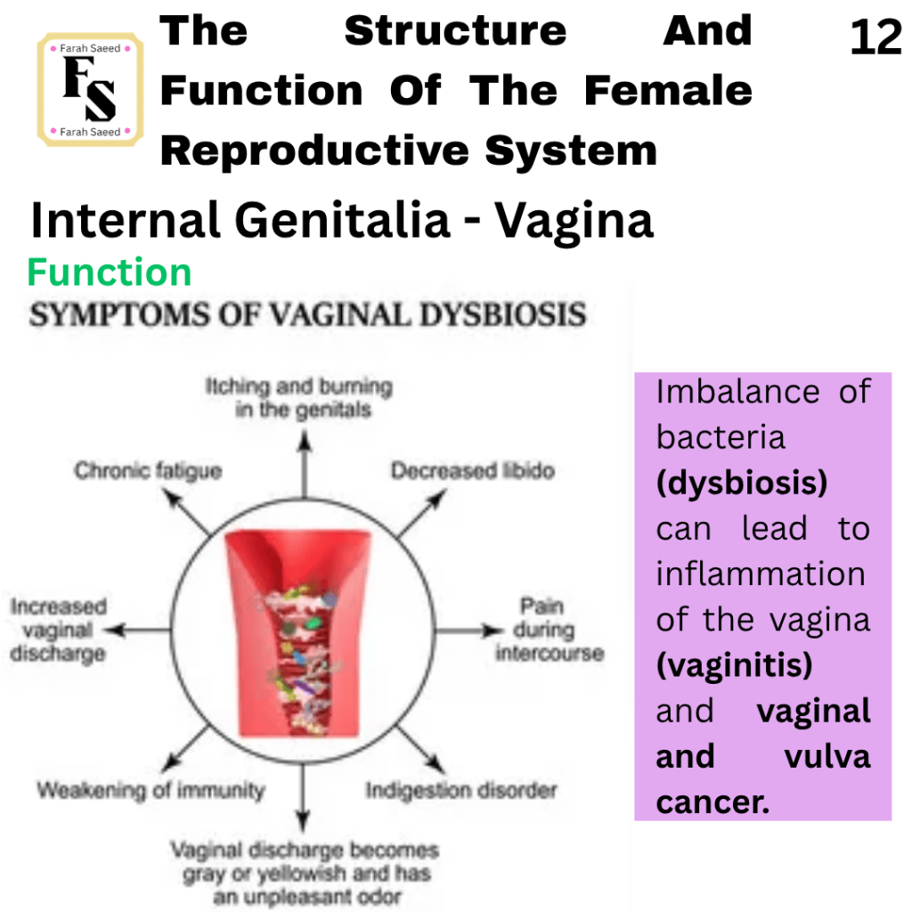
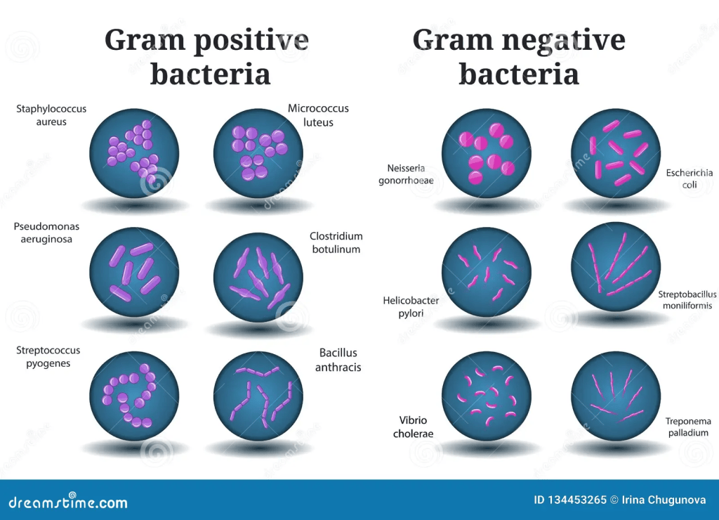
There is a difference between bacterial vaginitis and vaginosis. Vaginitis is an inflammation of the vagina. However, vaginosis is the overgrowth of bacteria. One of the main organisms that causes bacterial vaginosis is Gardnerella vaginalis, a type of anaerobic bacterium that does not need oxygen. It can occur in women who are pregnant, experiencing late miscarriage, and premature labour. This type of organism is formed from a transition from good bacteria, Lactobacillus, to anaerobic bacteria. The bacterial shift has risen after interactions with Gardnerella vaginalis, Prevotella bivia, and Atopobium vaginae. A fishy smell or the presence of thin, grayish-white discharge.
The Amsel criteria are used for diagnosis and include:
• A positive whiff test – this tests for odour or smell
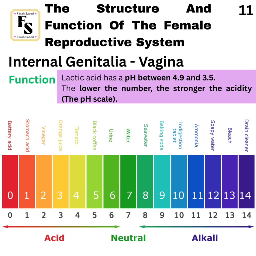
• Vaginal fluid pH greater than 4.5
• The presence of thin and homogeneous discharge
• Microscopic identification of “clue cells”
Whiff Test
A test can be done known as a Whiff test, where a cotton-tipped swab gets a sample of the discharge from the vagina, specifically the posterior fornix, and is added onto the glass microscopic slide.
A single drop of 10% potassium hydroxide (KOH) is added onto the sample. KOH is a strong alkali that increases the pH of the sample, where the non-volatile amine salts in the discharge convert into volatile, gaseos form. Volatile means it evaporates or vapourises easily.
Positive Whiff test – fishy smell when adding the KOH solution because the presence of volatile amines suggests a high amount of anaerobic bacteria.
Negative Whiff test – No change in odour
Clue cells
Clue cells are a type of squamous cell (flat) in the vaginal epithelial cells that the bacteria stick to. This can be indicated under the microscope, where overgrowth of anaerobic bacteria can appear stippled or granulated. This can be observed under a microscope in two main types of examinations. Wet mount examination is where a drop of the sample of vaginal discharge is added onto the microscopic slide. It is then mixed with saline and viewed under the microscope. The Gram stain is a discharge on a slide, fixed and stained. Clue cells can appear under the stain.
The clue test forms part of the Amsel criteria. A positive result suggests bacterial vaginosis because the test is highly specific, but sensitivity varies, which may miss cases as a single test alone.
Trichomonas
This is a type of parasite and protozoan that has a flagellum (protein-like structures) to move and affect the vagina, causing inflammation and vaginal discharge. The presence of trichomonads, poor odour and pH > 4.5.
It can affect moist areas of the body and lead to a condition called thrush, such as the mouth (oral) and vagina. In relation to vaginitis, it can affect the labia (skin folds) and vagina, causing cheese-like discharge and itchiness (pruritus).
Candida albicans (yeast/fungi)
Pseudohyphae have a tube-like filament structure which they begin from a single cell to a longer form. They create increased growth or sequential budding of yeast cells. They have an uneven, chain-like appearance in comparison to hyphae that have parallel walls. The presence of pseudohyphae and a pH outside the normal range (3.8 to 4.2).
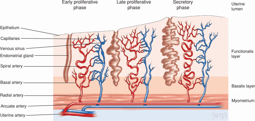
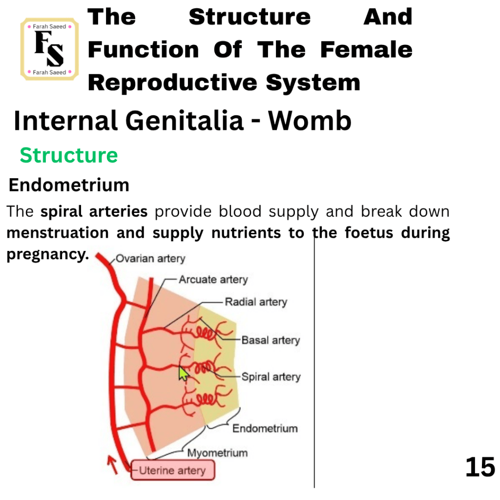
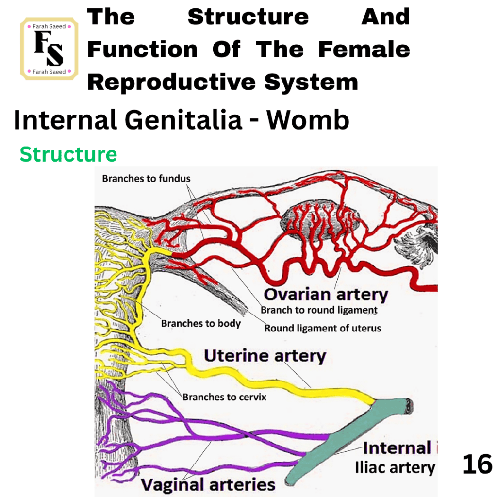
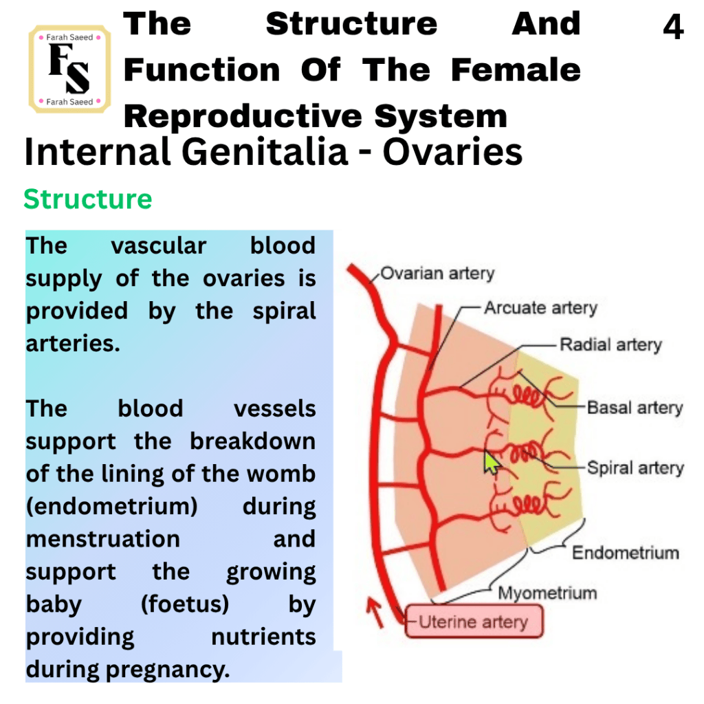
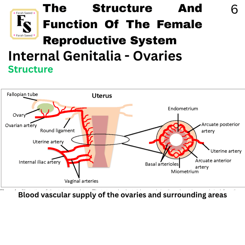
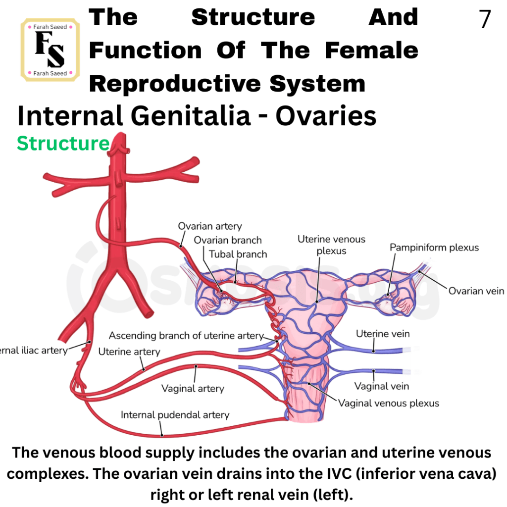
The two main blood supplies are provided by the arteries present in the uterus (womb) and the ovary. In the endometrium, there are coiled-shaped blood vessels called spiral arteries in the endometrial surface that provide the upper surface of the endometrium. The spiral arteries are formed in the following manner: The uterine arteries enter the muscle layer of the womb (myometrium), the middle layer of the womb. These branch out into arcuate arteries, then further into radial arteries, then basal arteries that provide nutrients to the basal layer of the endometrium, which is needed to recover and regenerate after its shedding during the period (menstruation). The spiral arteries are in the endometrium, specifically the subepithelial capillary plexus, where each spiral artery provides 4 to 9 millimetre squared of the endometrial surface.
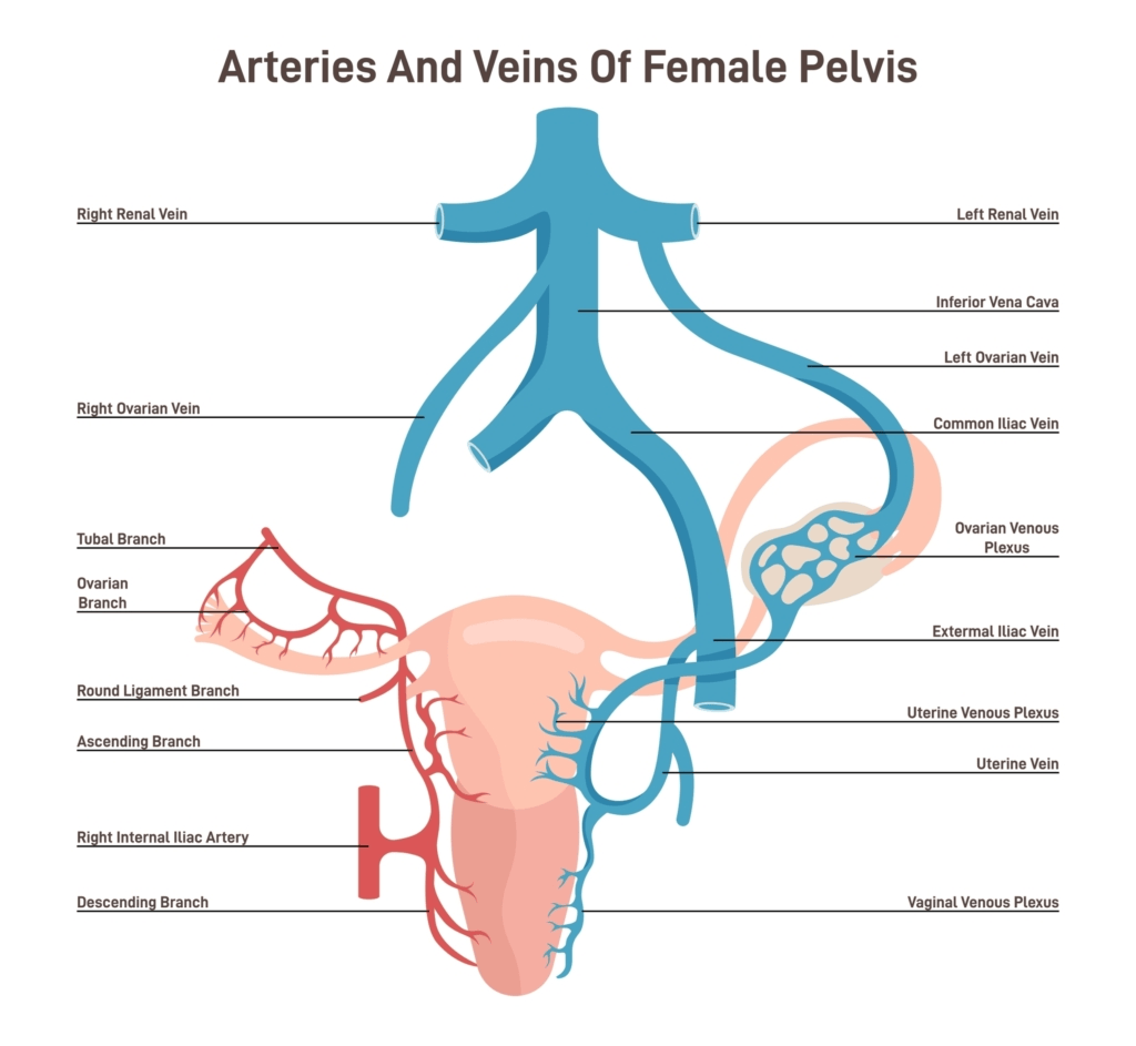
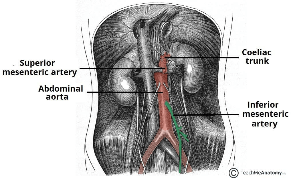
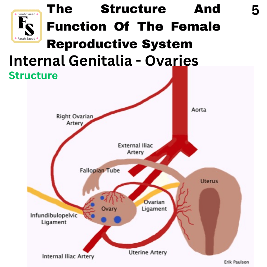
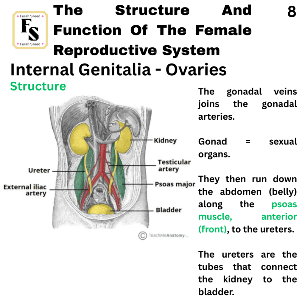
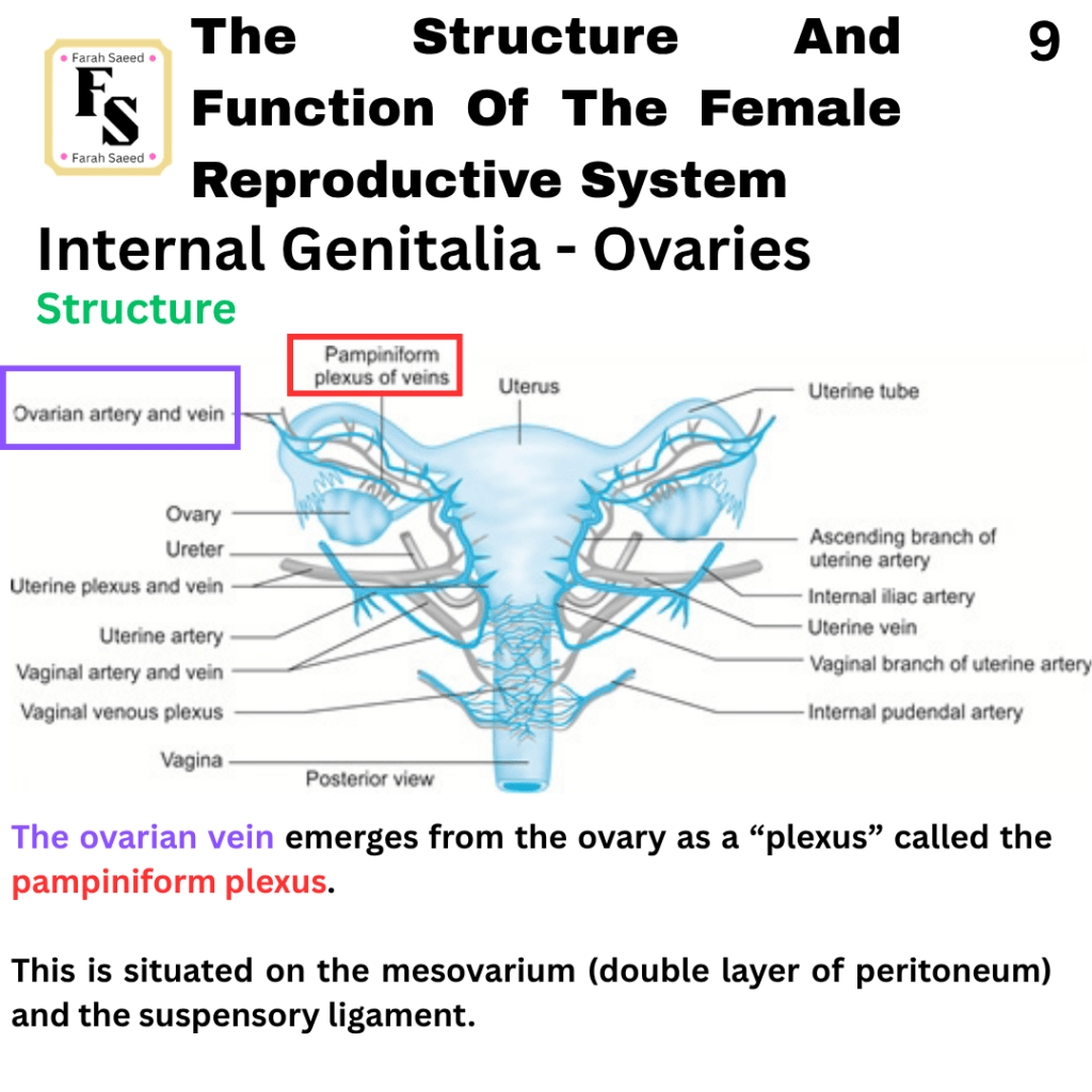
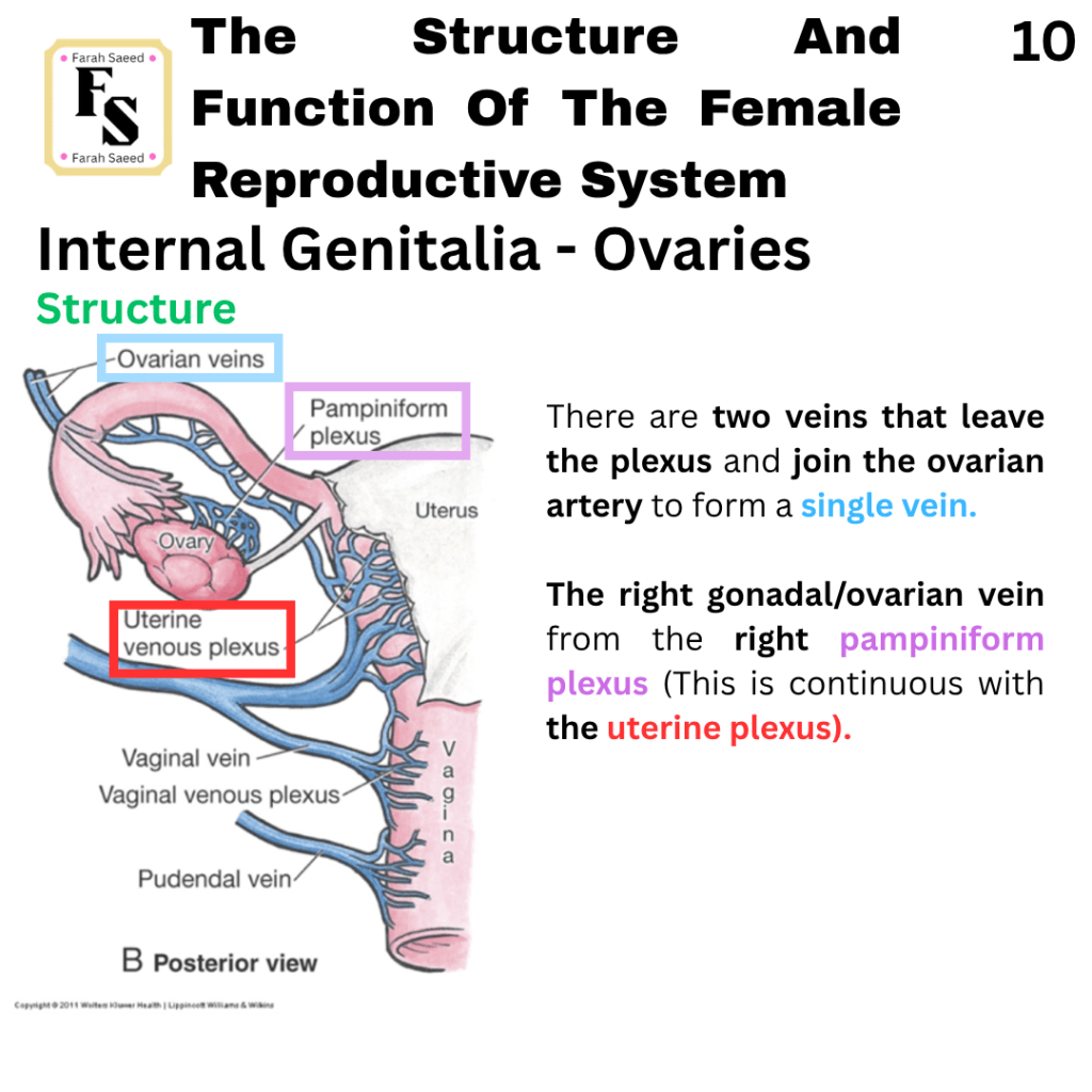
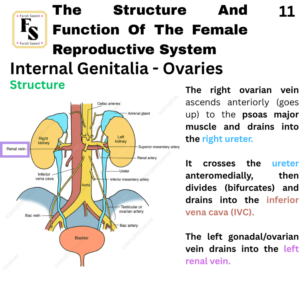
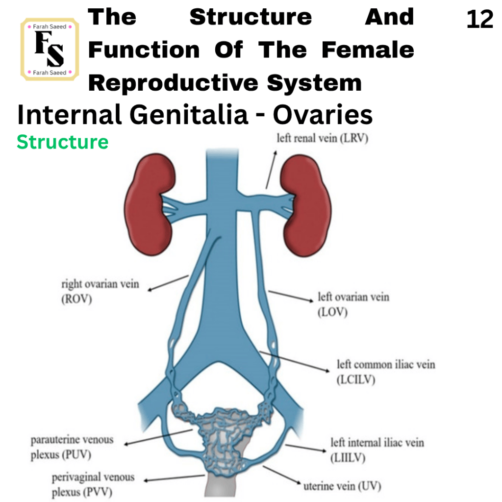
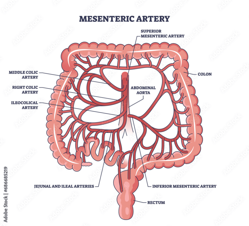
Through computed tomography (CT scan), the ovarian vein can be indicated through the inferior mesenteric artery, which is surrounded by fat in the retroperitoneum (behind the peritoneum [double layer]) adjacent to the abdominal aorta, and in the pelvis that is medial to the external iliac blood vessels.
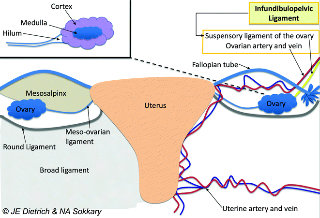
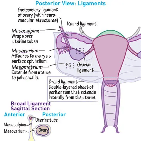
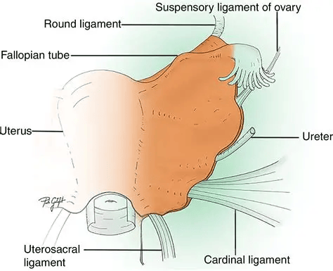
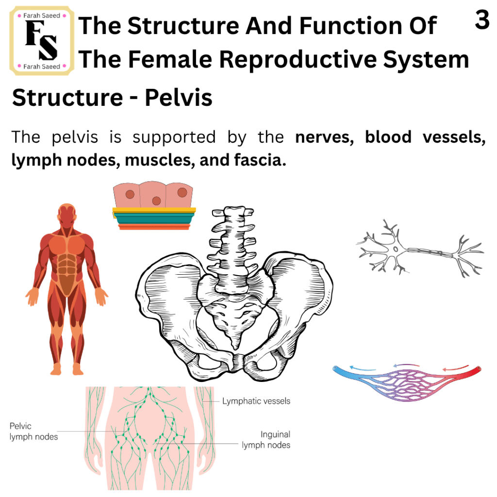
Pelvis, Fascias, Muscles, Blood Supply And Nerves
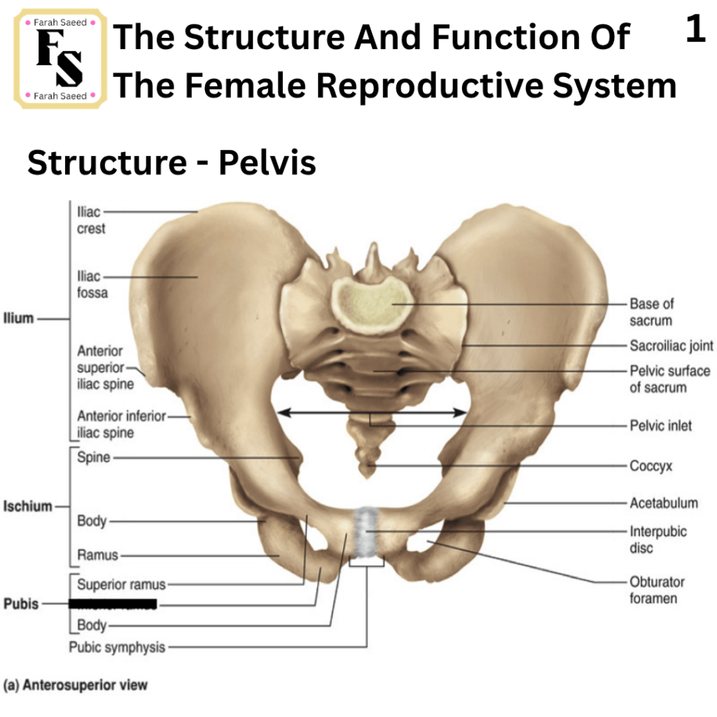
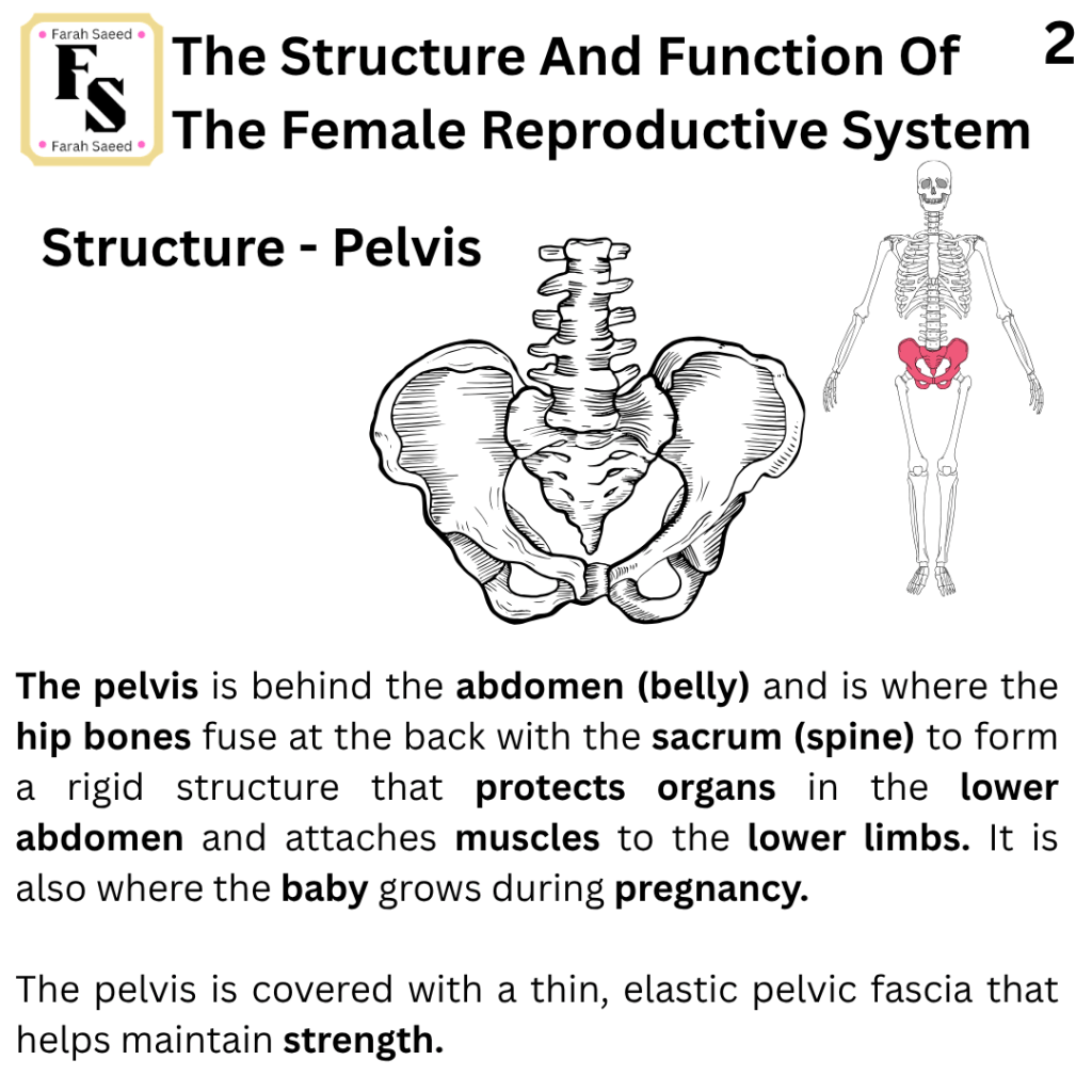
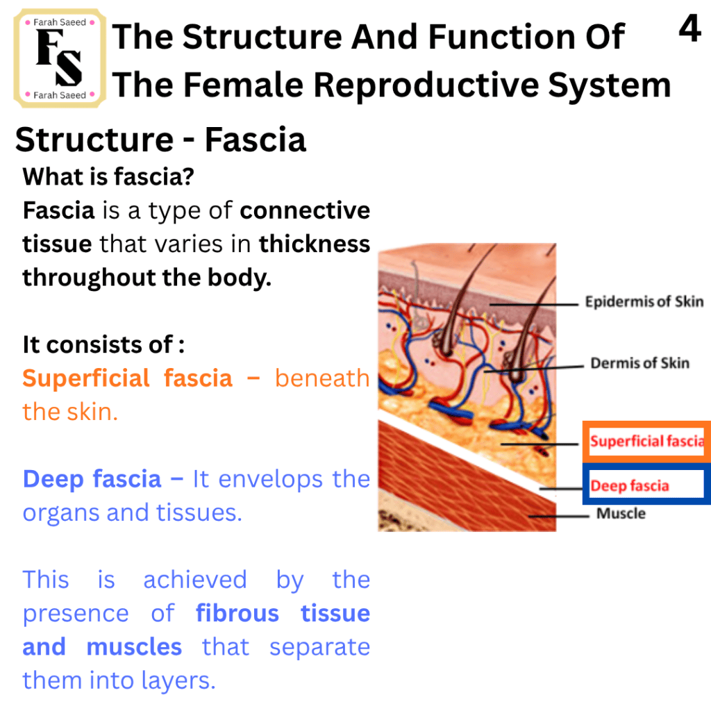
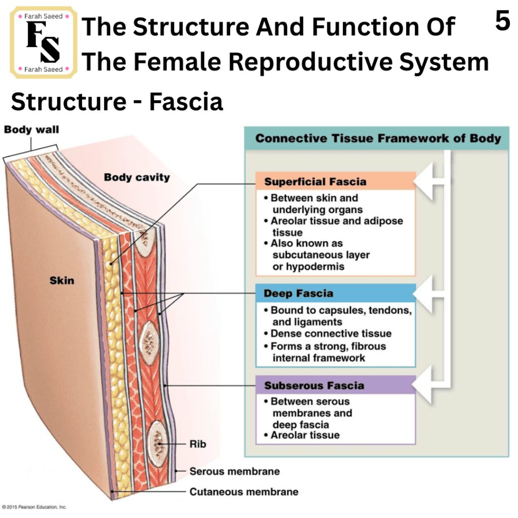
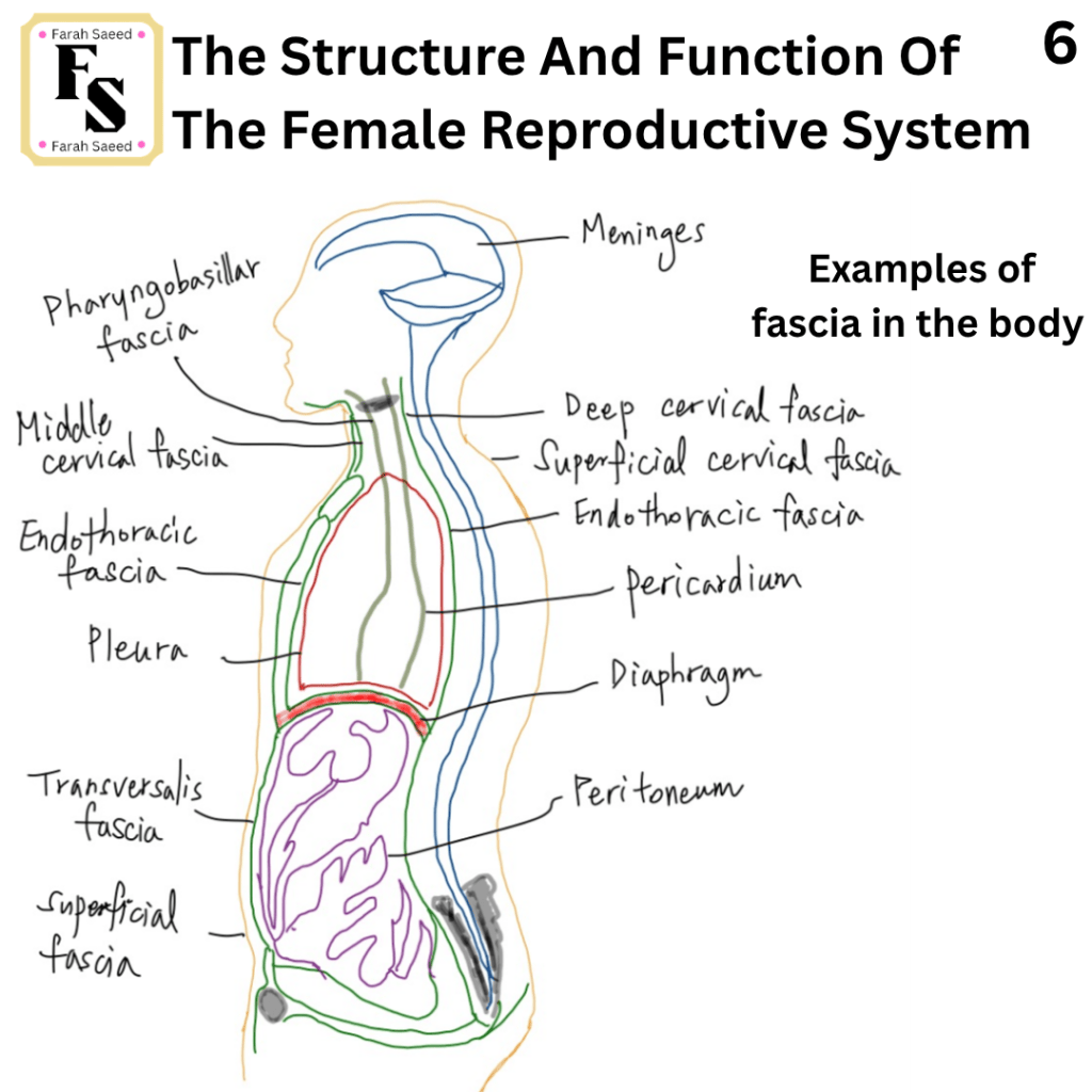
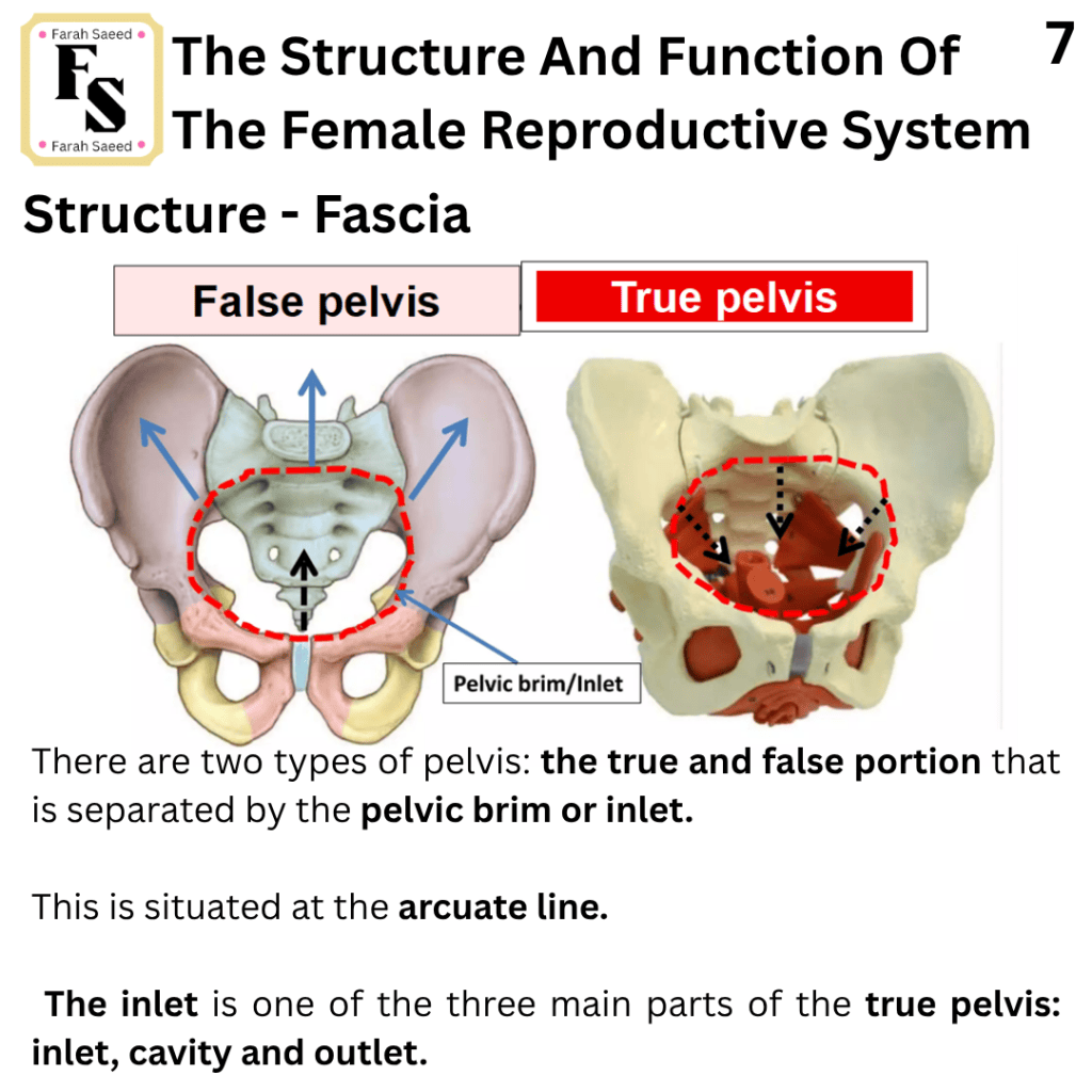
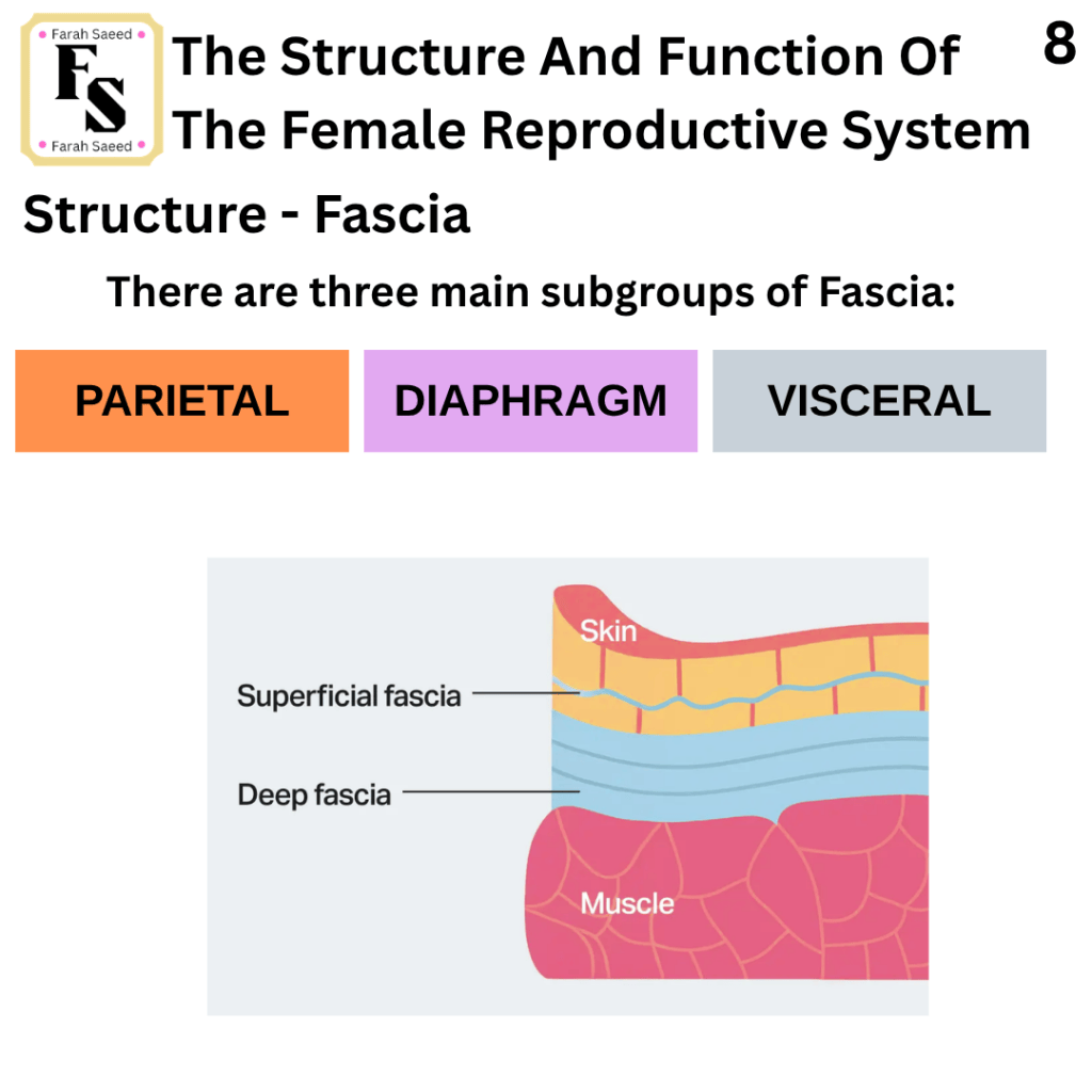
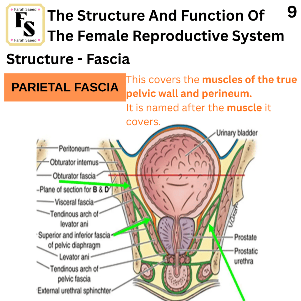
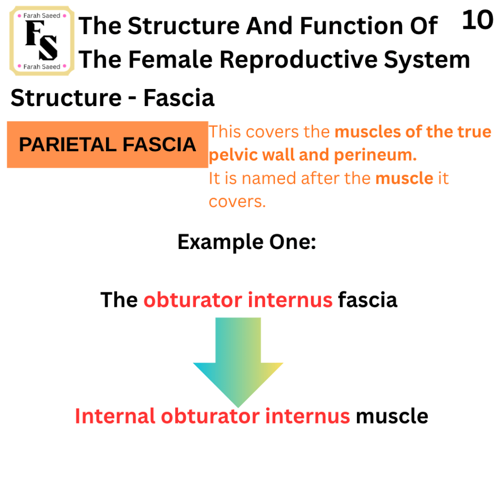
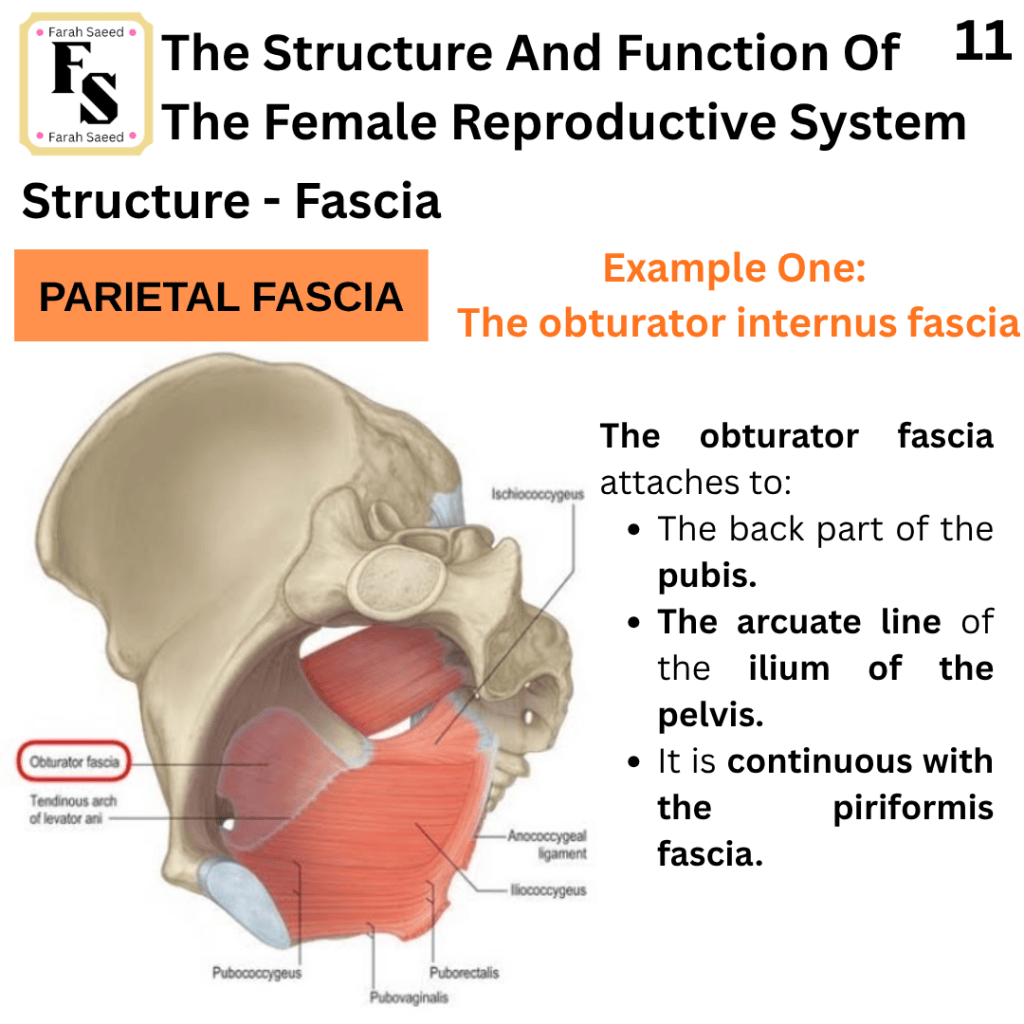
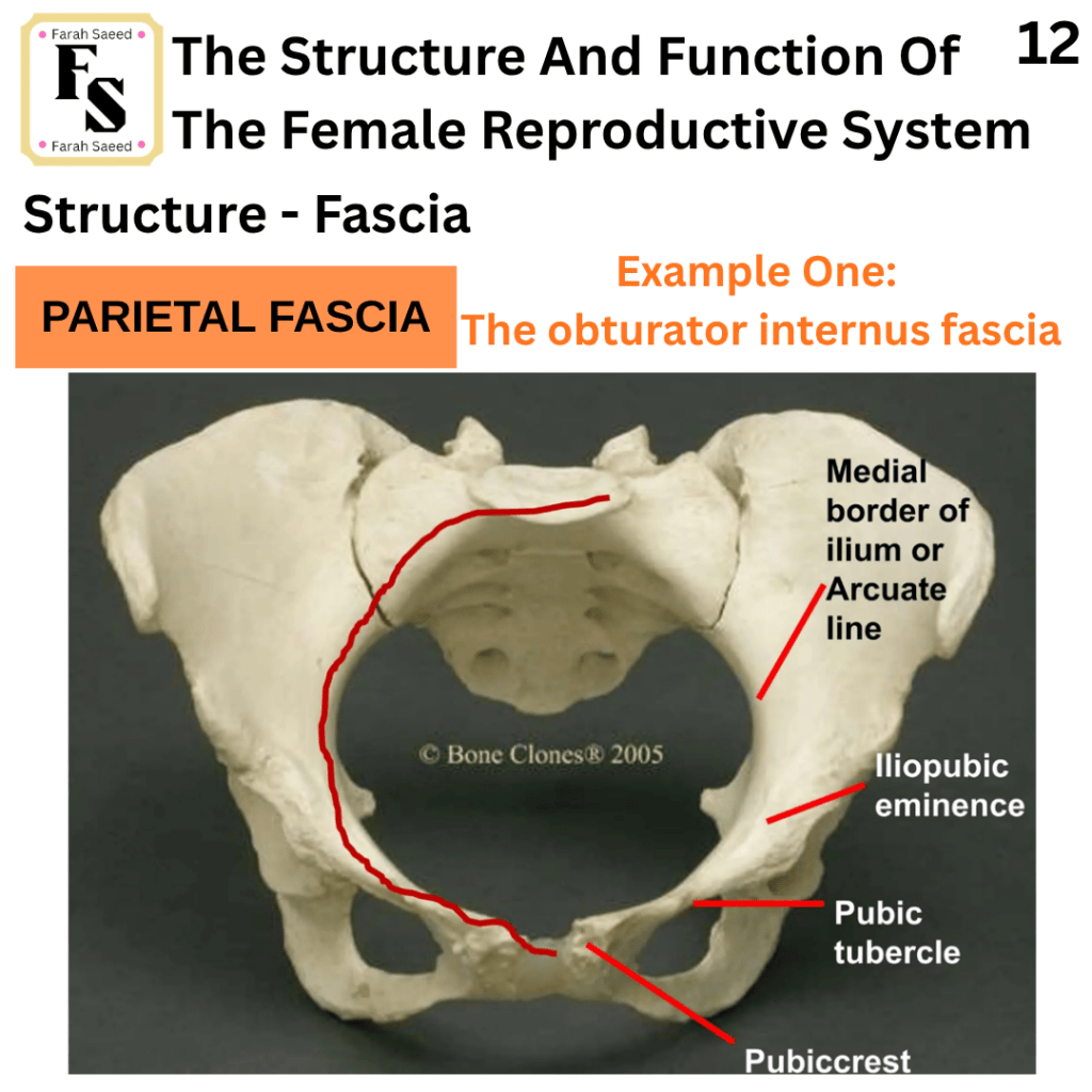
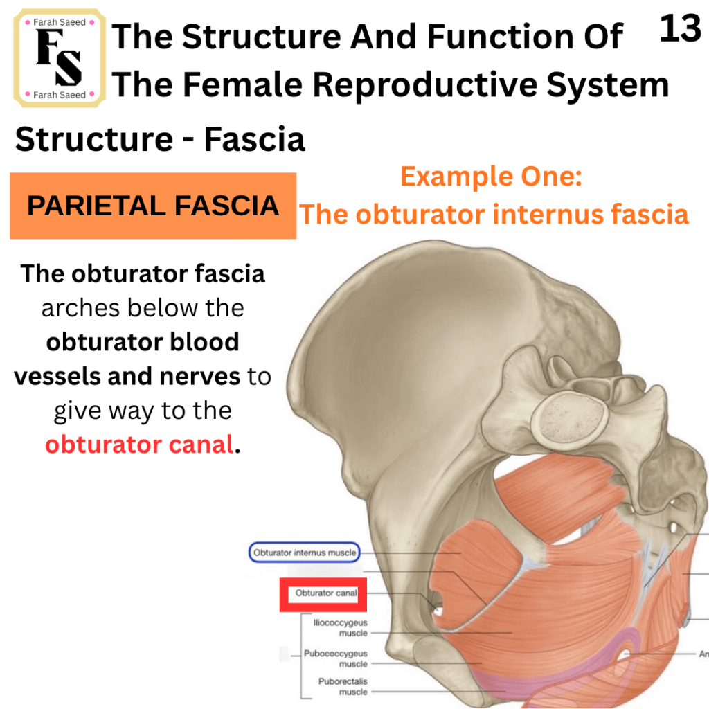
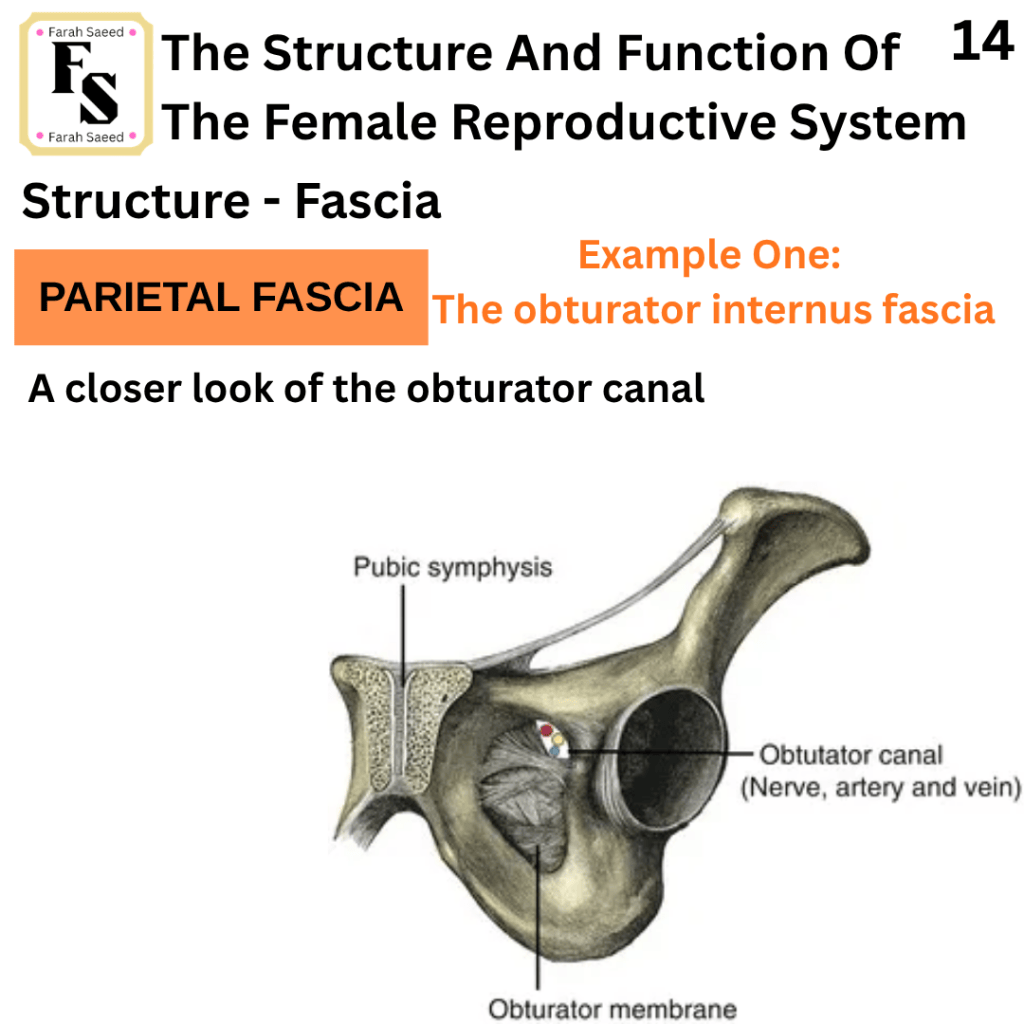
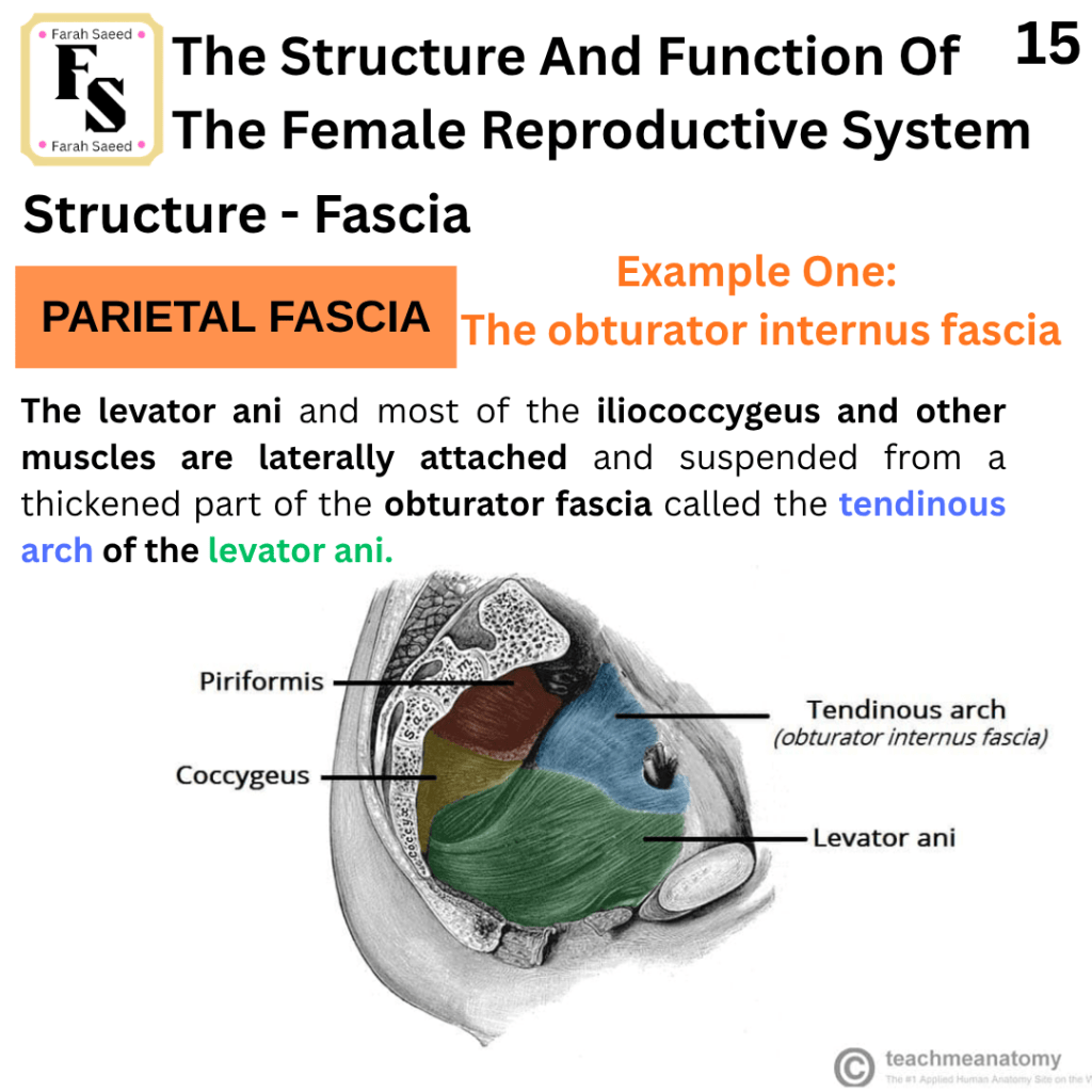
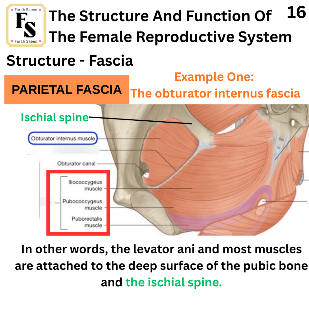
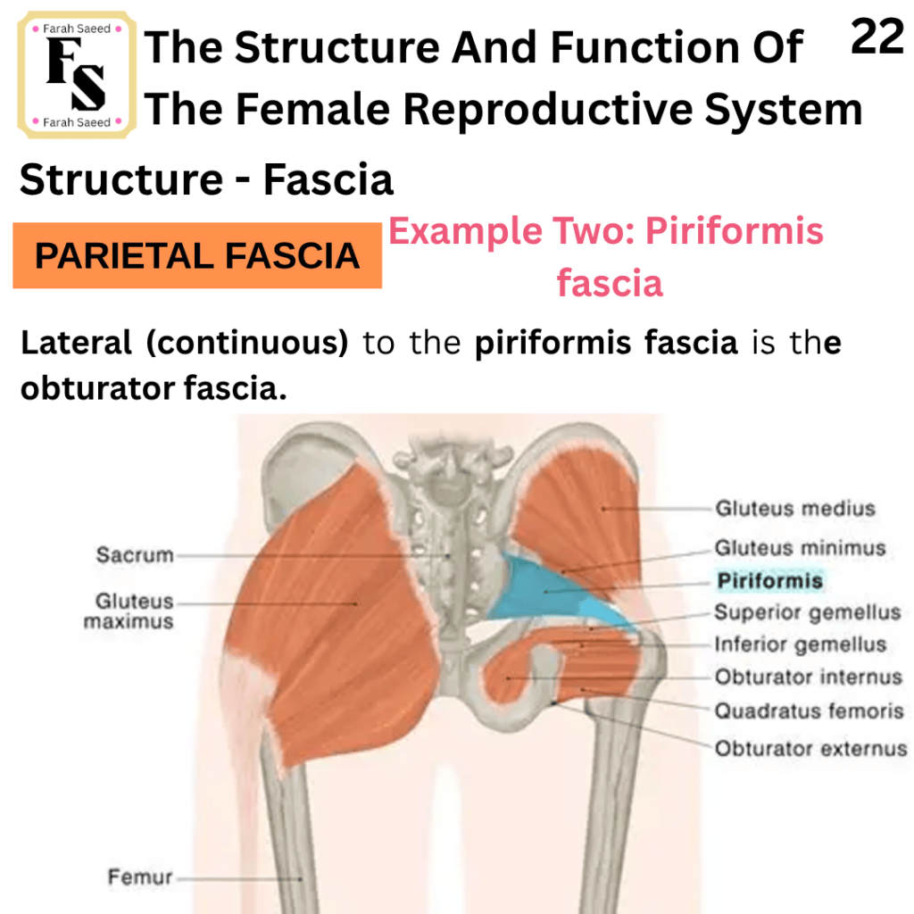
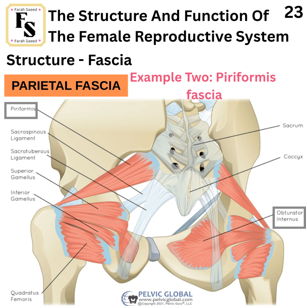
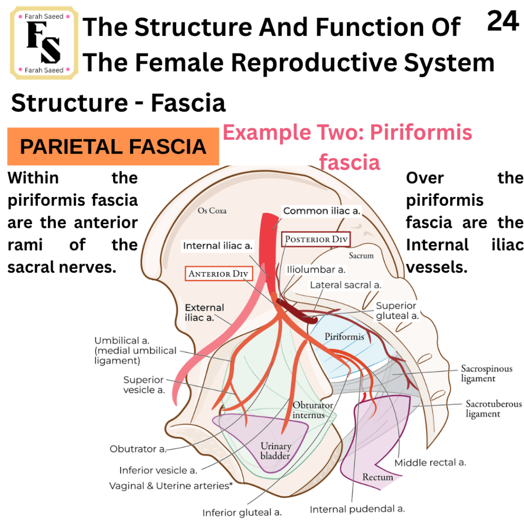
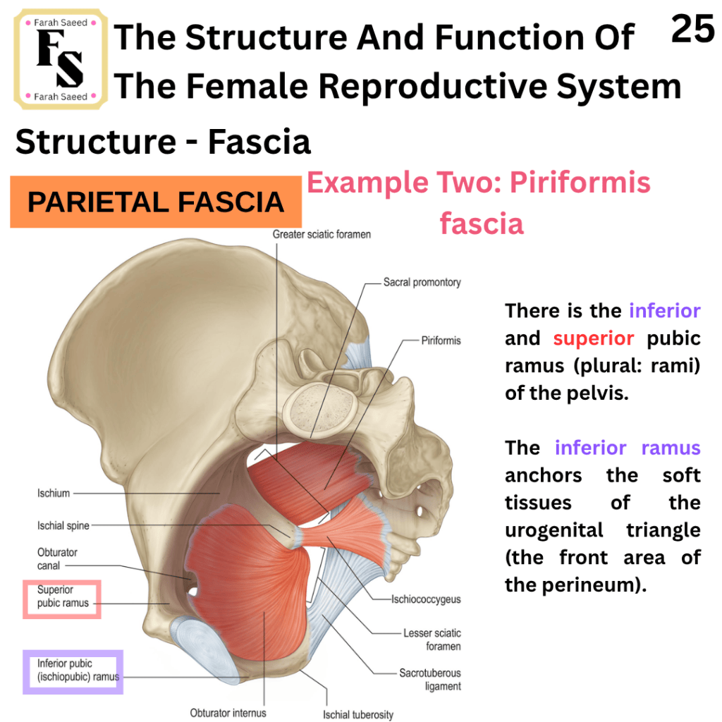
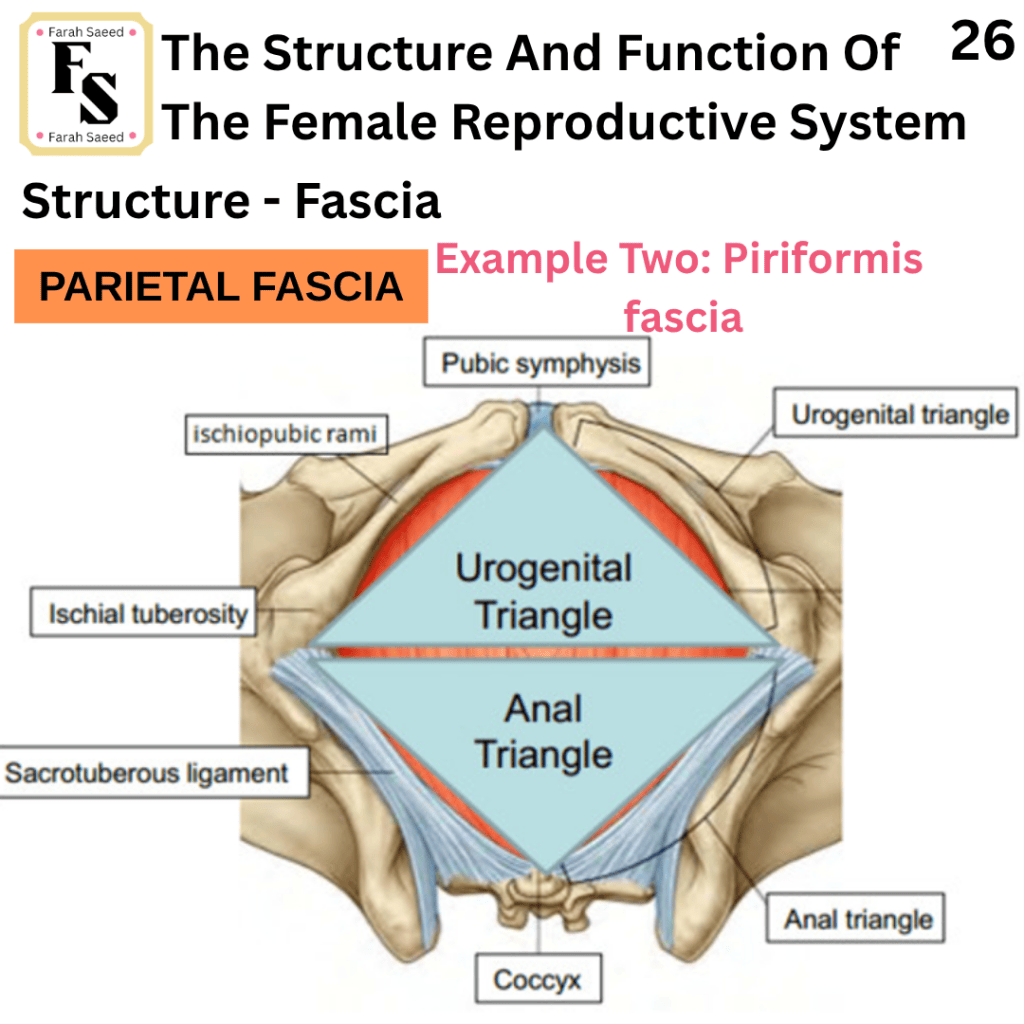
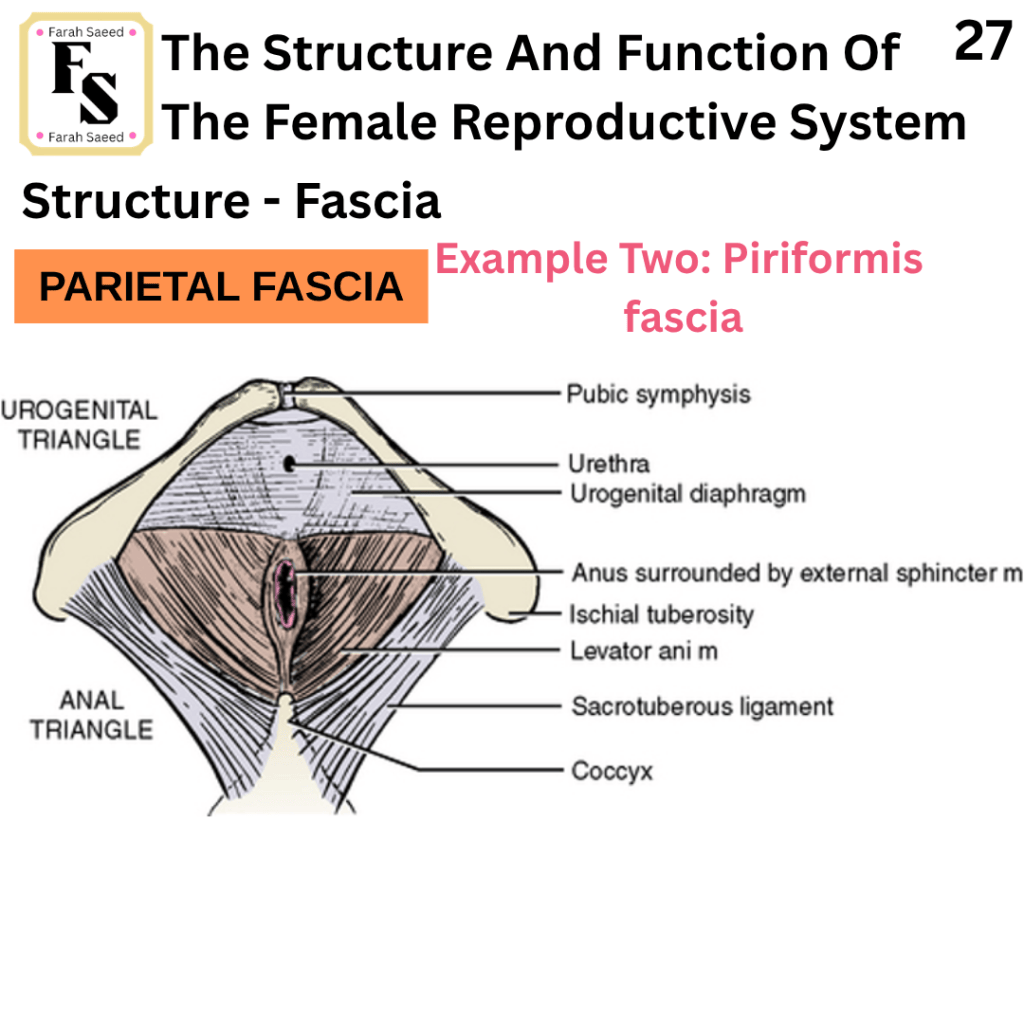
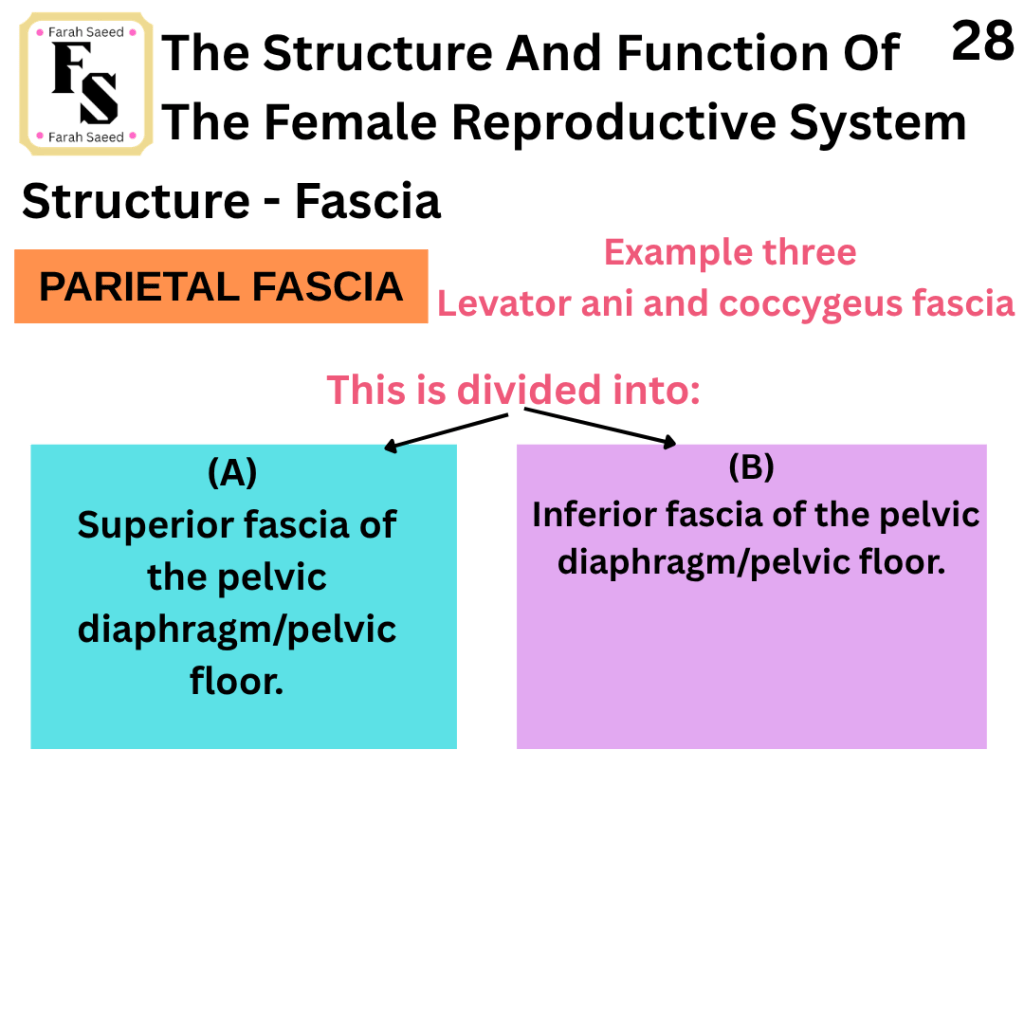
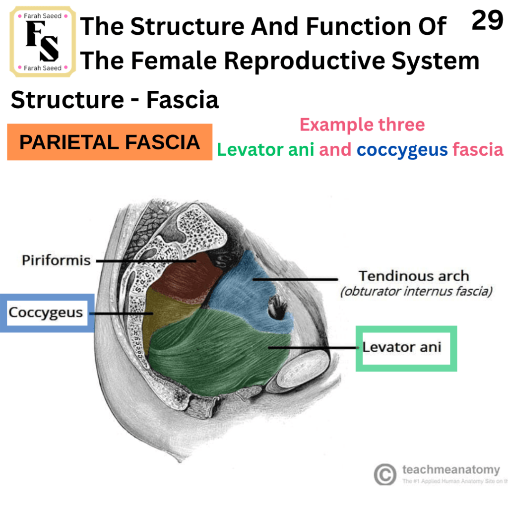
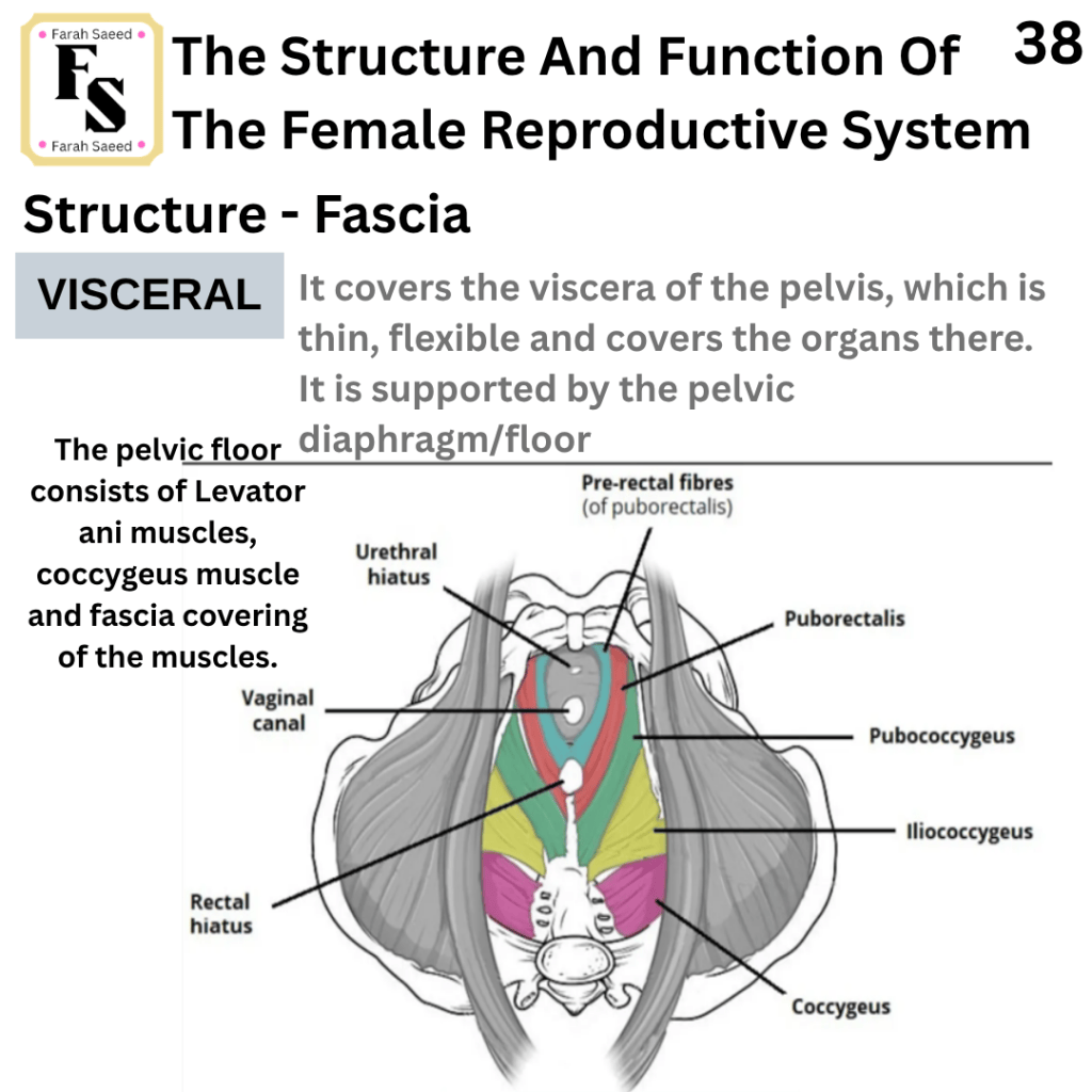
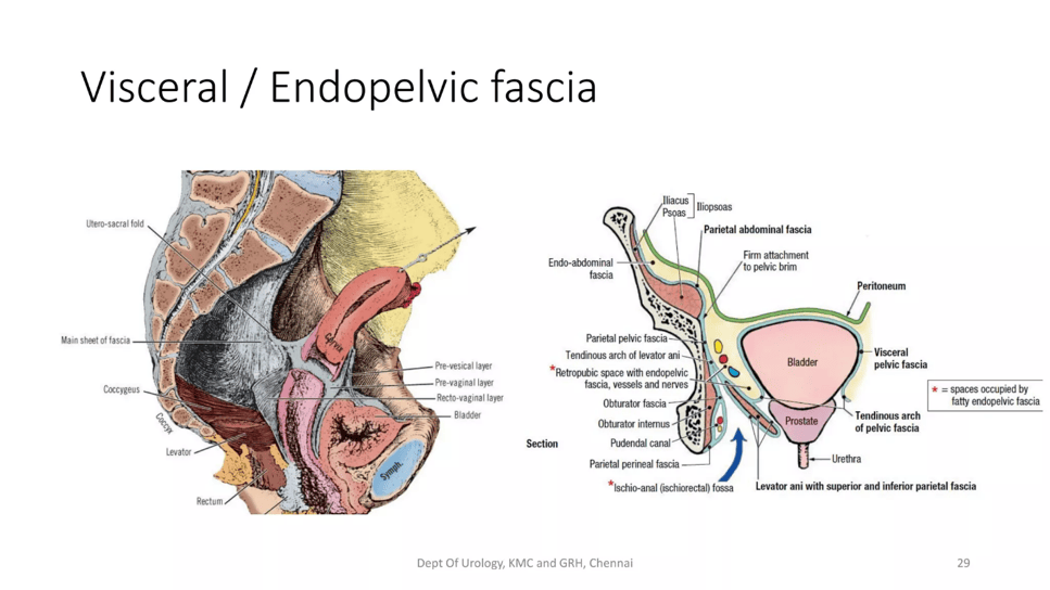
Pelvis and the Fascia
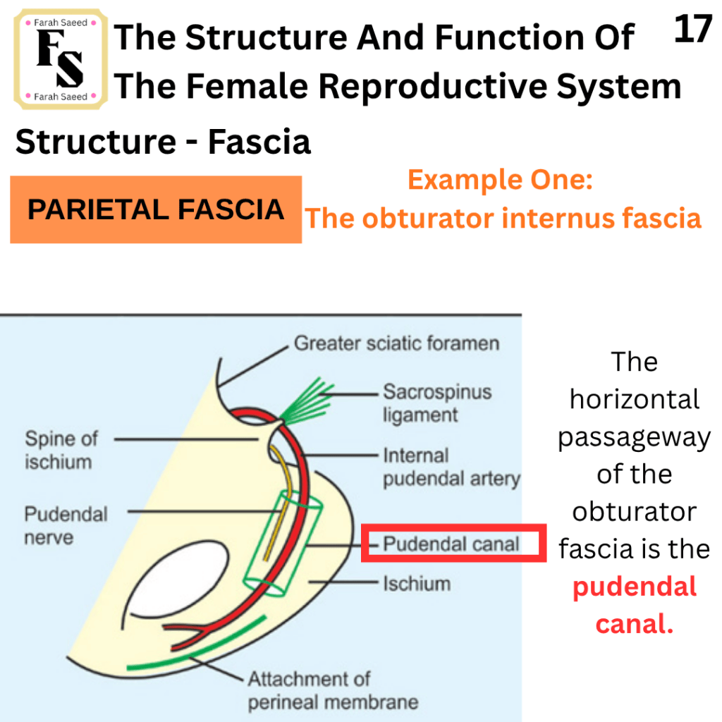
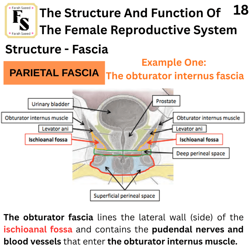
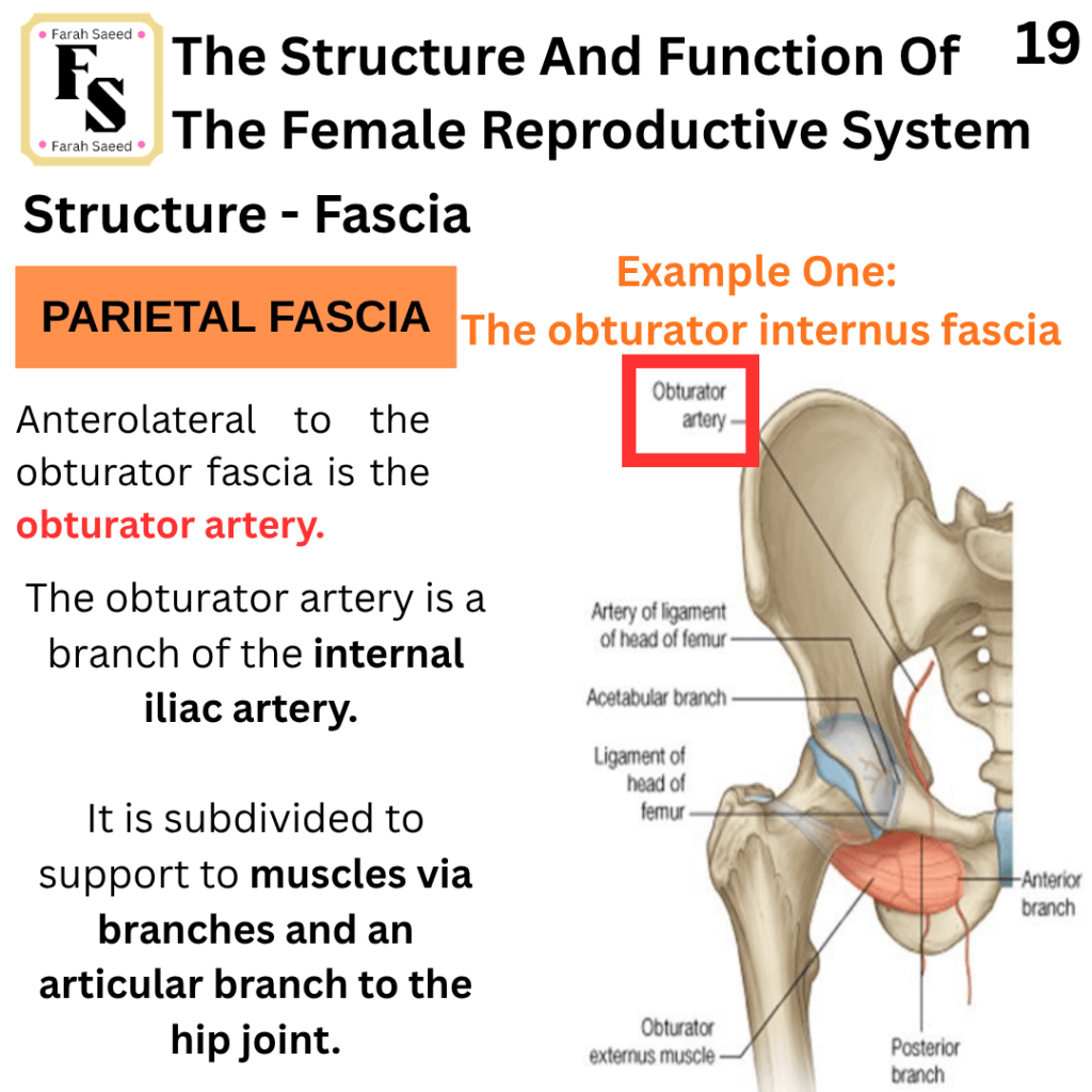
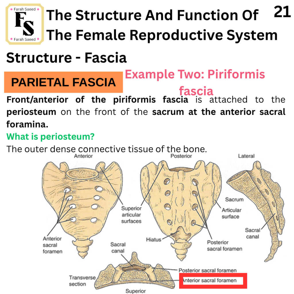
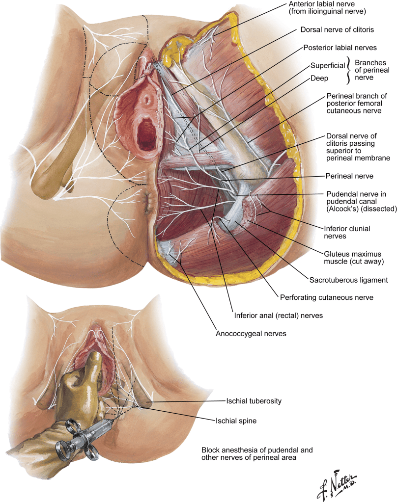
Nerves and Pudendal Blood Vessels
To learn more, visit:
Nerve Supply of female genital tract By Dr. Shivani Siddhpura
https://www.slideshare.net/slideshow/nerve-supply-of-female-genital-tract-edited-pptx/272141564
References
Anatomy.co.uk (2025) Fimbriae. Available at: https://anatomy.co.uk/fimbriae. Accessed (2nd September 2026)
Basic Medical Key (2016) 5. Gynecologic and Obstetric Surgery. Available at: https://basicmedicalkey.com/5-gynecologic-and-obstetric-surgery/ (Accessed: 1st September 2026)
Biology Insights (2025) What Is a Whiff Test and How Is It Performed?. Available at: https://biologyinsights.com/what-is-a-whiff-test-and-how-is-it-performed/ (Accessed: 29th August 2026)
Cassidy, J., Bissett, D., Spene, R. and Payne, M. (2010) Oxford Handbook of Oncology. Oxford: Oxford University Press.
CDC (2024) Gynecologic Cancers Basics. Available at: https://www.cdc.gov/gynecologic-cancer/about/index.html (Accessed: 10th August 2026)
Chumduri, C. and Turco, M.Y. (2021). Organoids of the female reproductive tract. Journal of Molecular Medicine, 99(4), pp.531–553. doi:10.1007/s00109-020-02028-0.
Ditki (n.d.) Vulva and Superficial Perineal Pouch. Available at: https://ditki.com/course/gross-anatomy/glossary/gross-anatomic-microscopic-structure/superficial-perineal-pouch-pudendum-female (Accessed: 17th August 2026)
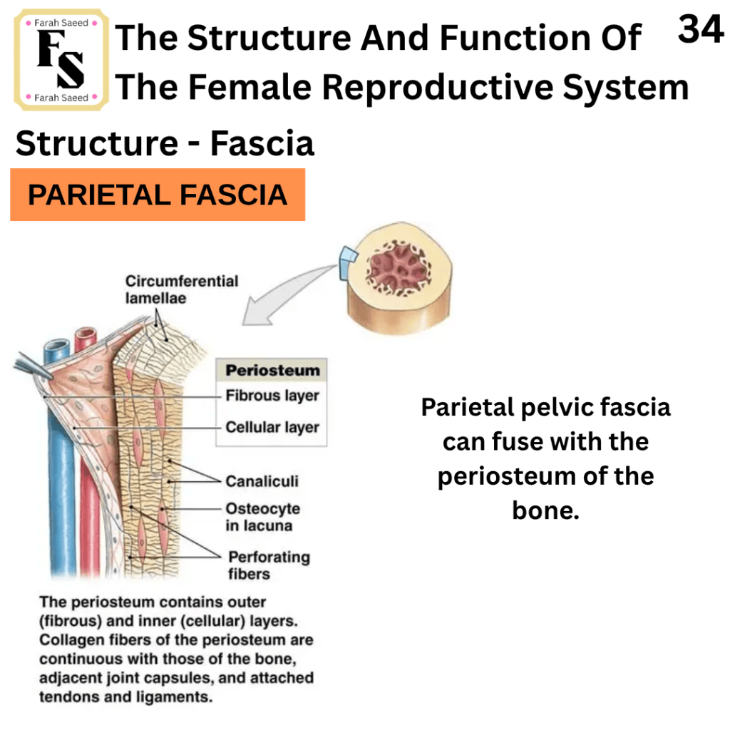
Elsevier (2026a) Parietal Pelvic Fascia. Available at: https://www.elsevier.com/resources/anatomy/connective-tissue/fasciae/parietal-pelvic-fascia/20727 (Accessed: 1st September 2026)
Elsevier (2026b) Obturator Fascia (Left). Available at: https://www.elsevier.com/resources/anatomy/connective-tissue/fasciae/obturator-fascia-left/17054 (Accessed: 1st September 2026)
Elsevier (2026c) Piriformis Fascia (Left). Available at: https://www.elsevier.com/resources/anatomy/connective-tissue/fasciae/piriformis-fascia-left/20712 (Accessed: 1st September 2026)
Elsevier (2026d) Tendinous Arch of Levator Ani. Available at: https://www.elsevier.com/resources/anatomy/connective-tissue/fasciae/tendinous-arch-of-levator-ani/20558 (Accessed: 1st September 2026)
Elsevier (2026e) Presacral Fascia. Available at: https://www.elsevier.com/resources/anatomy/connective-tissue/fasciae/presacral-fascia/19086 (Accessed: 1st September 2026)
Elsevier (2026f) Rectosacral Fascia. Available at: https://www.elsevier.com/resources/anatomy/connective-tissue/fasciae/rectosacral-fascia/23799 (Accessed: 1st September 2026)
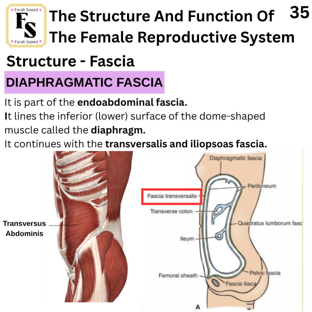
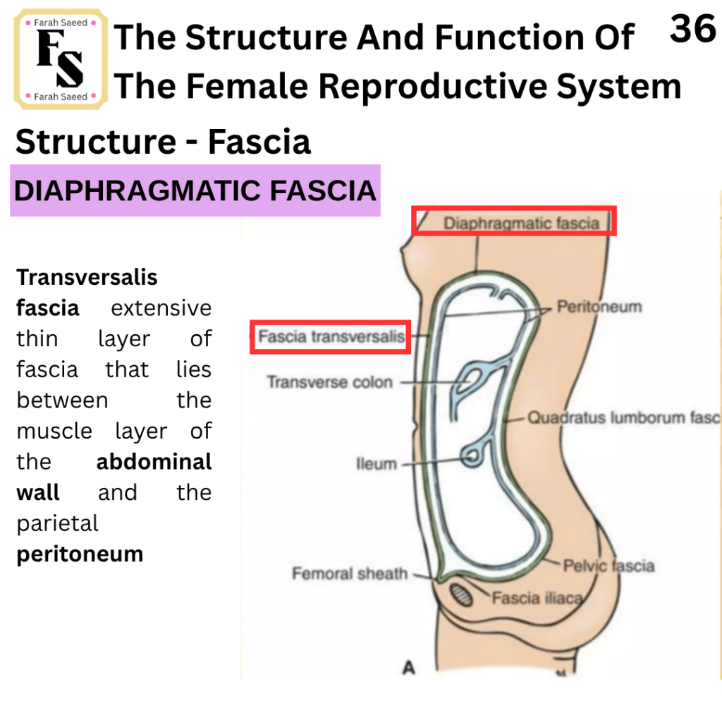
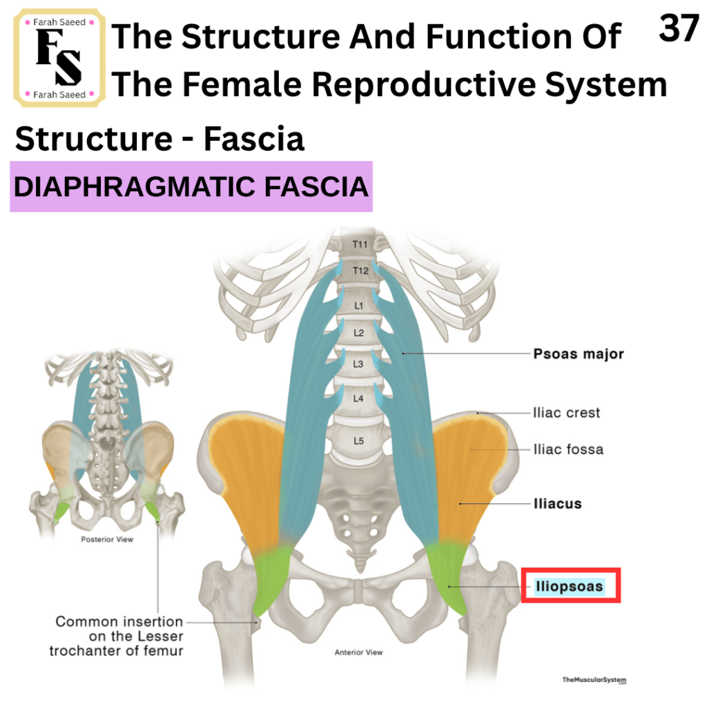
Elsevier (2026g) Diaphragmatic Fascia. Available at: https://www.elsevier.com/resources/anatomy/connective-tissue/fasciae/diaphragmatic-fascia/20666 (Accessed: 1st September 2026)
Elsevier (n.d.) Vaginal Nerves (Right). Available at: https://www.elsevier.com/resources/anatomy/nervous-system/peripheral-nervous-system/vaginal-nerves-right/22047#section-contributing-nerves-1 (Accessed: 8th September 2026)
Hacking, C. (2022) Gonadal Vein Available at: https://radiopaedia.org/articles/gonadal-vein (Accessed: 7th August 2026)
Jones, O. (n.d.) Bartholin’s Cyst and Abscess. Available at: https://teachmeobgyn.com/gynaecology/vaginal-vulval/bartholins-cyst-abscess/ (Accessed: 17th August 2026)
Karki, K. (2023) Clue Cells and Bacterial Vaginosis. Available at: https://microbenotes.com/clue-cells/ (Accessed: 29th August 2026)
Mónica Brauer, M. and Smith, P.G. (2015). Estrogen and female reproductive tract innervation: Cellular and molecular mechanisms of autonomic neuroplasticity. Autonomic Neuroscience, 187, pp.1–17. doi:10.1016/j.autneu.2014.11.009.
Morrison, J., Baldwin, P., Hanna, L., Andreou, A., Buckley, L., Durrant, L., Edey, K., Faruqi, A., Fotopoulou, C., Ganesan, R., Hillaby, K. and Taylor, A. (2024). British Gynaecological Cancer Society (BGCS) vulval cancer guidelines: An update on recommendations for practice 2023. PubMed, 292, pp.210–238. doi:10.1016/j.ejogrb.2023.11.013.
Namwanje, M. and Brown, C.W. (2016). Activins and inhibins: Roles in development, physiology, and disease. Cold Spring Harbor Perspectives in Biology, 8(7), p.a021881. doi:10.1101/cshperspect.a021881.
National Cancer Insitute (2024) What is Vulvar Cancer? Available at; https://www.cancer.gov/types/vulvar/what-is-vulvar-cancer (Accessed: 7th August 2026)
National Cancer Institute (2024) Vulvar Cancer Causes and risk factors. Available at; https://www.cancer.gov/types/vulvar/causes-risk-factors (Accessed: 7th August 2026)
National Cancer Institute (n.d) endocervical canal. Available at: https://www.cancer.gov/publications/dictionaries/cancer-terms/def/endocervical-canal (Accessed: 29th August 2026)
Rosner, J., Samardzic, T. and Sarao, M. (2024) Physiology, Female reproduction. Available at: https://www.ncbi.nlm.nih.gov/books/NBK537132/ (Accessed: 14th August 2026)
Science Insights (2025) Hyphae vs. Pseudohyphae: Key Differences in Medical Contexts. Available at: https://scienceinsights.org/hyphae-vs-pseudohyphae-key-differences-in-medical-contexts/ (Accessed: 29th August 2026)
Seladi-Schulman, J. (2026) Everything to Know About Female Reproductive Organs. Available at: https://www.healthline.com/health/womens-health/female-reproductive-organs (Accessed: 7th August 2026)
The Eve Appeal (2026) Gynaecological cancers. Available at: https://eveappeal.org.uk/information-and-advice/gynaecological-cancers/ (Accessed: 7th August 2026)
The Eve Appeal (2026) Vaginal Cancer. Available at: https://eveappeal.org.uk/information-and-advice/gynaecological-cancers/vaginal-cancer/ (AccessedL 7th August 2026)
The Eve Appeal (2026) Vulval cancer Available at; https://eveappeal.org.uk/information-and-advice/gynaecological-cancers/vulval-cancer/ (Accessed: 7th August 2026)
The Eve Appeal (n.d.) Womb Cancer Available at: https://eveappeal.org.uk/information-and-advice/gynaecological-cancers/womb-cancer/ (Accessed: 7th August 2026)
Thompson, L. (n.d.) The Vulva. Available at; https://teachmeanatomy.info/pelvis/female-reproductive-tract/the-vulva/ (AccessedL 7th August 2026)
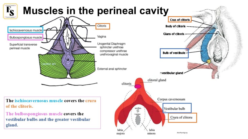
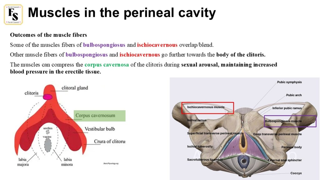
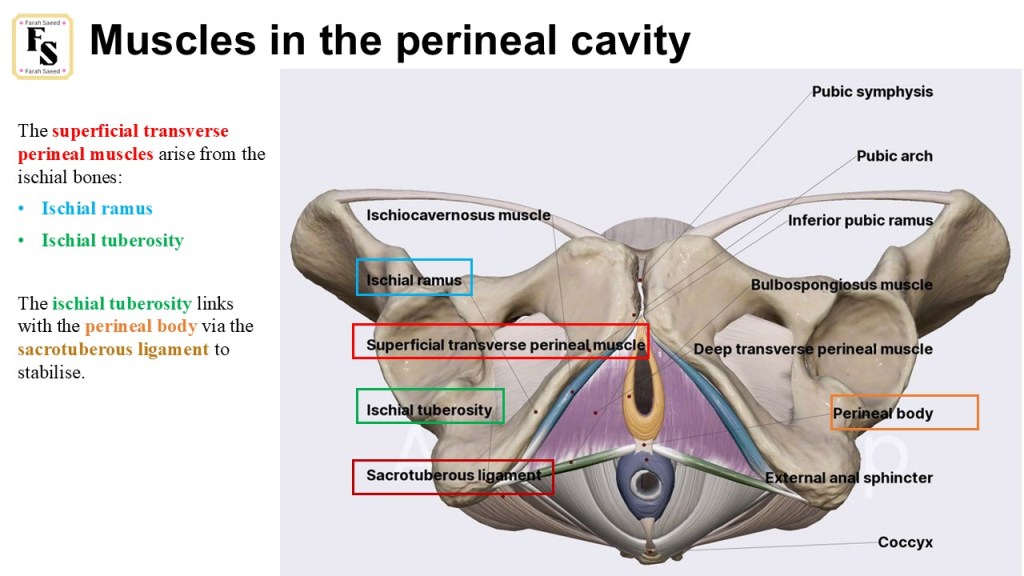
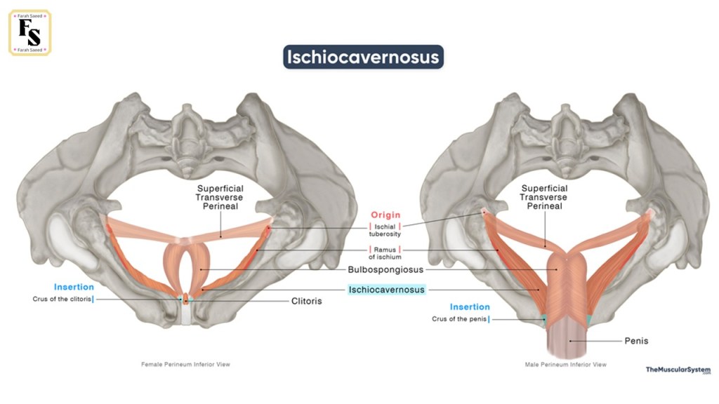
Supporting Images And Videos Of Pelvis, Fascia, Muscles, Blood Supply And Nerves
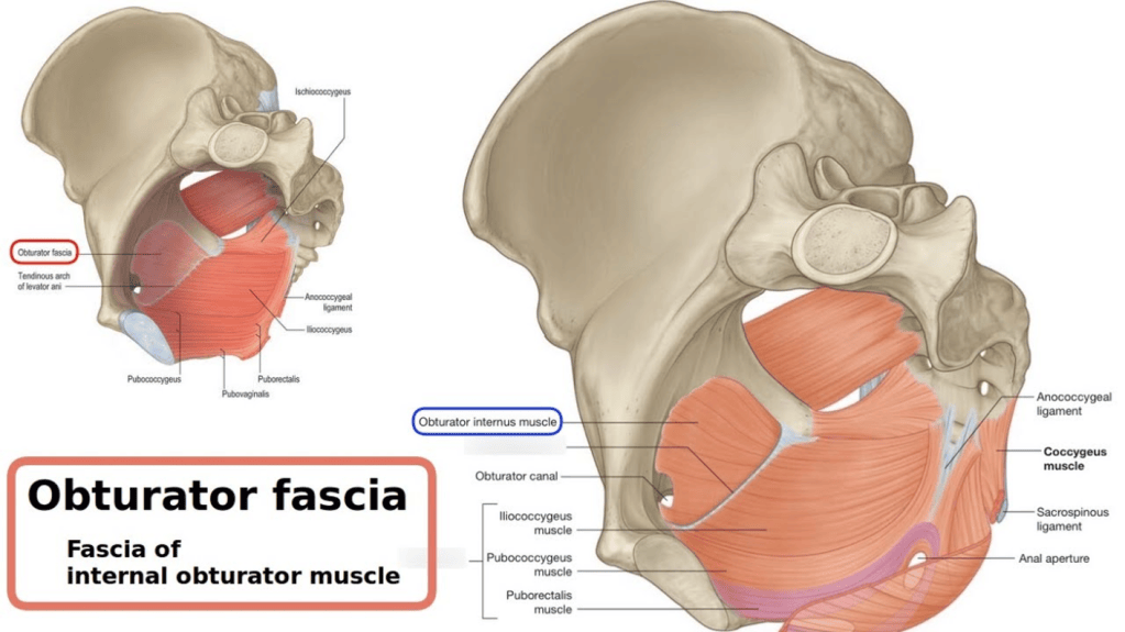
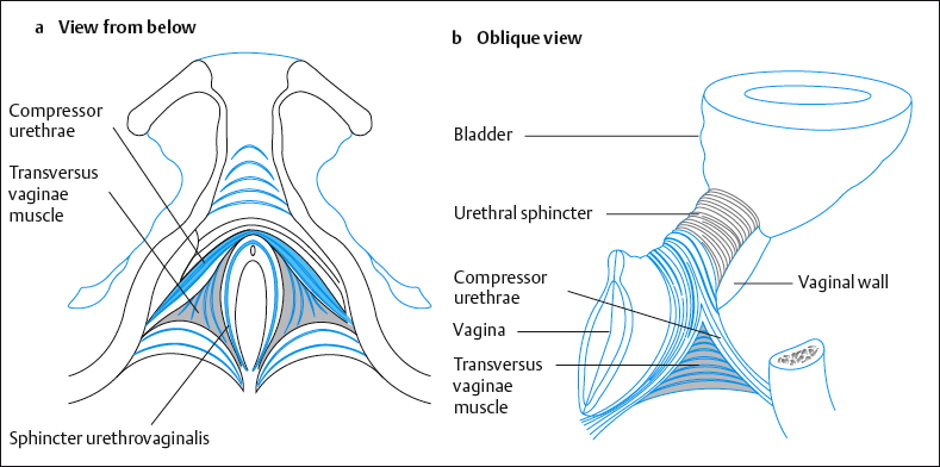
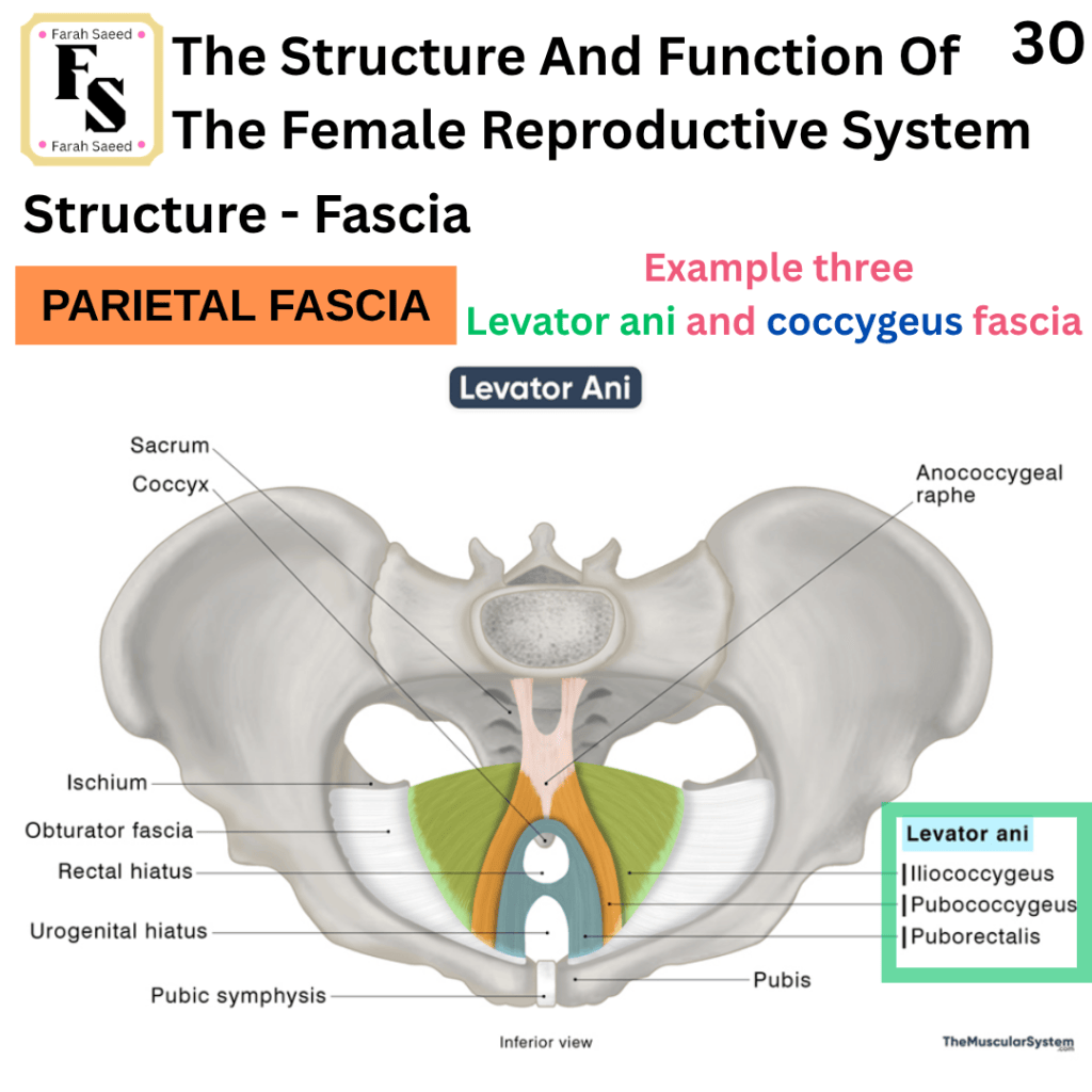
The pubovaginal fibers are directly below the bladder. This helps to release the urine/pee/wee from the body, and this process is known as micturition.
The pubococcygeal fibers help to control the movement of the coccyx by pulling it forward to help close the pelvic outlet. The pelvic outlet is further closed by the contraction of the pubococcygeal fibers, where it pulls the rectum, vagina, and bladder neck up towards the symphysis pubis. Other roles of the pubococcygeal fibers is the flexure at the rectum-anus junction (anorectal).
The removal of faeces/poo and straightening of the anorectal junction is achieved when the pubococcygeal fibers relax.
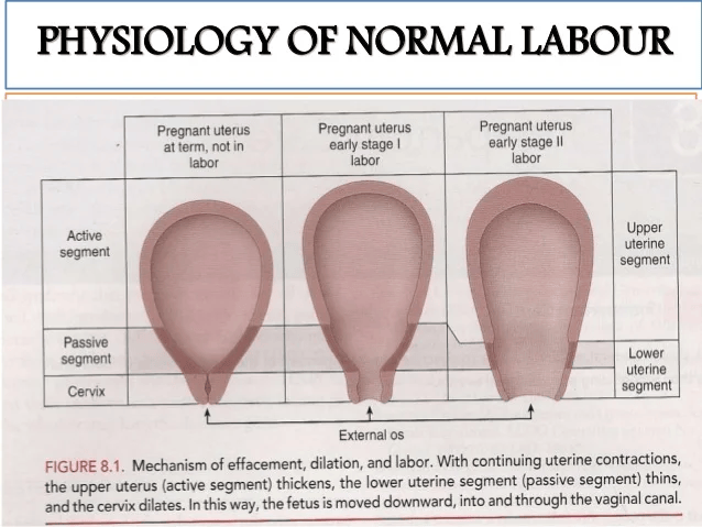
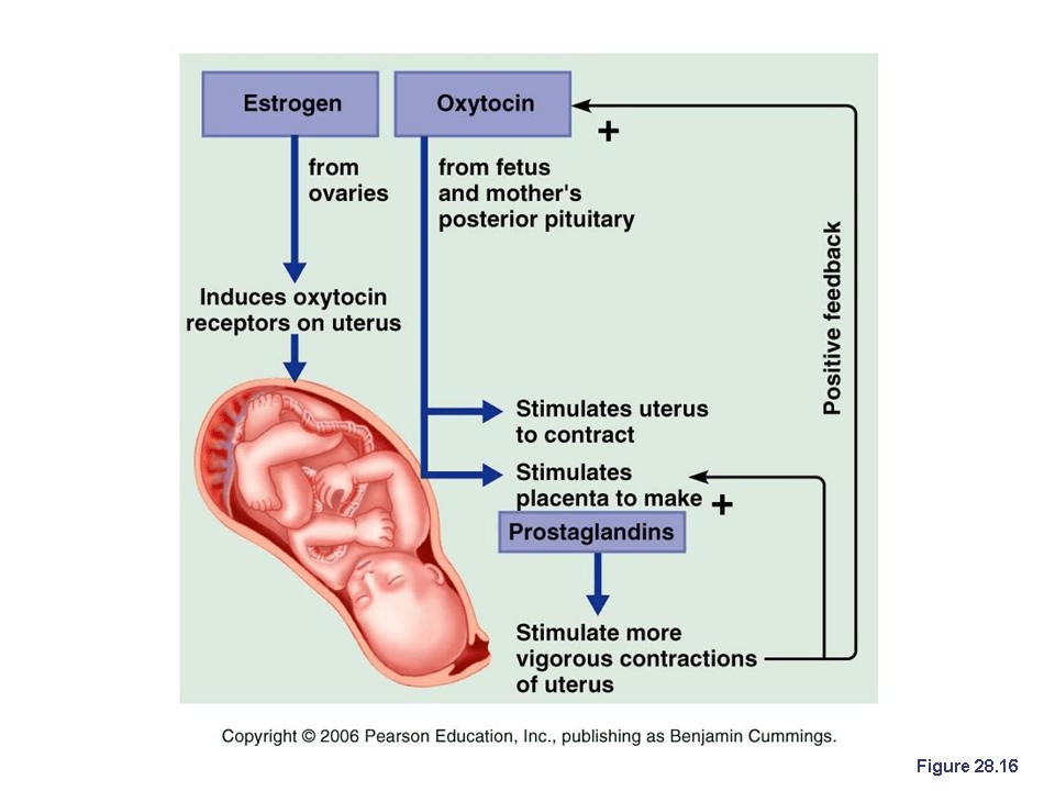
During childbirth/parturition, the levator ani muscles together help the foetal head to the lower part of the passageway (cervix/vagina).
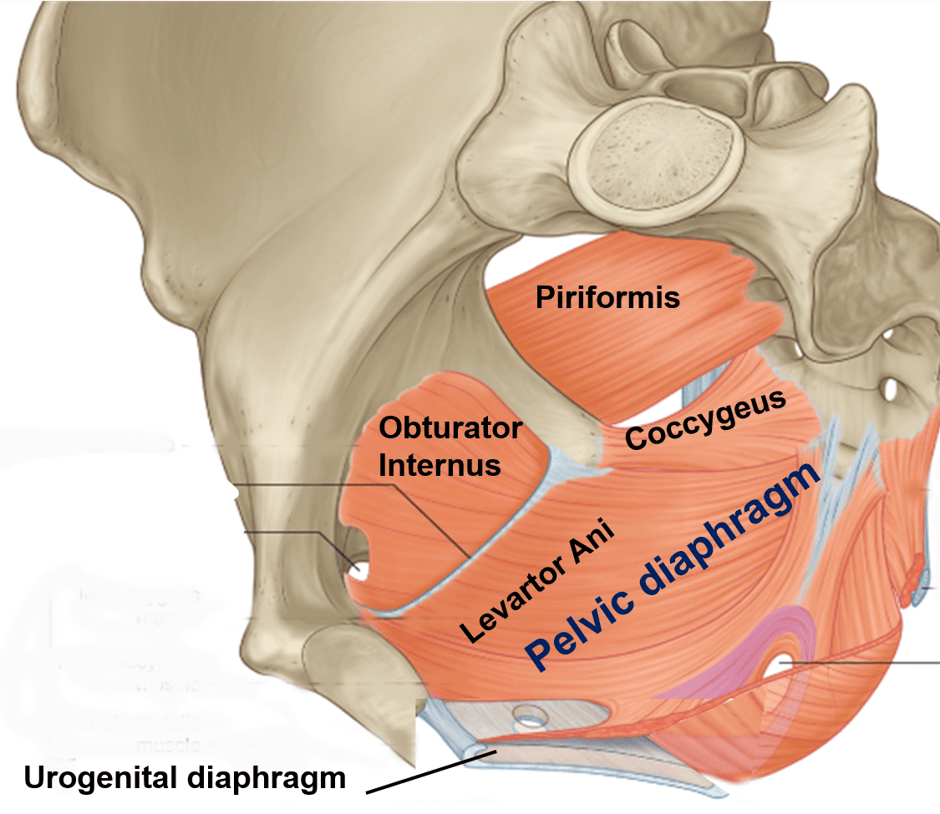
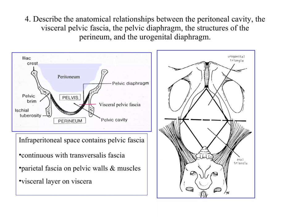
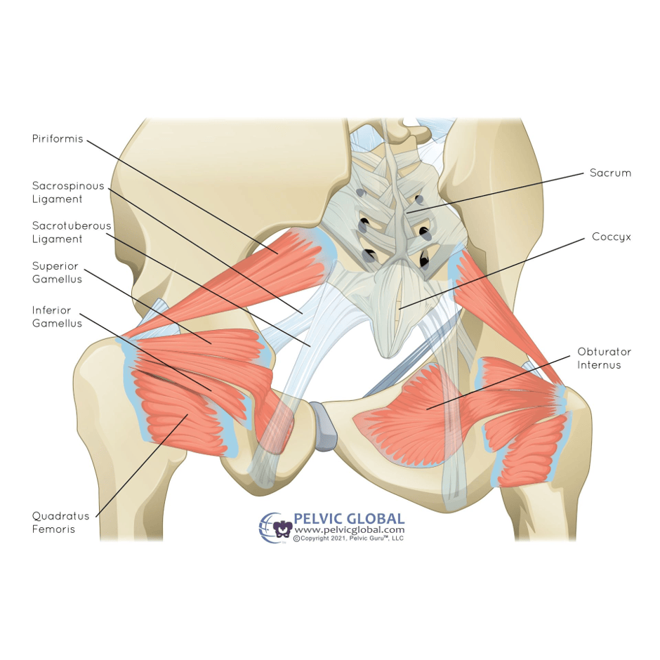
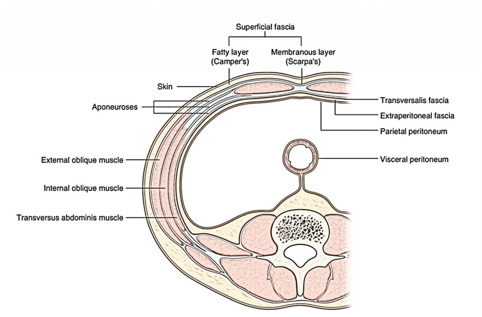
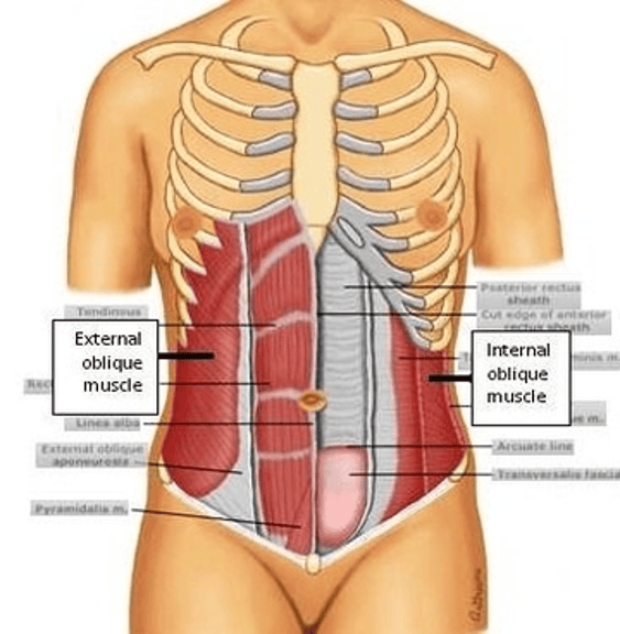
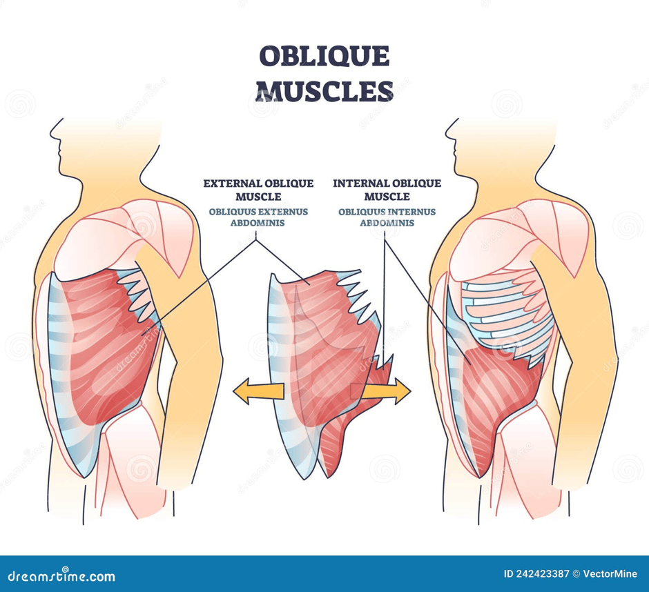
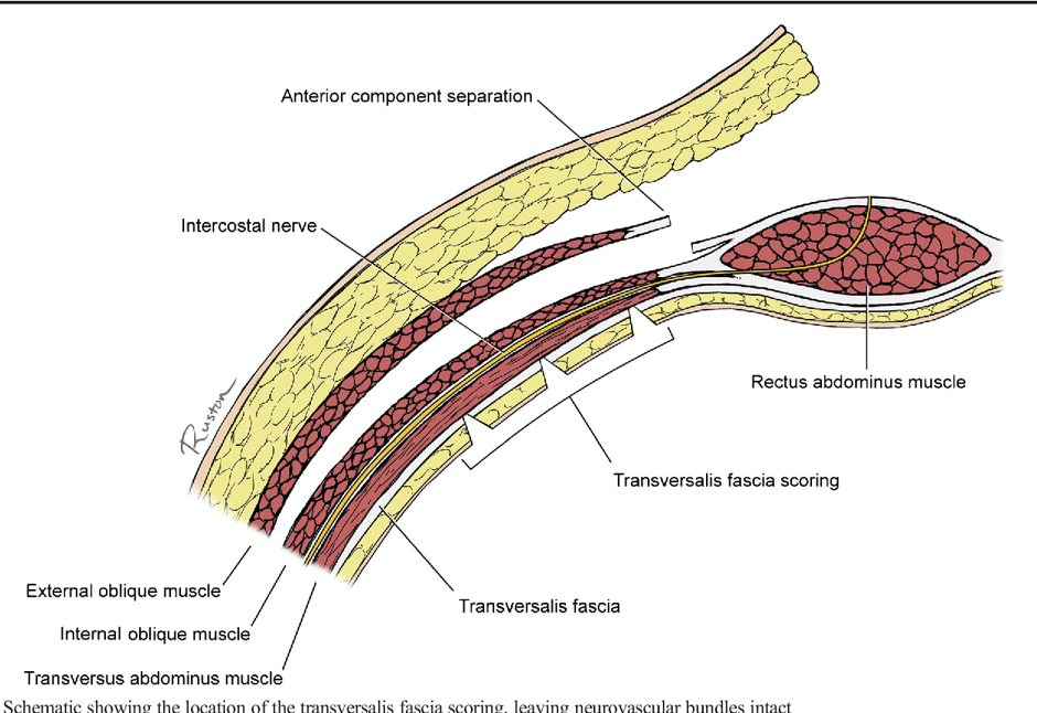
Lateral view of the oblique muscles. There are two types: external and internal. The external oblique covers some of the ribs, the anterior half of the iliac crest, the inguinal ligament, the pubic tubercle, and the aponeurosis of the rectus sheath. It is present on the side and front of the abdomen. The internal oblique is under the external oblique in a different direction. The transvers abdominance are under the obliques and consists of abdominal muscles that help with movement and protection.
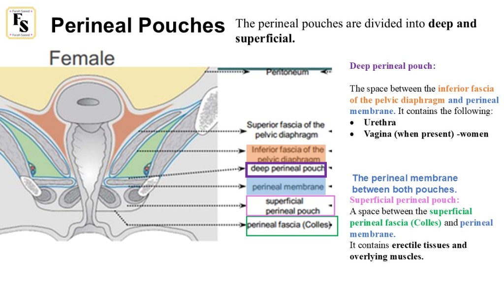
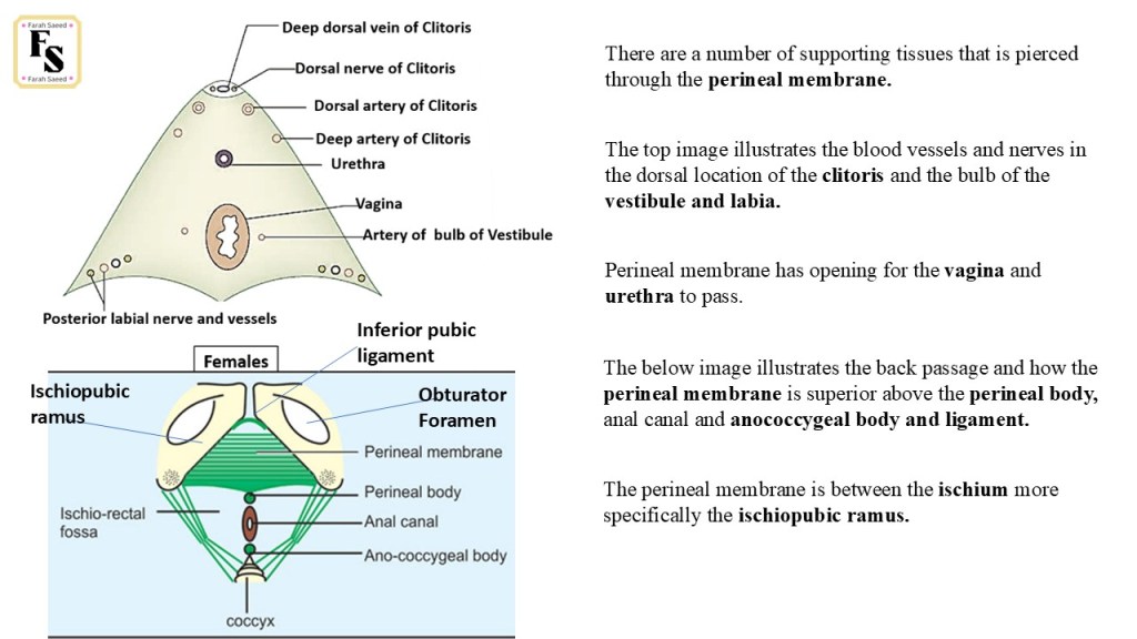

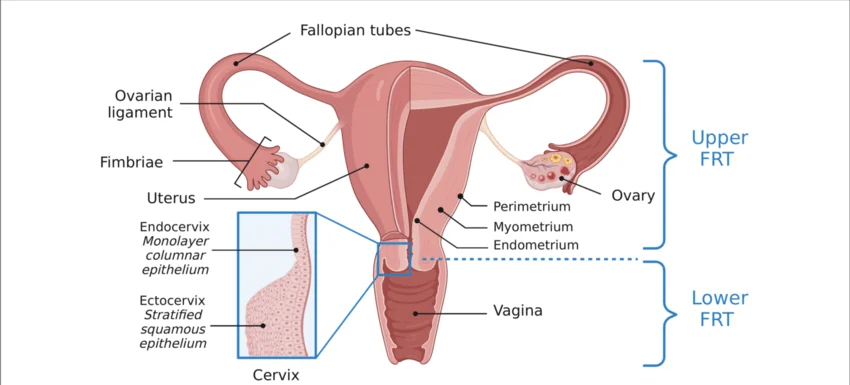
Summary of the Female Reproductive System


Leave a comment