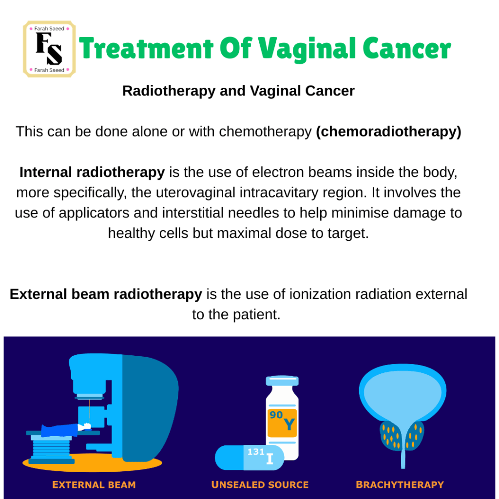
Give an example of a common chemoradiotherapy method used to treat vaginal cancer?
According to the guidelines,
Brachytherapy with a platinum-based chemotherapy is performed but does not exceed 7 to 8 weeks.
“45 to 46 Gy (1.8 to 2.0 Gy per fraction) and single- agent radiosensitizing chemotherapy, preferably cisplatin (weekly 40 mg/m²)”
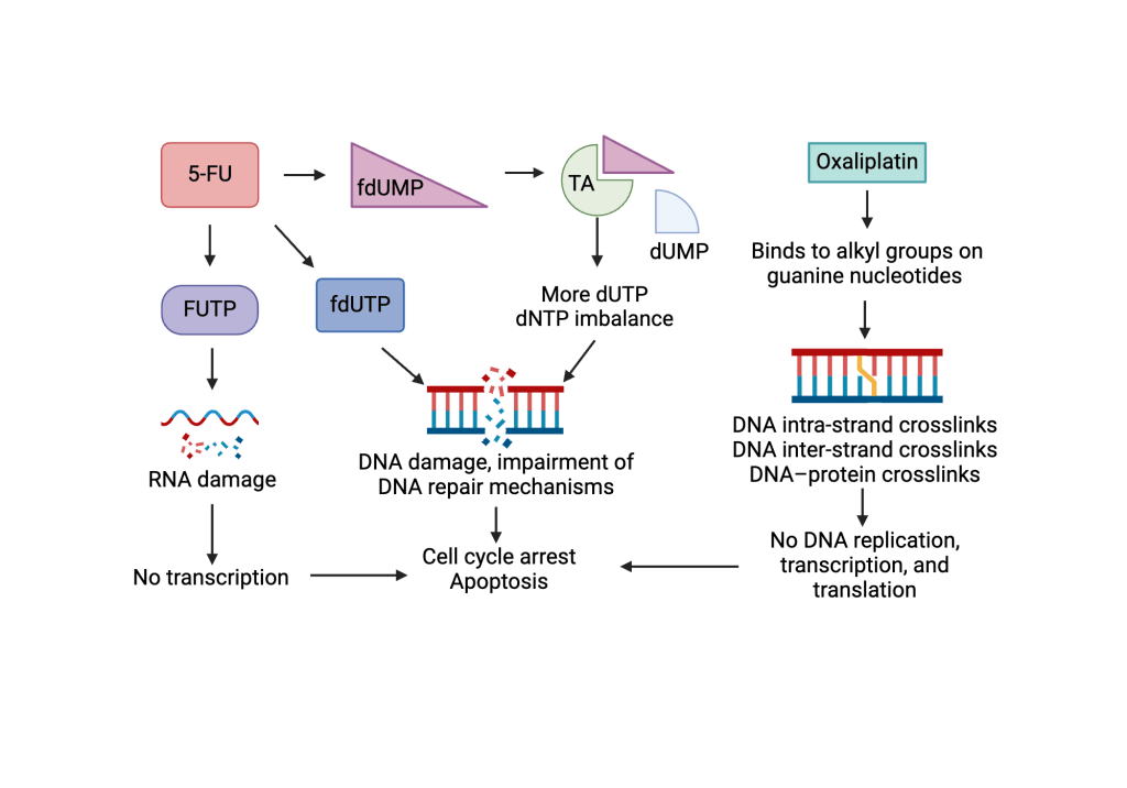
Other options are 5-Fluouracil (5-FU) and carboplatin.
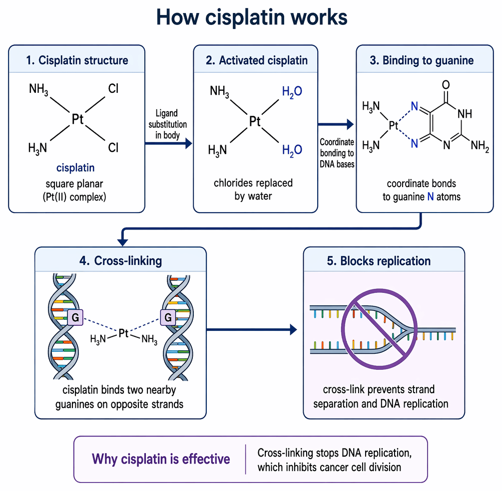
Carboplatin and cisplatin are DNA alkylating agents made of platinum.
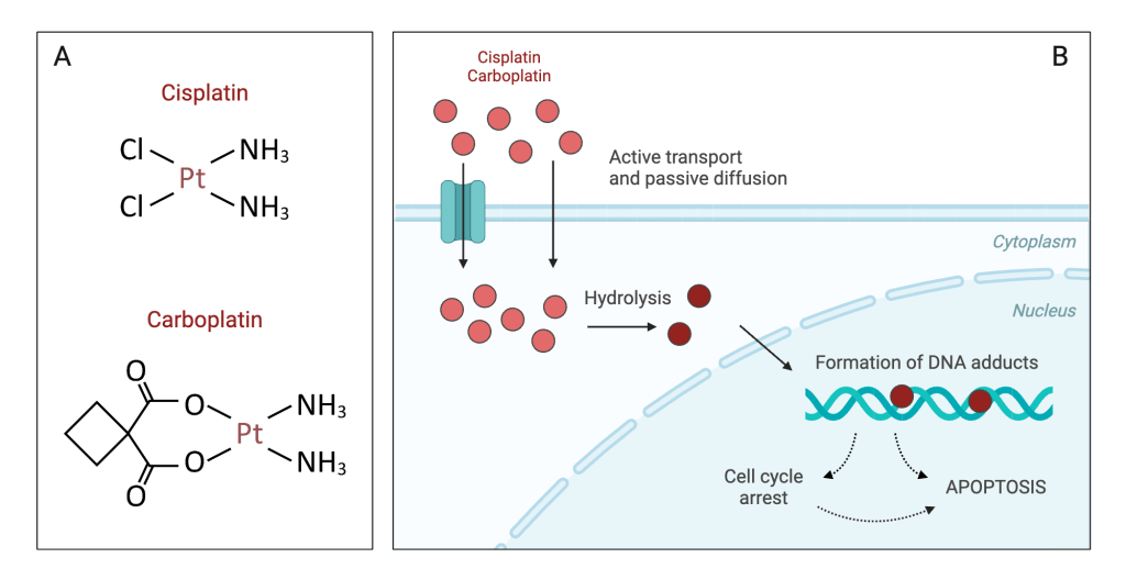
How do DNA Alkylating agents function?
- It forms crosslinks with N7 guanine residues (type of base found in DNA and RNA).
- They intercalate DNA and produce toxic reactive oxygen species)
- They cleave the transcription factor (CREB1) – this affects the transcription of target genes involved in cellular processes.
- This causes interstrand and intrastrand breaks in the DNA molecule.
It affects how cells divide, copy (replicate), grow, and die (apoptosis).
What is 5FU and how does it work?
5-FU (antimetabolite) – inhibits the enzyme thymidylate synthase, which is involved in the production of thymidylate. This is a critical step in folate metabolism. This inhibits DNA synthesis.
Did you know there are other options without the use of chemotherapy?
Regional Hyperthermia
However, it is more effective when combined with chemotherapy and radiotherapy.
Other names: Thermal therapy, thermal ablation, or thermotherapy.
Increasing the temperature to 113 Fahrenheit (45 degrees Celsius).
Moderate hyperthermia can be non-lethal (39 to 42°C) or lethal (>42°C).
Aim: To kill cancer cells over a larger area (regional – body, organ, or limb) with minimal harm or none to normal tissues.
Why?
REASON ONE FOR HYPERTHERMIA
There are several changes within the physiological systems that cause the sensitivity of cells and tissues to moderate hyperthermia.
- Oxygen levels
- pH
- Blood flow.
Cancer cells are more sensitive to heat energy than non-cancerous cells. This reflects the phase in the cell cycle. S phase, where DNA replication takes place, and mitosis (the process by which cells grow and divide) are the most sensitive.
Additional factors that hyperthermia can help:
- Increase sensitivity to heat.
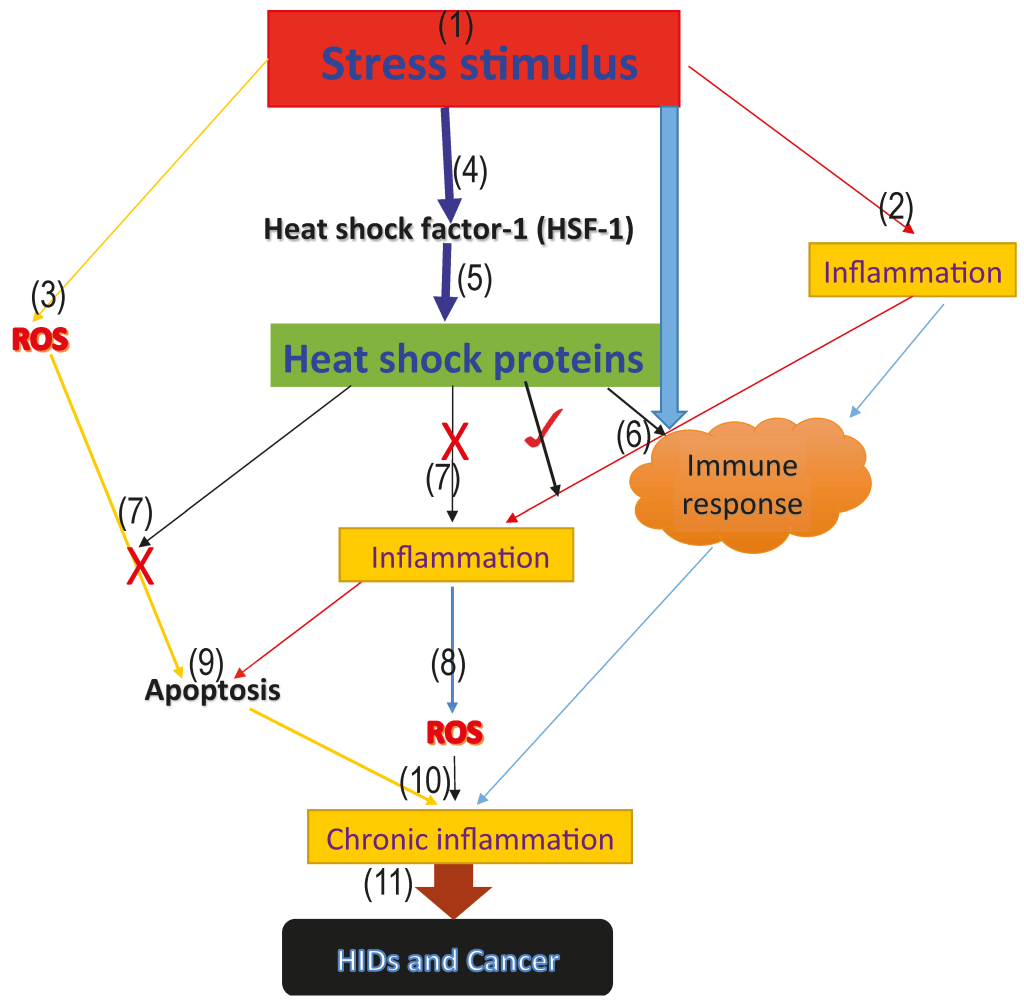
- Release of heat shock proteins.
- Cellular defence mechanism
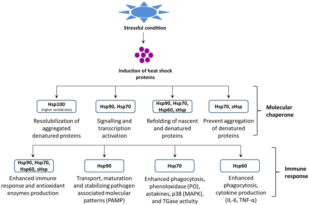
What Are Heat Shock Proteins (Hsp)?
Main roles: regulate cell death (apoptosis)
Subtypes:
Regulation of the intrinsic pathway (apoptosis) in mitochondria and the extrinsic pathway (Fas death receptor): Hsp70 and Hsp27. It prevents t-Bid translocation to mitochondria, preventing cytochrome release.
Survival protein: Hsp70.
Negative regulation of activating molecular scissors (caspase-2): Hsp90.
Protection against oxidative stress that promotes cell death: Hsp70 and Hsp27 (phosphorylated)
Overexpression of Hsp70 increases cell growth, poor differentiation and poor response to cancer treatment, poor activation of caspases and proteases, evading cell death.
Resistant to chemotherapy (doxorubicin and cisplatin): overexpression of Hsp27
What is apoptosis?
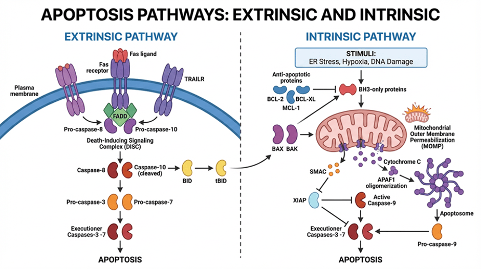
In an extrinsic pathway, a death ligand like Fas or tumour necrosis factor (TNF) binds specifically to its receptor protein found on the cell surface. The ligand-receptor complex then undergoes a conformational change and several complexes bind together (oligomerise). The signal is then transduced into the cell. Due to the conformational change, a part of the complex is revealed and is called a death domain. An adaptor protein like FADD or TRADD, depending on the receptor activated; e.g if it is a Fas receptor-ligand complex, then it is FADD (Fas-associated adaptor death domain). If it is the TNF receptor-ligand complex, then it is TRADD (TNF-associated adaptor death domain). The reason for the adaptor proteins is to transduce the death signal from the receptor to the caspases. The first caspase to be activated is pro-caspase 8. As it’s the first enzyme, it self-cleaves as several become close together. The activated caspase 8, receptor, ligand, and adaptor protein form the Death-inducing Signalling Complex (DISC). This leads to a cascade or a domino effect where other caspases are activated: 3, 6 and 7. This then results in the cleavage of protein targets to induce cell death (apoptosis). Amongst the targets are laminin to shrink the nucleus, actin and intermediate filaments that facilitate the cytoskeleton and muscle movement. The process can be halted by a protein called C-Flip, which binds to the death domain and prevents the binding of caspase 8 and its activation.
On the other hand, intrinsic pathway is stimulated by damage by the DNA and oxidative stress. It relies on the Bcl-2 family of proteins. Some promote apoptosis, and others prevent apoptosis. They differ in the number of BH domains (Bcl-2 homology domains). Some have one BH domain (BH3-only domain), whilst others have a few. Examples of proteins that promote apoptosis are Bax, Bak, and Bcl-x5. BH3-only proteins also promote apoptosis, like Bad, BID, Bim, Noxa, Puma, Hrk/DP5, and Bik. Proteins that evade apoptosis are Bcl-2, A1, Mcl-1, and Boo. The mitochondria, where respiration takes place to provide energy, have a double membrane, inner and outer, separated by the intermembrane space. Proapoptotic proteins regulate the release of mediators. This is referred to as mitochondrial outer membrane permeabilization (MOMP).
Upon activation of apoptotic signal, Bid and Bim bind to activate Bim. This leads to a conformational change in Bax as it translocates from the cytoplasm to the mitochondria and inserts into the outer mitochondrial membrane. Oligomerization of six to eight molecules takes place. It increases permeability of the outer membrane to form a membrane channel and release mediators of apoptosis. Cytochrome c important part of aerobic respirstion lots of oxygen is released into the cytoplasm and assemble complex to form an apoptosome along with ATP and Apaf-1. Activation of Apaf-1 leads to recruitment of procaspase-9. There are protein domains like CARD present on Apaf-1 and procaspase-9. This leads to activation of caspase 3, 6, and 7. Apoptosis can be prevented by inhibitors of apoptosis proteins IAP prevents aopotosis by binding and inhibit caspase 3 and 7. XIAP, a type of protein, can bind to caspase 9 and inhibit it. Second, mitochondrial-derived activator (Smac/Diablo) is also released from mitochondria and modulates the process of apoptosis. It has a similar domain to caspase 9 and competes to bind to XIAP. NFKB transcription is activated and promotes inflammation.
Bid is also involved in the crosstalk between intrinsic and extrinsic pathways, as the extrinsic pathway caspase 8 cleaves Bid, and the activated Bid binds to Bax and Bak to facilitate the release of cytochrome c and the downstream caspase cascade.
REASON TWO FOR HYPERTHERMIA
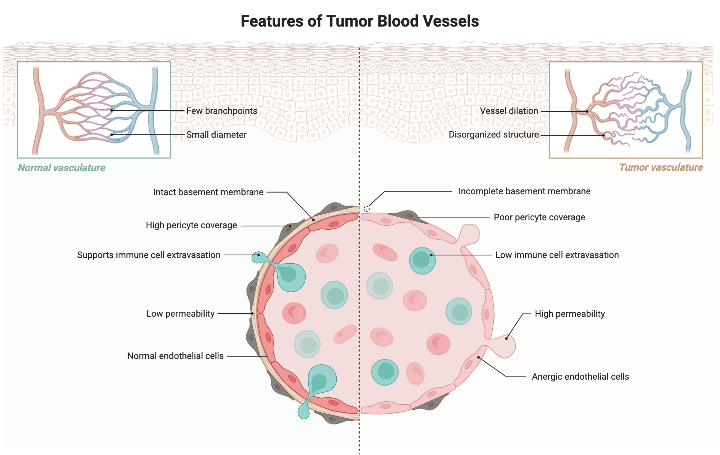
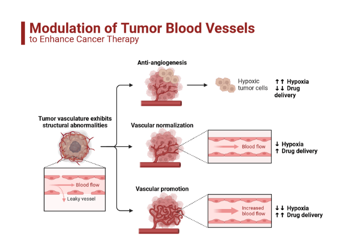
Tumours have fewer vascular cells compared to normal cells (poor vascularisation).
Important Notice
Tumours that have low levels of oxygen (hypoxia) become resistant to radiotherapy and chemotherapy due to poor drug delivery and relying on oxygen to make free radicals to cause toxicity in tumours. Hypoxic tumours have lower proliferation. Hyperthermia can destroy multidrug-resistant tumours by increasing sensitivity.
On the other hand, exposure to cells at temperatures to 43 to 45 decreases for about 10 to 30 minutes. This increases tolerance, or “thermotolerance,” which is a survival response allowing cells to become resistant to heat shock, reactive oxygen species, heavy metals, and environmental stressors.
If tumour cells exhibit low levels of stress, cells survive by activating stress responses to protect and induce DNA repair, protein folding, and remove damaged proteins.
However, ongoing stress can cause cells to die.
What is the method for hyperthermia?
There are several instruments and techniques used to create heat.
The area at first is numbed and small probes with thermometers is added into the tumour to detect the temperature during treatment.
CT scans can help ensure the probes are placed correctly.
Non-ionizing radiation: probes that make energy from microwaves, ultrasound, radio waves (also called radiofrequency) and laser.
Conductive heat sources: heating fluids such as blood or chemotherapy drugs and putting them into the body (called perfusion). Placing the entire body in a heated chamber or hot water bath or wrapping with heated blankets
One or more of these devices is placed around the vagina to be treated using the deep tissue.
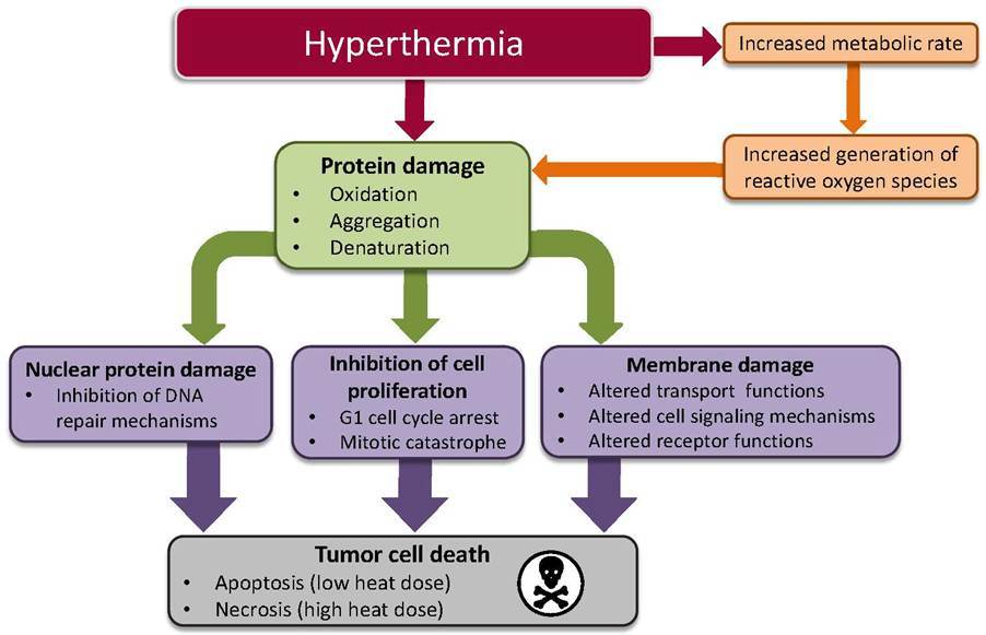
Hyperthermia can affect cellular homeostasis, causing direct protein damage through oxidation, aggregation (joining together), and breakdown (denaturation), or indirect damage by increasing the metabolic rate and production of reactive oxygen species to cause protein damage at lethal (≥42°C) and non-lethal (40°C) temperatures. Examples of free radicals include the formation of superoxide and hydrogen peroxide (H2O2), and the breakdown or malfunction of the mitochondria. The enzymes that produce the superoxide are NADPH oxidase and xanthin oxidase and were found to be of high levels of activity during hyperthermia. Defenses against hydrogen peroxide, like the pentose phosphate pathway, help to maintain the levels of the antioxidant reduced glutathione (GSH). This further exacerbates the production of ROS, causing damage to proteins, fats (lipids), and nucleic acids (DNA and RNA).
The increase in heat can cause the cell cycle to stop (G1 arrest), production of proteins to stop, prevent DNA synthesis and repair, and increase breakdown of misfolded proteins via the proteasome or lysosome (structures in the cell). It can affect the membrane cytoskeleton lowering the thickness (viscosity) and how the movement of ions and small molecules can pass through (permeability) leading to an increase in ions (sodium, hydrogen and calcium) and sodium-potassium pump that goes against the concentration gradient and metabolic changes where there is low levels of ATP. It can negatively affect post-translational modifications (after the production of proteins by the addition of sugar (glycosylation), acylation (acyl groups), phosphate groups (phosphorylation), and the addition of ubiquitin (ubiquitination). The addition of a farnesyl group from farnesyl diphosphate (farnesylation) to the protein is catalysed by farnesyltransferase (FTase), which helps with cellular communication and correct positioning of proteins, especially for protein families like Ras that are involved in signal transduction with Raf and MAPK.
On a molecular level, it can cause double-stranded breaks and fragmentation as a result of impairment of DNA repair mechanisms. Nuclear proteins can accumulate or join together (aggregation) and can affect DNA replication and transcription.
What Are The Types Of Internal Radiotherapy (Brachytherapy)?
Image-guided adaptive brachytherapy
Image-guided adaptive brachytherapy (IGABT) is the gold standard treatment for locally advanced cervical cancer and other gynaecological cancers.
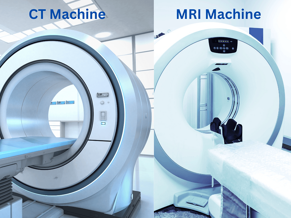
It is commonly linked with a scan, mainly Magnetic Resonance Imaging (MRI), for analysis.
Sometimes computed tomography or ultrasound is used if MRI is not available.
The purpose is help induce higher radiation dose to remaining tumour (residual tumour) while minimising expose to healthy organs like bladder and large intestine (colon, sigmoid, rectum).
This helps to shrink the tumour and reduce organ motion over a multi-fraction treatment course.
What dose is recommended for IGABT?
“A brachytherapy dose of 30 to 40 Gy (EQD2) to reach a total EBRT+brachytherapy dose of equal to or greater than 75 to 85 Gy EQD2 (D90) (assuming 45 Gy through) to the CTV- THR. Dose volume and point constraints for rectum, bladder, vagina, sigmoid, and bowel are recommended, and they have to be based on the published clinical evidence.”
Important notice:
The lower third of the vagina is sensitive; hence, brachytherapy should be delivered in several fractions at high dose rate (usually 3–4) or in 1 to 2 courses as pulse dose rate brachytherapy.
Can intravaginal brachytherapy be done without anaesthesia or with anaesthesia (sleepy drug)?
It can be done either with or without anaesthesia.
The purpose of intravaginal brachytherapy is to help remove residual tumours that are more than 7 mm.
There are intracavitary applicators as well as interstitial needles for cases that are bulky or complex, to help with high-risk clinical tumour volume (HR-CTV) – these are areas that contain gross and adjacent tissue at highest risk of microscopic disease.
Moreover, not only HR-CTV but also:
The residual gross tumor volume (GTV- Tres) after chemo- radiotherapy
The intermediate- risk clinical target volume (CTV- TIR).
Important notice
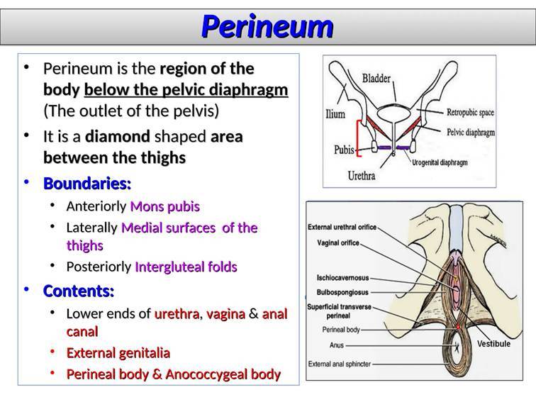
The type of applicator determines whether interstitial needles are positioned either intravaginally through the applicator or through the perineum, with or without a template.
What are the types of applicators in central and peripheral source positions used in intravaginal brachytherapy?
Mold
3D-printed applicator
What other factors can affect internal radiotherapy (brachytherapy)?
The surface and texture of the applicator.
The tissue depth for the dose point (5 mm)
“Radiography- based dose point constraints plus 3D dose volume constraints as available for rectum, bladder, vagina, sigmoid, and bowel are recommended, and they have to be based on the published clinical evidence.”
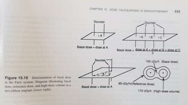
The use of the system, e.g., Paris, for interstitial – small space between parts of the organ.
What is the Paris system?
The aim is to optimise the interstitial placement and dose for the use of iridium wire and hair implants to give an even distribution of the dose.
Through this system, low doses are commonly given, e.g., 0.5 Gray per hour, to the prescribed reference dose.
What are the principles of use of wires or catheters?
Placed near the area to be treated or through the area.
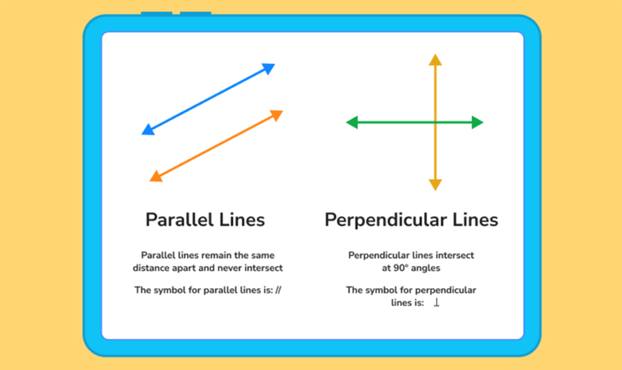
Sources straight and parallel.
Equal length.
Separation between sources: 5 and 20 mm, and depends on volume—equal linear activity.
An insight into the Paris system.
The sources should be perpendicularly positioned along the midway. If not equal length, centrally placed close to the mean mid-length of wires. The dose rate between the wires is due to length and is at its maximum. If wires are not parallel to the central plane, they should be perpendicular to the main direction of sources that lines through the implant.
Perpendicular means an angle of 90 degrees, similar to a right angle or a corner of a square.
For more info, please click the link below:
Bownes, OP., Richardson, C. and Lee, C. (2021) The Paris system (for interstitial treatments) The Paris system (for interstitial treatments). Available at: https://1library.net/article/paris-interstitial-treatments-paris-interstitial-treatments.y8n7vg4y (Accessed: 9th October 2026)
Brachytherapy And Primary Tumour
ESTRO/ESGO/SIOPe Guidelines for the management of patients with vaginal cancer states as follows:
“Brachytherapy is preferred over EBRT for treatment of the
primary tumor. A total dose of 50–60 Gy EQD2 is prescribed.”
A gentle reminder: brachytherapy is applied to precisely reach the target area (primary tumour) and spare organs at risk (gonads, urinary, rectum, and bone structure). A low dose is also given for the uterus.
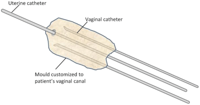
Here, we will explain the use of an intravaginal applicator for the brachytherapy procedure and the key factors associated with this is: the anatomy, or how the vagina looks, and the shape of the tumour.
This is why a vaginal mould is needed and is generally done under anaesthesia.
If external irradiation is however required (eg, pelvic lymph node involvement), proton beam therapy is preferred.
Gentle reminders
Wash hands in all cases and appliances.
The procedure may be uncomfortable, and medications may be needed to relieve pain afterward, e.g., also be discharge or cramps or diarrhoea
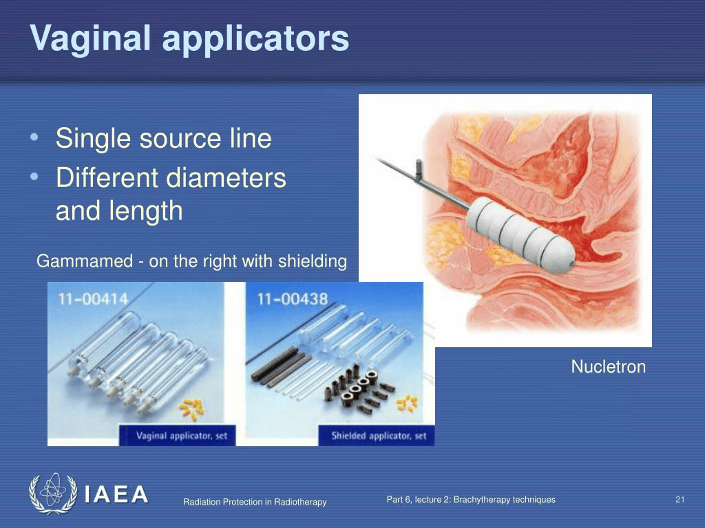
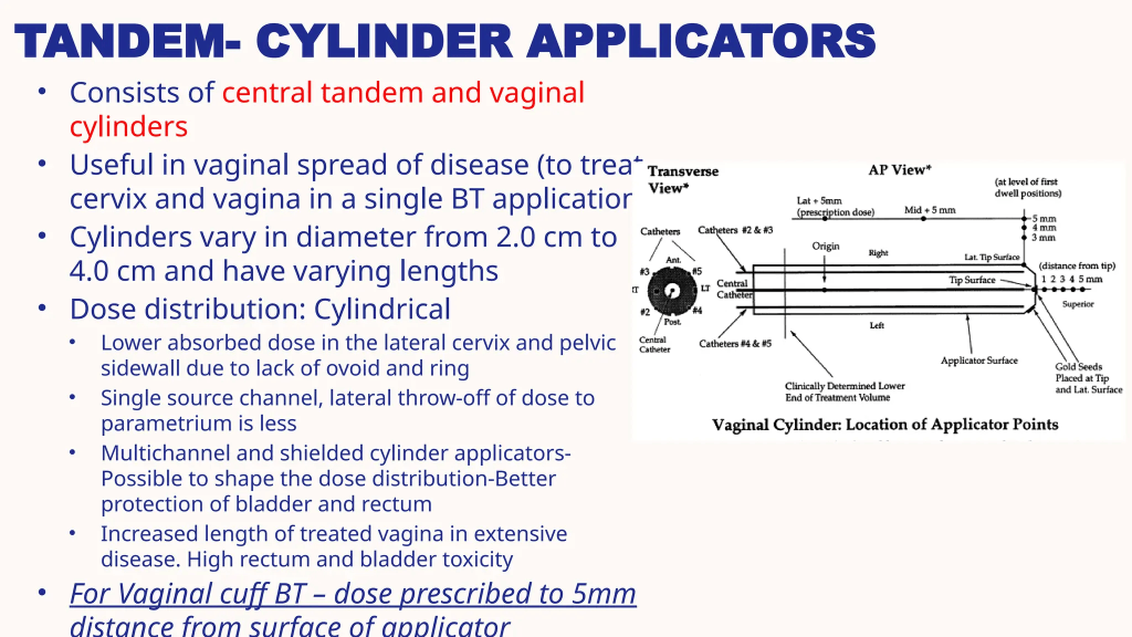
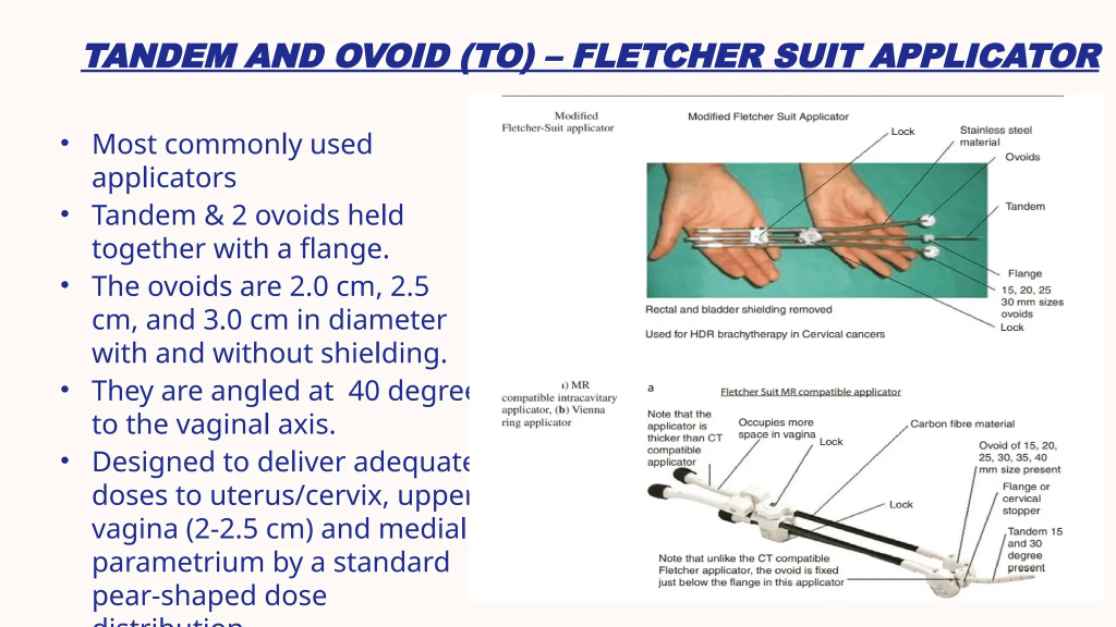
Remaining polyps (residual) – surgical debulking (removal of the polyps – polypectomy) – to allow applicator.
If there is growth beyond the womb and vagina (parametrial and/paravaginal extension) – large, odd, or lower vagina, applicators do not fit correctly – interstitial catheter is then applied.
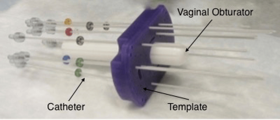
- It enters via vagina and perineum
- Obturator
- Template – plastic, stitched outside the body. It has holes where catheters are in and kept in a stable position.
- General anaesthesia, epidural space (back in spine between dura mater and the vertebral canal) The aim is to numb the spinal nerve roots, or Sedation (calm the mind and relieve anxiety) can be performed.
- Foley catheter – collect urine
- Laparoscopy, ultrasound, CT or MRI help with placement
- Applicator in place and template sutured in skin.
- There are several holes in the template.
- Catheter will be added into the templates and into where the tumour is placed.
- Uncomfortable and pain; pain relief given.
- Bed rest applied in place
- Lift head to a 20-degree angle.
- Not able to move in bed apart from toes and upper body.
- Medications can be given to prevent any bowel movement.
- 1 to 2 times per day for several days
- A radiation therapist will attach the brachytherapy treatment machine to the catheters in your template.
- Geiger counter to check the amount of radiation before treatment.
- Monitor treatment
- Radioactive material moves into catheters for 10 to 20 minutes.
- Radioactive source returned back into the machine.
- Full step-by-step procedure is in the link below. The aim is to provide an overview of what the procedure involves:
Misher, C. (2026) Interstitial Brachytherapy for Gynecologic Cancers. Available at: https://www.oncolink.org/cancer-treatment/radiation/types-of-radiation-therapy/brachytherapy-for-gynecologic-cancers/interstitial-brachytherapy-for-gynecologic-cancers (Accessed: 8th October 2026)
New Article About Improving The Efficiency Of Brachytherapy-associated catheters.
What other types of radiotherapy are available?
External beam radiotherapy (EBRT)
This is where there is an external source of beams of ionizing radiation. Common examples are superficial X-ray therapy, where subatomic particles called electrons are emitted from a heat cathode (negative electrode) and accelerated across an X-ray tube. It then strikes a tungsten-metal-based anode (positive electrode) and undergoes interactions to produce X-rays. This is commonly used for the skin in a range of 80 to 300 kV (kilovolts). To treat deeper skin layers, some linear accelerators produce electron beams with different energies (4 to 20 MeV (mega-electron volts).
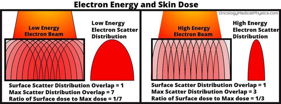
It is advised to deliver 45 Gray if the para-aortic lymph node is involved and spread to renal vessels (renal artery and vein). Hence, the target volume includes a minimum region from the vessels.
It can also be applied for inguinal-femoral lymph nodes for cancer in sensitive areas like the lower third of the vagina.
Overall treatment time for EBRT should not exceed 5 to 6 weeks.
External beam radiotherapy should entail:
IMRT: Intensity Modulated Radiation Therapy.
It involves small radiation beams with different intensities delivered to the tumour precisely and to critical organs. It creates a conformal dose.
The choice depends on the patient’s condition and the availability of the technique.
IMRT is MORE PRECISE than 3DCRT because it covers the planning target volume (PTV) with a better dose distribution. This minimizes damage to normal tissue, side effects, and secondary malignancies.
There is a boost to involved nodes that should improve outcomes.
IMRT (Intensity-modulated radiotherapy) is applied in the following areas:
Adjacent tissues of the vagina
Paravaginal space.
Parametrium
Uterine cervix if in situ.
Pelvic lymph nodes (obturator, internal, external, common iliac and presacral).
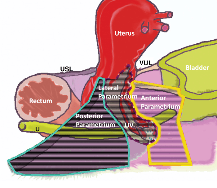
Prior to dosing, imaging is needed for safety, lower margins and avoid uncertainty.
“The total dose, including the contribution from brachytherapy, should be 55 to 60 Gy (equivalent dose to 2 Gy per fraction [EQD2]).” See ESTRO/ESGO/SIOPe Guidelines.
What to do if IMRT or IGABT is unavailable?
3D Conformal EBRT or 3D CRT
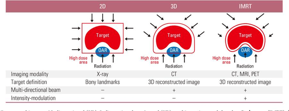
OAR = Organs at risk
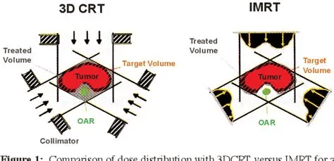
Important notice: 2025 update on radiotherapy – see Cancer of the vagina: 2025 update.
“The optimal or lower threshold dose is 70 Gy, which has been shown to improve outcomes. Doses higher than 70 Gy result in significant grade 3 and 4 toxicities.”
References
Adams, T.S., Maluleke, J.C. and Cuello, M.A. (2025). Cancer of the vagina: 2025 Update. International Journal of Gynecology & Obstetrics, 171(S1), pp.48–59. doi:10.1002/ijgo.70325.
Ball, T.L. and Douglas, R.G. (1949). Cryptomenorrhea. The American Journal of Surgery, 78(1), pp.40–49. doi:10.1016/0002-9610(49)90183-x.
Cassidy, J., Bissett, D., Spene, R. and Payne, M. (2010) Oxford Handbook of Oncology. Oxford: Oxford University Press.
Misher, C. (2026) Interstitial Brachytherapy for Gynecologic Cancers. Available at: https://www.oncolink.org/cancer-treatment/radiation/types-of-radiation-therapy/brachytherapy-for-gynecologic-cancers/interstitial-brachytherapy-for-gynecologic-cancers (Accessed: 8th October 2026)
National Cancer Institute (2026) Vaginal Cancer Treatment (PDQ®)–Health Professional Version. Available at: https://www.cancer.gov/types/vaginal/hp/vaginal-treatment-pdq (Accessed: 17th August 2026)
Nout, R.A., Calaminus, G., Planchamp, F., Chargari, C., Lax, S., Martelli, H., McCluggage, W.G., Morice, P., Pakiz, M., Schmid, M.P., Stunt, J., Timmermann, B., Vokuhl, C., Orbach, D. and Fotopoulou, C. (2023). ESTRO/ESGO/SIOPe Guidelines for the management of patients with vaginal cancer. International Journal of Gynecologic Cancer, [online] 33(8). doi:10.1136/ijgc-2023-004695.
Bettaieb, A., Wrzal, P.K. and Averill-Bates, D.A. (2013). Hyperthermia: Cancer Treatment and Beyond. Cancer Treatment – Conventional and Innovative Approaches. [online] doi:10.5772/55795.
National Cancer Institute (n.d) Hyperthermia To Treat Cancer. Available at: https://www.cancer.gov/about-cancer/treatment/types/hyperthermia (Accessed: 2nd November 2026)
Lowell, L. (2025) Farnesylation: A Key Process in cell function and disease. Available at: https://biologyinsights.com/farnesylation-a-key-process-in-cell-function-and-disease/ (Accessed: 4th November 2026)
Ikwegbue, P., Masamba, P., Oyinloye, B. and Kappo, A. (2017). Roles of heat shock proteins in apoptosis, oxidative stress, human inflammatory diseases, and cancer. Pharmaceuticals, [online] 11(1), p.2. doi:10.3390/ph11010002.
Nature Index (2026) Image-Guided Brachytherapy Techniques in Cervical Cancer Management. Available at: https://www.nature.com/nature-index/topics/l4/image-guided-brachytherapy-techniques-in-cervical-cancer-management (Accessed: 4th November 2026)


Leave a Reply