Below summarizes the main points for the treatments in accordance to the European Society of Gynaecological Oncology (ESGO), European Society for Radiotherapy and Oncology (ESTRO) and the European Society of Paediatric Oncology (SIOPe) 2023 guidelines. A 2025 update of Vaginal cancer has been provided by experts from the following reference:
Adams, T.S., Maluleke, J.C. and Cuello, M.A. (2025). Cancer of the vagina: 2025 Update. International Journal of Gynecology & Obstetrics, 171(S1), pp.48–59. doi:10.1002/ijgo.70325.
Please read the guidelines attached for further details.
Childhood and Yolk Sac Tumours
First treatment is chemotherapy – they are chemotherapy-sensitive.
Vaginal Rhabdomyosarcoma (RMS)
Chemotherapy
Chemotherapy depends on stage, age, size of tumour, whether it has spread to the nodes, and whether there are molecular or pathological findings under the microscope.
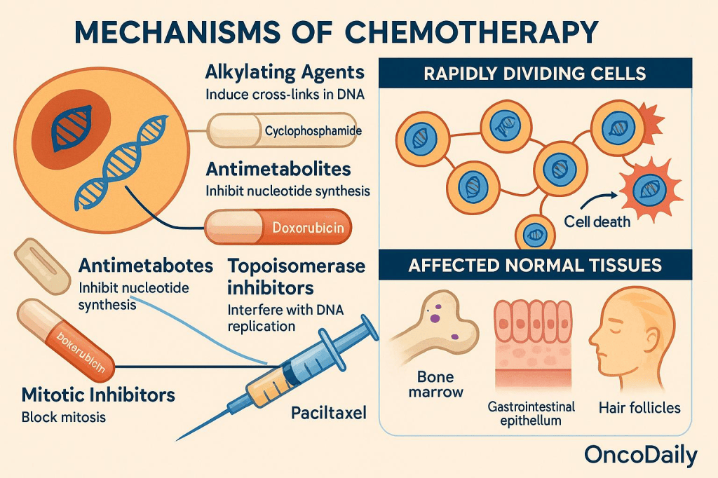
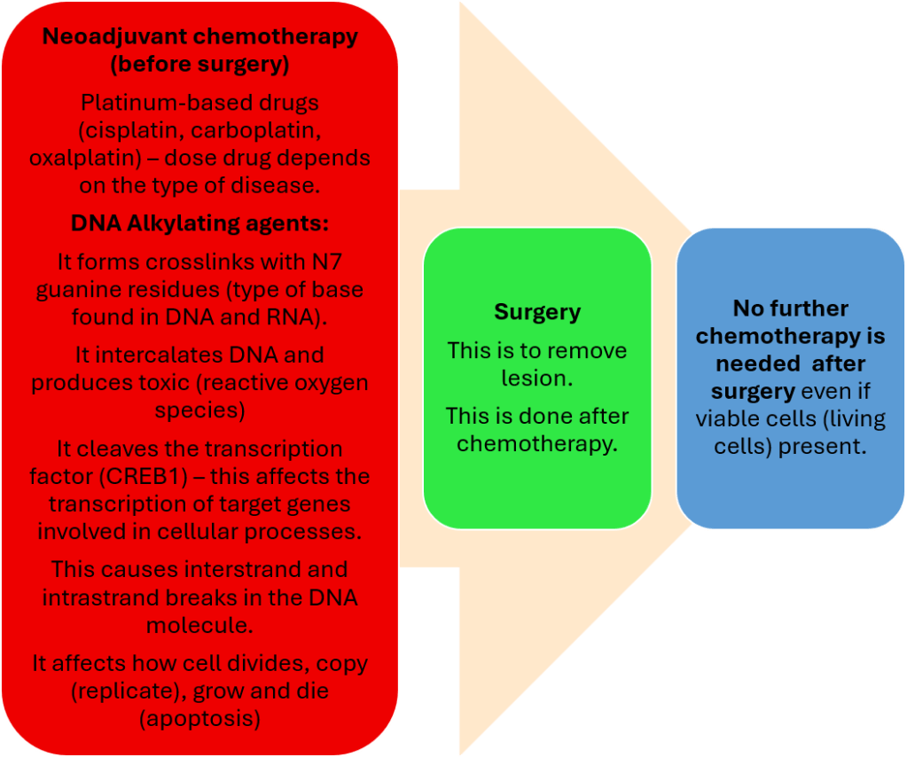
Before (neoadjuvant) and after surgery (adjuvant), there is combination chemotherapy that includes DNA alkylating agents, for instance, cyclophosphamide and ifosfamide.

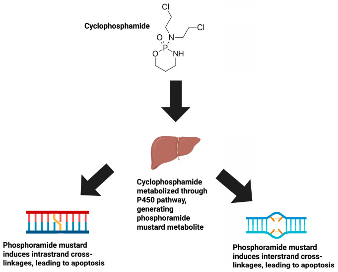
How does cyclophosphamide and ifosfamide work?
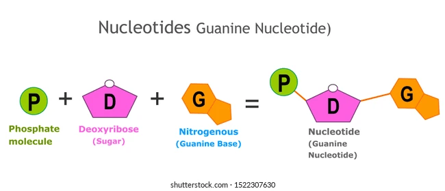
It forms crosslinks with N7 guanine residues (a type of base found in DNA and RNA).
It intercalates DNA and produces toxic reactive oxygen species.
It cleaves the transcription factor (CREB1) – this affects the transcription of target genes involved in cellular processes.
This causes interstrand and intrastrand breaks in the DNA molecule.
It affects how cells divide, copy (replicate), grow, and die (apoptosis)
Did you know?
In rare cases, regionally involved lymph nodes and/or fusion-positive RMS requires maintenance strategies after adjuvant therapy.

Local Therapy
Aim: preserve organs.
This commonly begins after 3 and 6 courses of neoadjuvant chemotherapy. This depends on the tumour response and histological type.
Biopsy
Second-line chemotherapy
Proton Beam Therapy
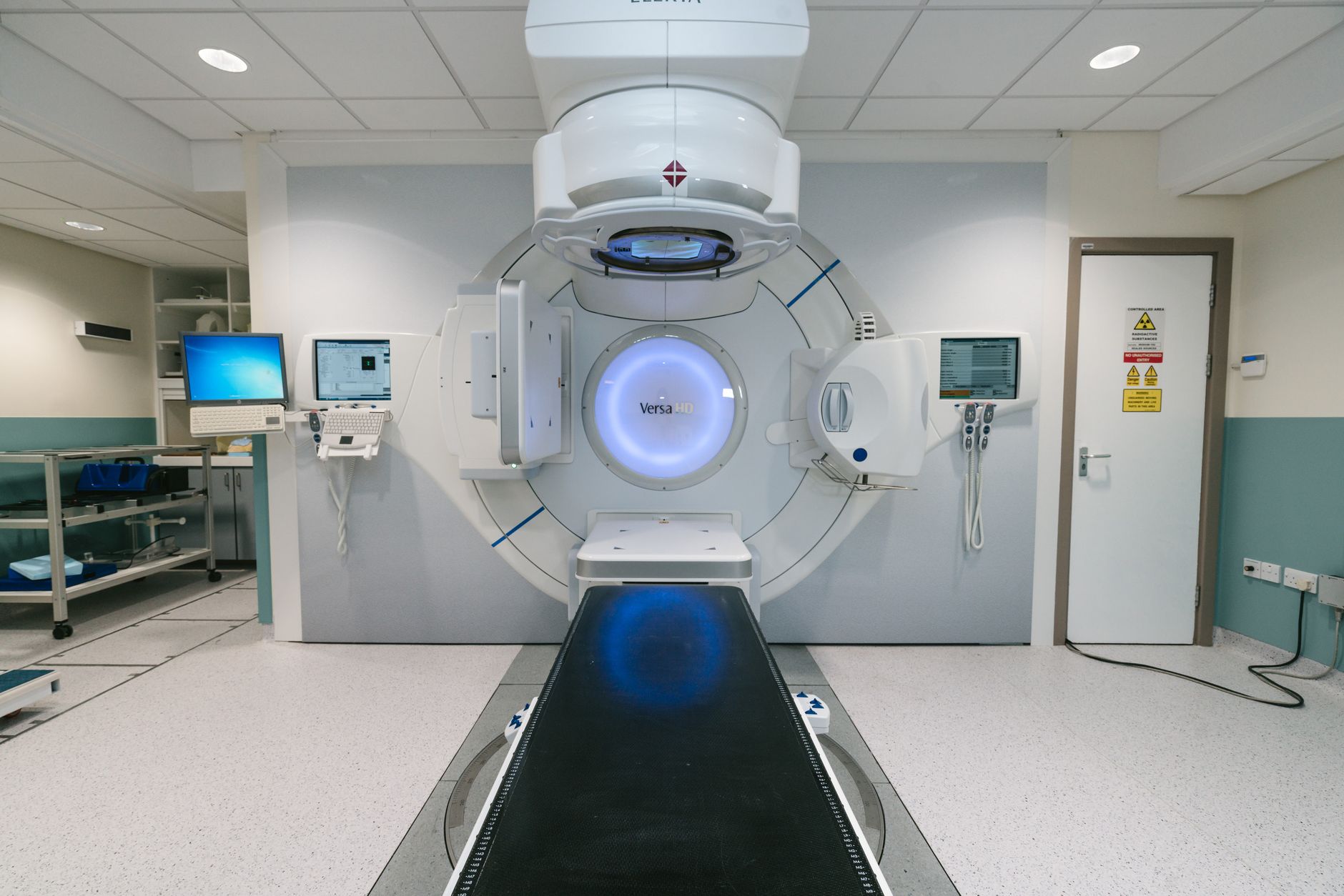
Further Insight About Proton Beam Therapy
It uses high-energy protons rather than photons to precisely meet the patient target volume (PTV) (millimetre precision) and has little to no effect on surrounding areas. The protons are transmitted via an ion source where hydrogen is subdivided into protons and electrons. They are then passed through a cyclotron, where they are accelerated. Thereafter, they enter through an energy selection system and adjust their energy accordingly. This allows the beam to transfer the right amount of energy through the track and then the irradiation centre in the gantry. This then goes round, adjusting the dose of protons depending on the size and shape of the tumour.
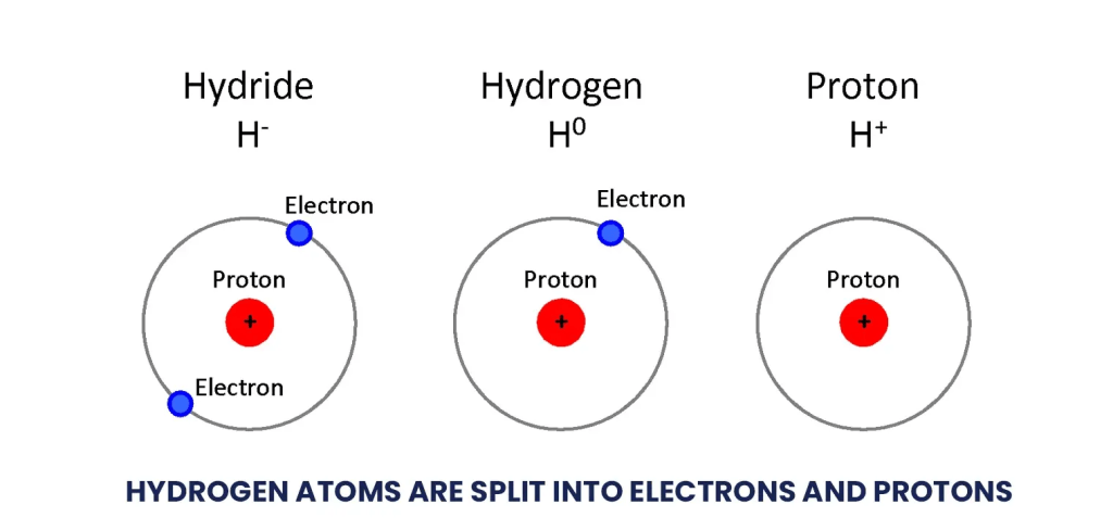
How Does Proton Therapy Work By Proton Therapy Centre
Brachytherapy
This is performed if the following is present:
No complete response after chemotherapy.
Conservative surgery with free margins is not possible.
There are lymph nodes discovered by observing tissue under the microscope (histology), cells under the microscope (cytology), or through scanning (radiology).
Normal tissue in children is radiosensitive and has a choice to undertake pulse dose rate (PDR) or high dose rate (HDR) brachytherapy
- PDR – This is performed on an hourly basis as pulses. The estimated PDT is between 0.42 and 0.5 Gray per hour. This gives a total dose of 50 to 60 Gray.
- HDR – This form of brachytherapy utilises an estimated total dose of 27.5 to 36 Gray. This is delivered in 5 to 12 fractions over 3 to 10 days.
In most patient cases, bilateral ovarian transposition is needed.

Bilateral Ovarian Transposition
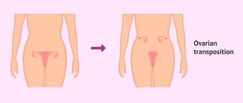
What is bilateral ovarian transposition?
This is necessary in most cases, using temporary transposition in patients who are young or before puberty, as protection against radiation. The 3D imaging helps to guide treatment planning. The target volume is defined by residual tumour sites, as per clinical and radiological assessment. Relevant radiation dose to the ovary is estimated.
What is ovarian transposition?
This process is also known as ovarian suspension, oophorectomy, or ovariopexy.
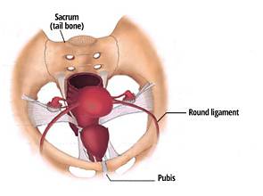
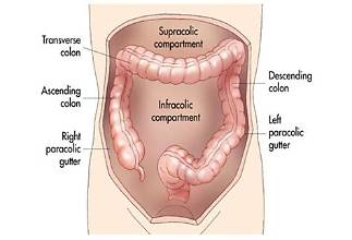
It is performed above the pelvic brim and the side (lateral). This helps to minimise ovarian dose of radiation and improve patient response. There are several lateral locations: base of the round ligament, the level of the lower kidney pole, and the paracolic gutters.
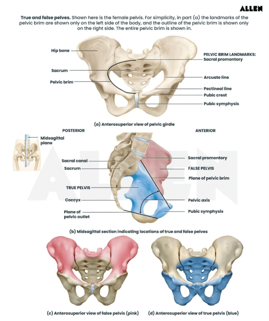
What Is The Difference Between Ovarian Transplantation and Ovarian Transposition?

The main difference between ovarian transplantation and ovarian transposition is that the vascular pedicle remains intact.
This helps to preserve the ovarian blood supply (artery and vein) and is important to see via imaging techniques to help detect the transposed ovary and risks of ovarian torsion. Ovarian transposition is where the ovaries are relocated in pelvic radiation laterally (side) or superiorly (above) the pelvic brim to preserve functions of hormones and fertility.
This pedicle contains the gonadal artery and vein, which supply the ovary.
Ovarian torsion is the twisting of the ovary around the vascular pedicle and ligaments.
Torsion compresses the outflow of the vein (venous outflow). This causes congestion and enlargement, affecting the flow of blood in the arteries and obstructing blood flow (ischaemia).
Congestion is the build-up of blood in the organ due to back pressure in the vein. It may also be associated with oedema, which is the accumulation or build-up of fluid in the tissues.
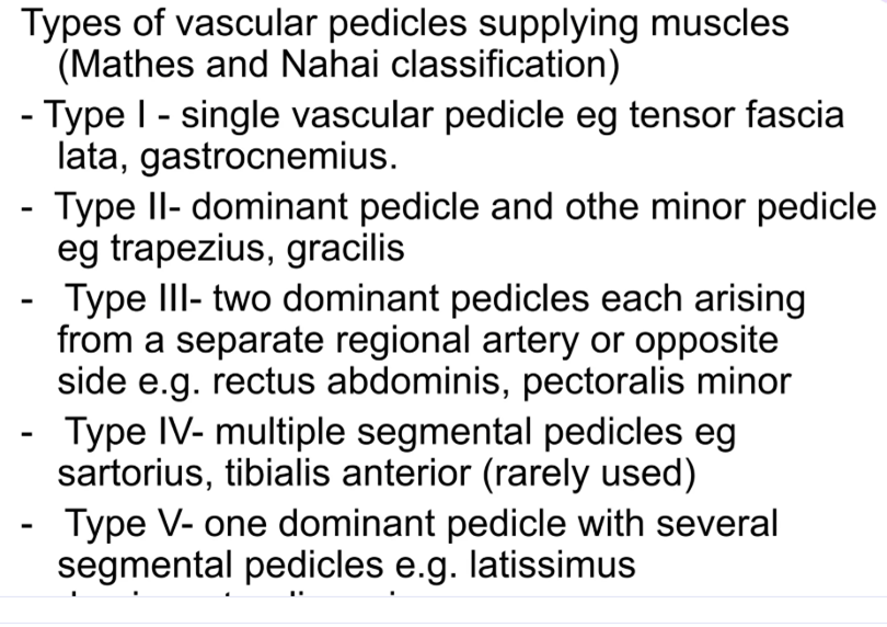
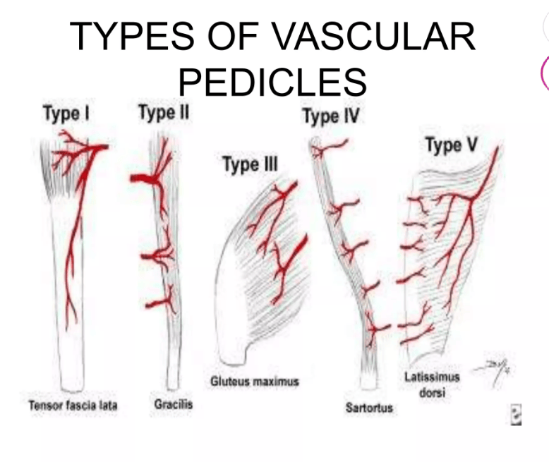
Source: HafeezAzeez
What are the causes of ovarian transposition?
Hypermobility of the ovary: <50%
Adnexal mass: ~50-80% – adnexa is the joining of parts with fallopian tubes and ovaries that link with the womb.
Most lesions are dermoid cysts or parovarian cysts
Large cystic ovaries undergoing ovarian hyperstimulation are at particular risk
Masses between 5-10 cm are at the highest risk
Recommended Lecture On Vascular Pedicles and Other Basic Surgical Procedures By Hafeez Azeez
https://www.slideshare.net/slideshow/basic-surgical-procedures/198359135
A Further Insight Into Ovarian Transposition
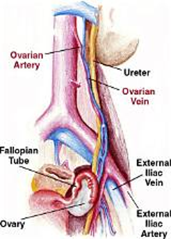
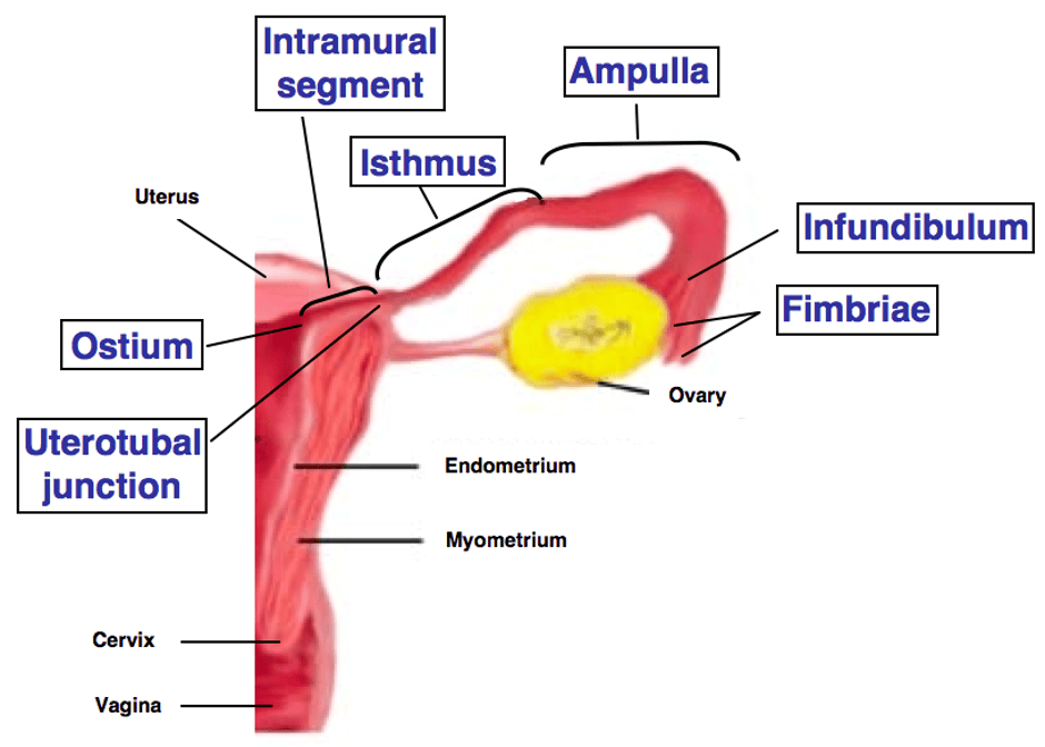
The ovary is supplied by two main blood vessels: an artery and a vein.
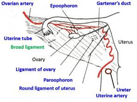
The ovarian veins form a plexus in the broad ligament.
The broad ligament is a round, cord-thickening area. It is also
This then communicates with the uterine plexus, providing dual blood supply.
If an ovarian mass is present, the ipsilateral ovarian vessels may be enlarged.
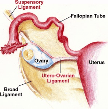
The round ligament is situated at the lower end of the broad ligament, where it connects to the uterus. It is particularly situated in the parametrium.
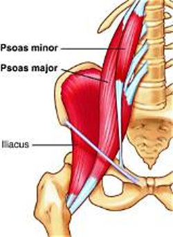
The gonadal veins are always anterior to the psoas muscle and the common iliac vessels.
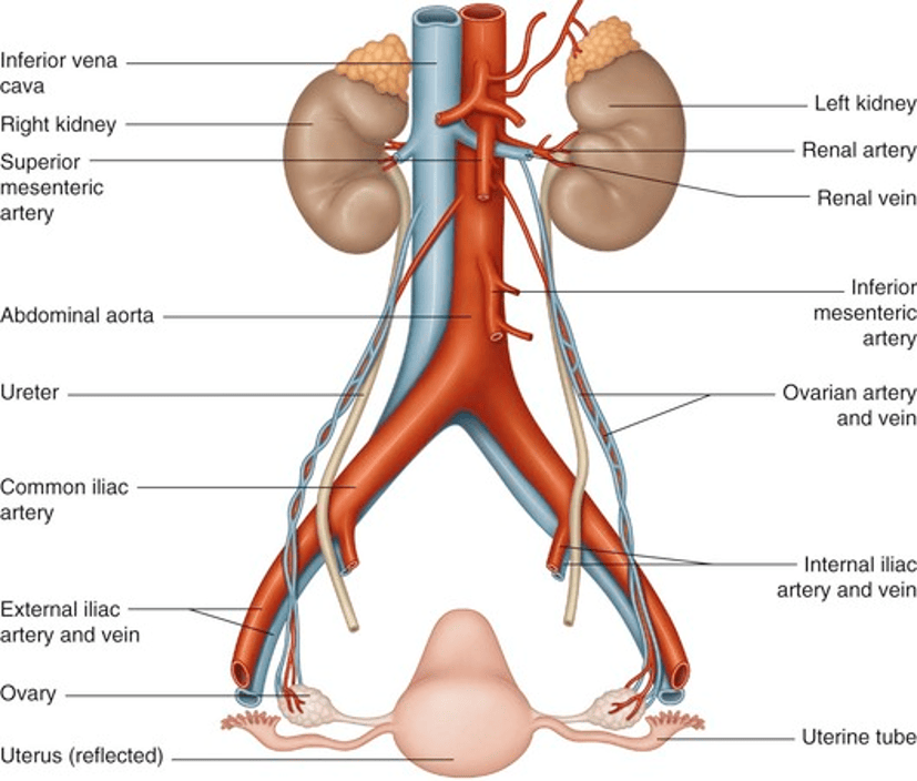
What was the technique before?
Previously, ovaries were relocated medially by suturing them to the posterior uterus to protect them from radiation injury.
They were further protected during radiotherapy with a lead shield placed centrally on the abdomen.
However, medially placed ovaries still absorb radiation from scatter and some transmission through the shield, reducing the efficacy of the procedure.
What imaging techniques are being used for ovarian transposition?
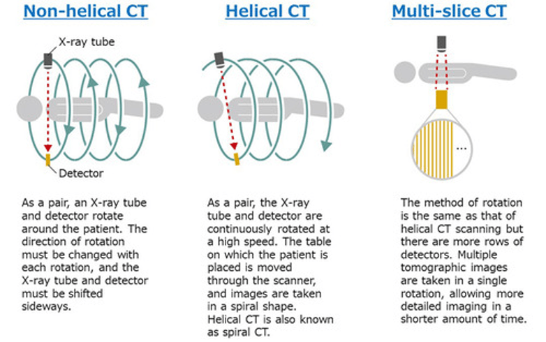
A single-detector helical CT scan with contrast enhancement and thin sections, and axial scanning, helps to examine the blood supply to the ovary via the ovarian veins.’ An example of a contrast agent is a non-ionic iodinated contrast agent (Ultravist [Iopromide], 300 mg I/mL.
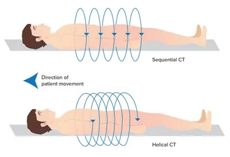
The helical CT allows contiguous volumetric data acquisition. This is a continuous, gapless 3D block of voxels. Voxels are how volumetric data is built. The 3D element encodes properties like density or intensity. The aim is to have no gaps between slices where there is accurate 3D reconstruction, segmentation and analysis.
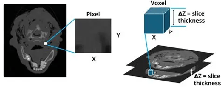
There are several advantages of helical CT, which aims to improve spatial resolution and optimise contrast enhancement and thin sections.
A Further Insight Of Vascular Pedicle In Imaging
If a large pelvic mass originating from the ovary, uterus, bowel, and retroperitoneum is noted on CT, differentiation of the organ of origin of the pelvic mass may occasionally be unclear.
The suspected origin of the mass is another important indication of a correct diagnosis obtained on imaging.
The ovarian vascular pedicle sign can be defined when the gonadal veins directly join the pelvic mass.
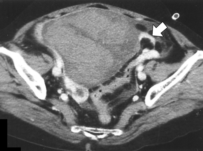
The patient has surgically confirmed torsion (twisting of the ovaries) with haemorrhagic infarction. Haemorrhagic infarction is blood released from an organ or tissue that is dead.
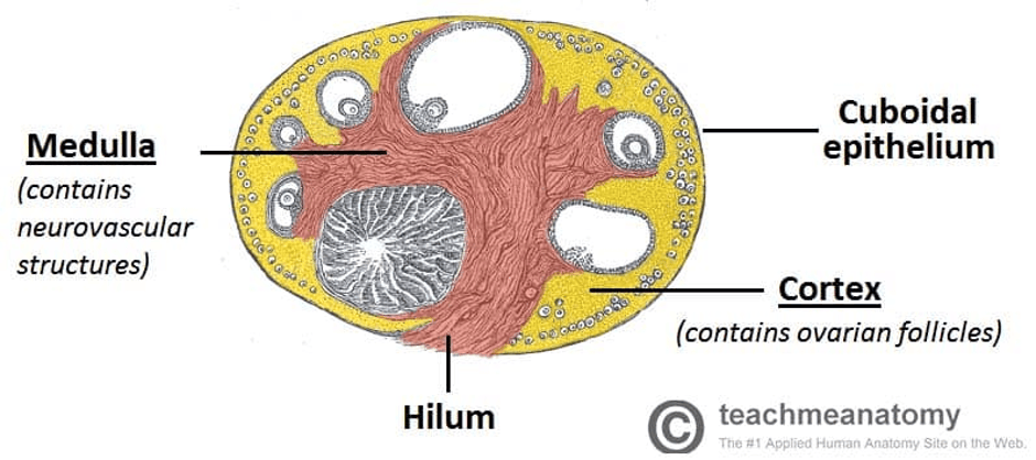
Axial single-detector helical CT was performed from the common iliac vessels to the left ovarian vascular pedicle. CT scan shows the left ovarian vascular pedicle (arrow) is discontinuous with the tumor in the ovarian hilum, as the ovarian pedicular vessels appear “floating” within the ovarian hilum. The hilum is the hollow area situated at the surface of the ovary
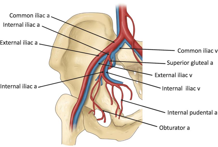
. The iliac blood supply supports the lower limbs and pelvic region. The left and right common iliac arteries form the terminal branches of the abdominal aorta. Each branches into the external iliac artery and the smaller internal iliac artery.
On the other hand, the right and left common iliac veins unite to form the inferior vena cava. They unite with the internal and external iliac veins.
This CT pelvic scan has been done with a nonionic iodinated contrast agent (Ultravist [Iopromide], 300 mg I/mL using IV at a rate of 4 mL/sec to help visualise the ovarian vascular pedicle sign. The scanning delay was 70 or 120 sec. The scanning parameters were 120 kVp, 220 mAs, a 5- to 8-mm collimation, an 8-mm/sec table speed, and a 5-mm reconstruction interval. mA (milliamp), mm (millimetre), kVp (kilovoltage peak).
The kVp is the maximum tube voltage applied across an X-ray tube, determining photon energy, beam penetration, image contrast, and patient dose. Typical values range from 50–60 kVp for extremities to 100–125 kVp for chest radiographs, and about 120 kVp for routine adult CT.
Oligometastatic Disease
Cases with Limited oligometastatic disease and response after chemotherapy; focal treatment of metastatic sites is considered.

Recommended Article About Ovarian Vascular Pedicle
The Intrauterine Pressure Catheter
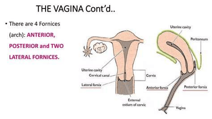
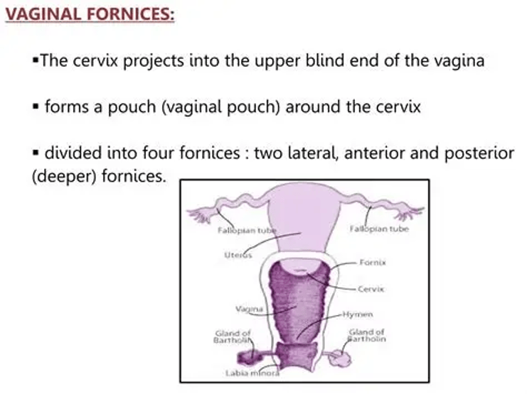
Purpose: if growth affects the exocervix or fornices in patients with vaginal cancers
Non-cancerous purposes of IUPC:
During pregnancy and labour (parturition) – They depend on the force of gravity to keep the foetal head down (descend) in the pelvis to see the cervix during labour.
It helps to shorten labour and decrease the need for forceps or vacuum-assisted delivery.
It relieves maternal anxiety.
It helps detect abnormalities in the womb.
It helps to monitor pressure in the uterus to know how much fluid is in the amniotic sac. Drains fluid from the uterus if too full.
Diagnose uterine abnormalities.
How does IUPC function?
Wash hands appropriately per normal procedure before and after.
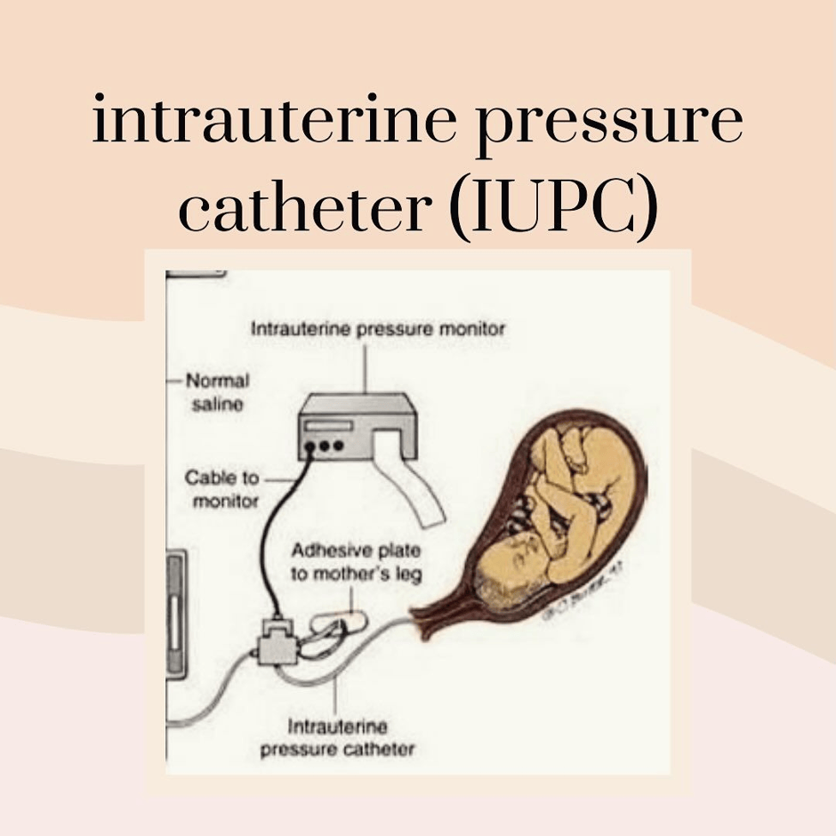
An intrauterine device (IUD) is a small, lubricated balloon catheter (small, thin tube) that enters the uterine cavity via the cervix, gently back and forth until it reaches its destination.
It then gets inflated with saline, which increases the intrauterine pressure.
The IUPC is connected to a monitor, which measures and monitors the uterine contractions.
To keep it simple, it measures uterine contractions, pressure, and heart rate.
The IUPC is in place for 24 hours.
After that, it will need to be removed and replaced with a new one.
IUPC and labour, how does it work?
If the patient is active in labour and this is indicated by 4 to 5 centimetres dilation, IUPC is added and positioned.
Once the IUPC is in place, it will be connected to a monitor. The woman will then be able to see her contractions on the monitor.
The IUPC can stay in place for up to 24 hours.
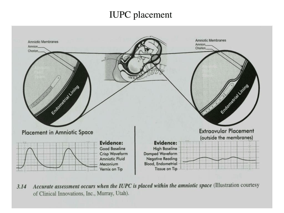
Like any procedure, there is a risk of infection; the delicate nature of the cervix may be easily damaged; premature labour.
For more info and full procedure details, visit:
Gupta, R. and Shunmugasundaram, S., Lamba, A. and Kumar, G. (n.d.) Understanding Intrauterine Pressure Catheter (IUPC): A Complete Guide. Available at: https://hidocdr.com/articles-details.php?artId=e6297dad-d455-4877-bfab-a29c3097c159 (Accessed: 8th October 2026)
How do you know if there are any remaining tumours?
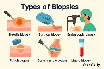
A biopsy (diagnostic) is taken, and the tissue is seen under the microscope for confirmation.
There may be exophytic (outer growth from epithelial surface) or polypoid (abnormal growth like a polyp) lesions, but this is not linked to vaginal wall resections.
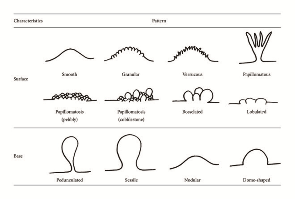
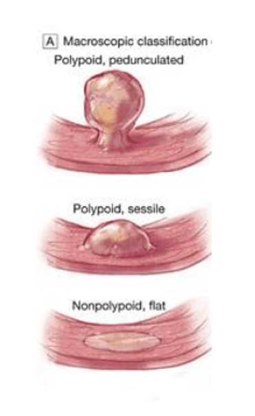
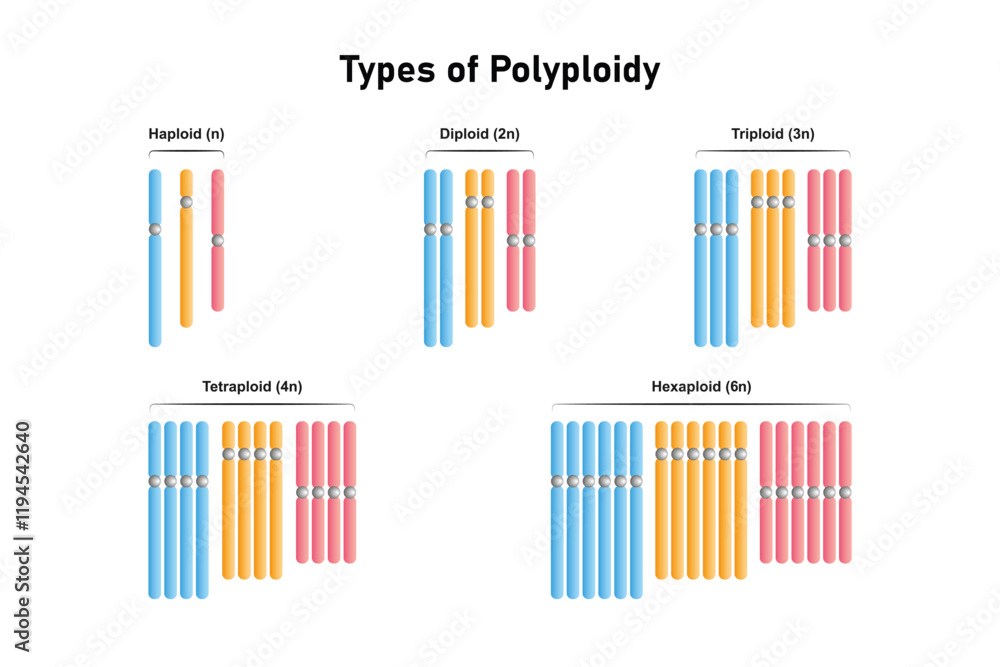
Did You Know The Difference Between POLYPOID and POLYPLOID?

Please note there is a difference between POLYPOID and POLYPLOID
Polypoid = POLYPS
Polyploid = nothing that can be seen on a lesion; it’s three or more sets of CHROMOSOMES (thread-like structures that store genetic material)
What if there is residual tumour in the fornix and cervix?
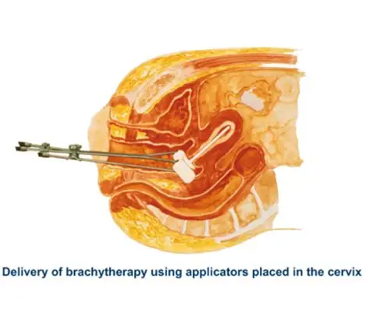
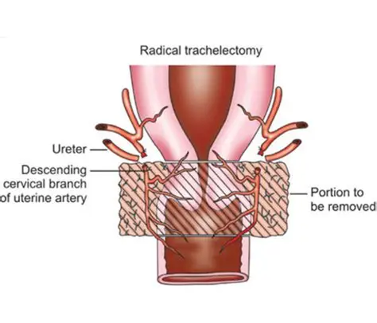
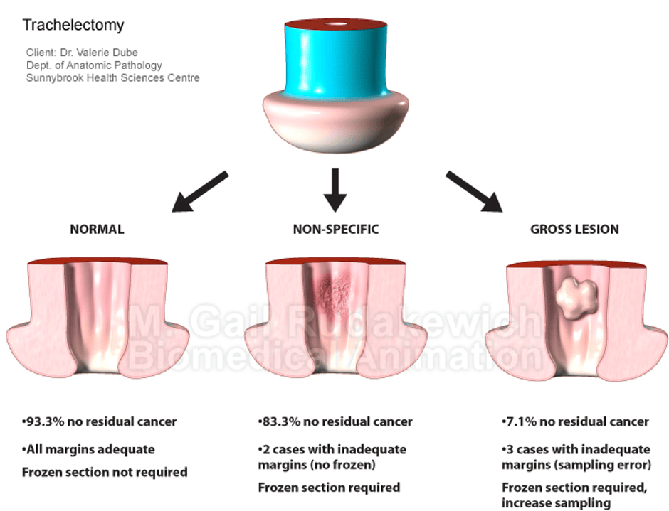
Brachytherapy for cancer of the cervix by NHS
What if there is residual tumour that involves more than half of the vagina, or it is multifocal?
What if there is residual tumours and has spread through the regional nodes?
Surgery is not recommended to remove lymph nodes.
If the choice is to remove the lymph nodes, it must be to the extent of the EBRT target volume.
Image-guided adaptive brachytherapy.
“A brachytherapy dose of 30 to 40 Gy (EQD2) to reach a total EBRT+brachytherapy dose of equal to or greater than 75 to 85 Gy EQD2 (D90) (assuming 45 Gy through) to the CTV- THR.
ESTRO/ESGO/SIOPe Guidelines for the management of patients with vaginal cancer.
Dose volume and point constraints for rectum, bladder, vagina, sigmoid, and bowel are recommended, and they have to be based on the published clinical evidence.”
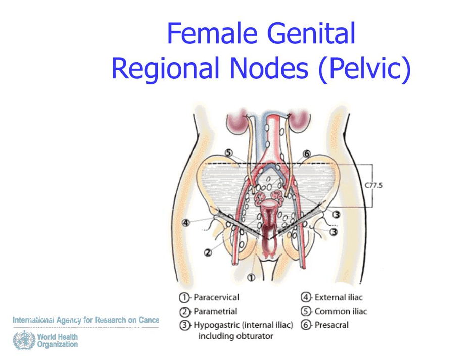
Rhabdomyosarcoma – Sarcoma botyroides
This is the most common soft tissue sarcoma that affects youngsters, especially the vagina and cervix. This varies based on the patient’s case.
Less radical surgery as first-line treatment.
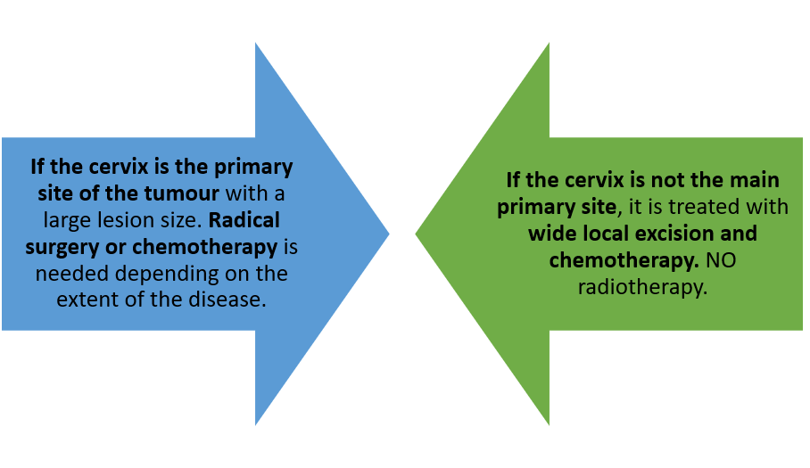
Please see Cancer Update 2025 guidelines.
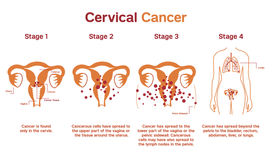
Granular cell tumour (GCT)
Status: Rare
Modes of treatment:
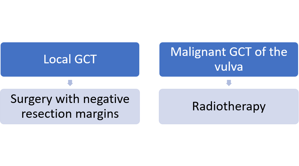
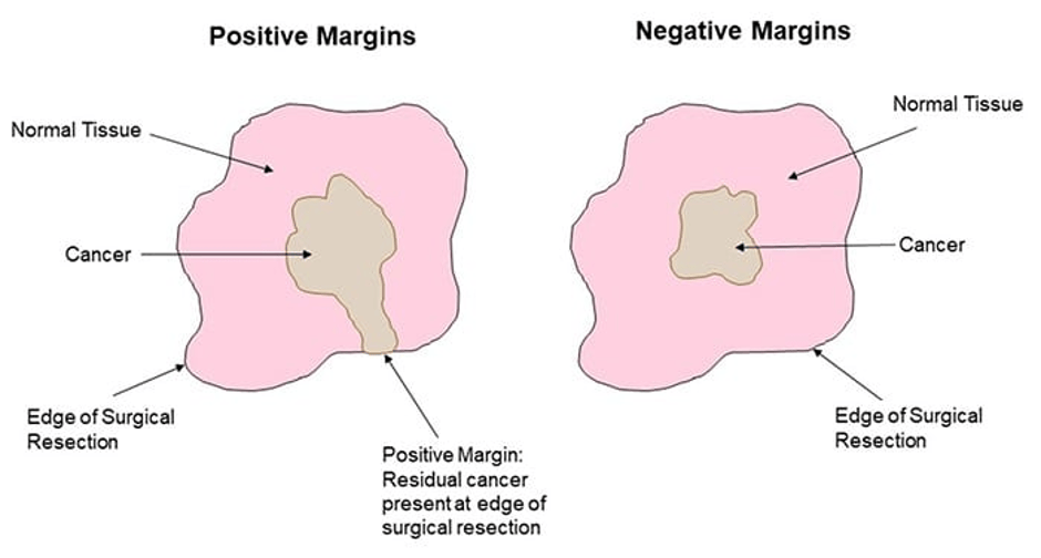

Identify type of GCT before planning treatment. This requires biopsy or evaluation of margins intraoperatively (extended local excision) using frozen section.
Lymphadenectomy (removal of lymph nodes) and vulvectomy (removal of the vulva) help to improve prognosis.
Some researchers think radiation therapy is NOT suitable due to vascularity, and the neoplasm prefers vascular reduction over surrounding areas.
Other researchers need radiation therapy after operation with a total dose of 60.8 Gy, and no recurrence was found after 16 months of follow-up.
Imaging techniques
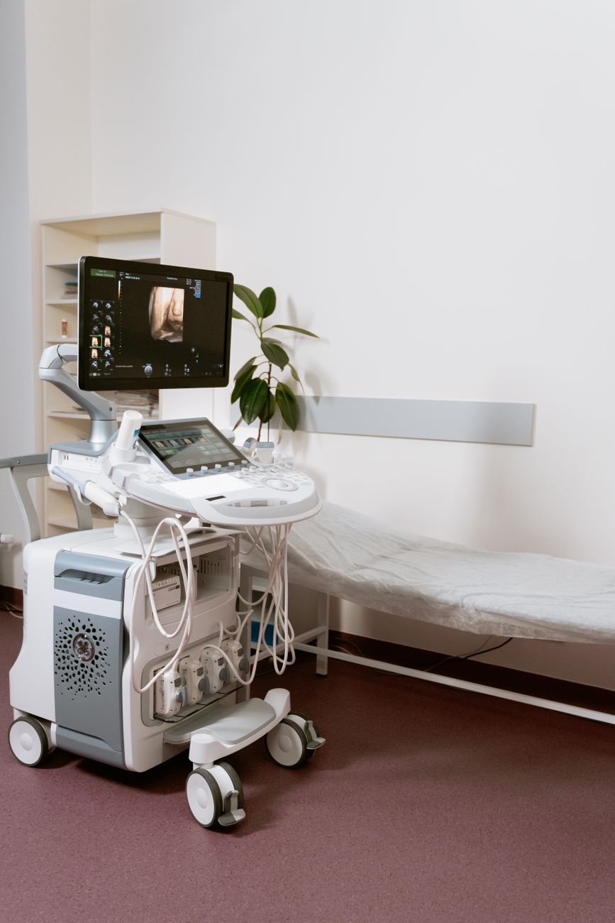
Ultrasound
It evaluates response during treatment.
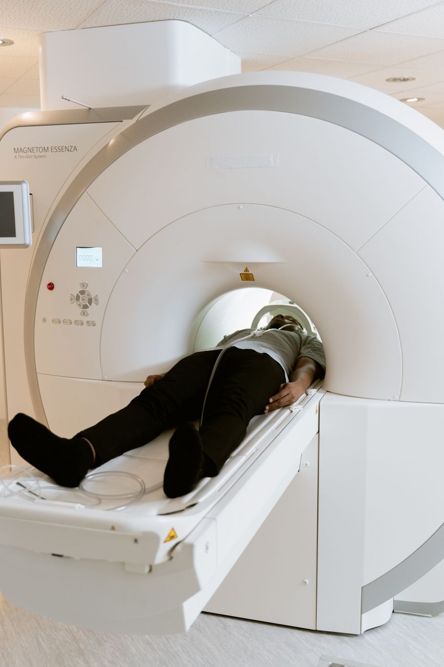
Magnetic Resonance Imaging
At the end of cytotoxic treatment, Pelvic MRI monitors response for at least three courses. Vaginal nodules are removed.
Other Methods Of Evaluation
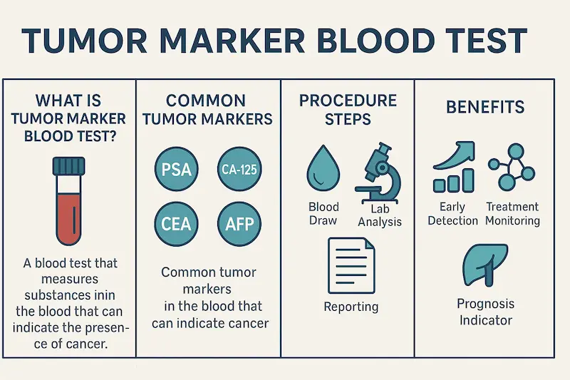
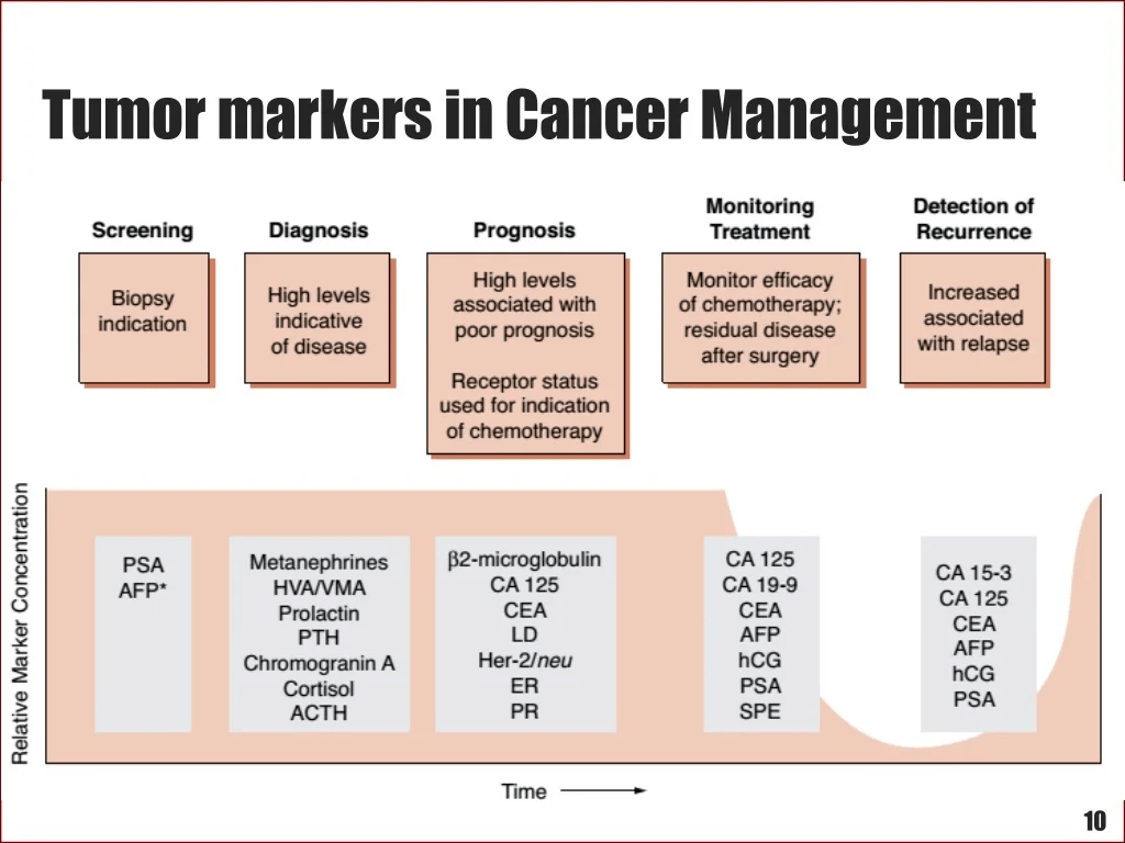
Tumour Markers
Proteins in blood where overexpression refers to cancerous and non-cancerous abnormalities.
Good news….what happens if there is complete remission (no cancer remains) after neoadjuvant chemotherapy.

Firstly, this is confirmed with:
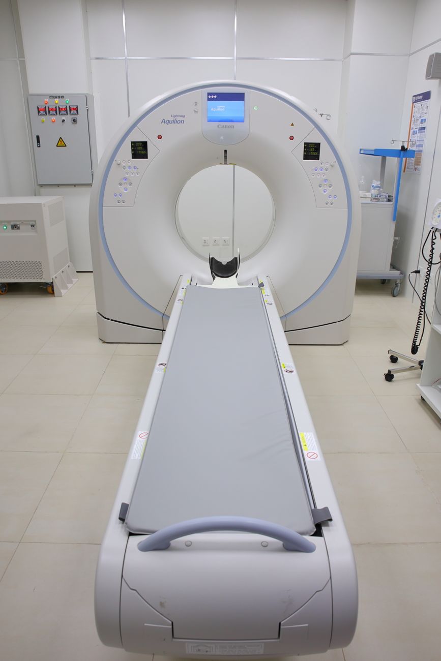
Negative Pelvic MRI
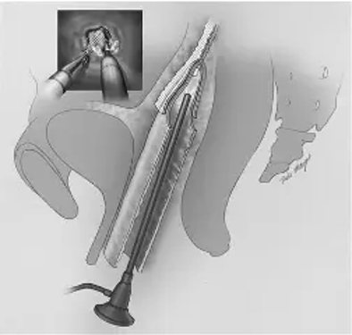
Negative Vaginoscopy
Light with tube detects vagina. The name of the device to detect vaginoscope.
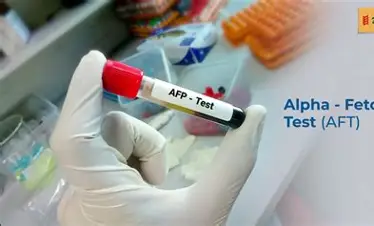
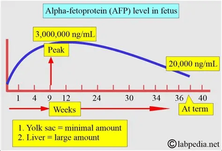
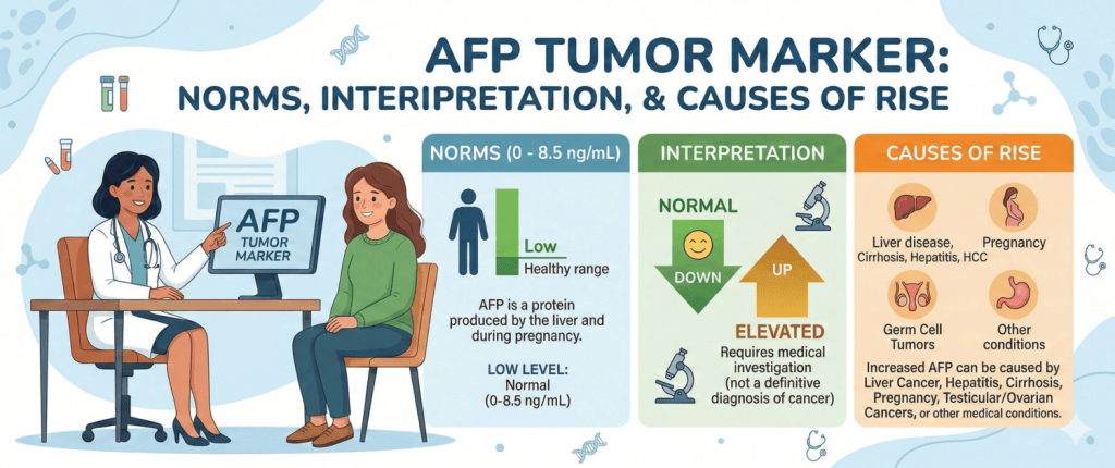
Normal Levels of AFP
What is AFP?
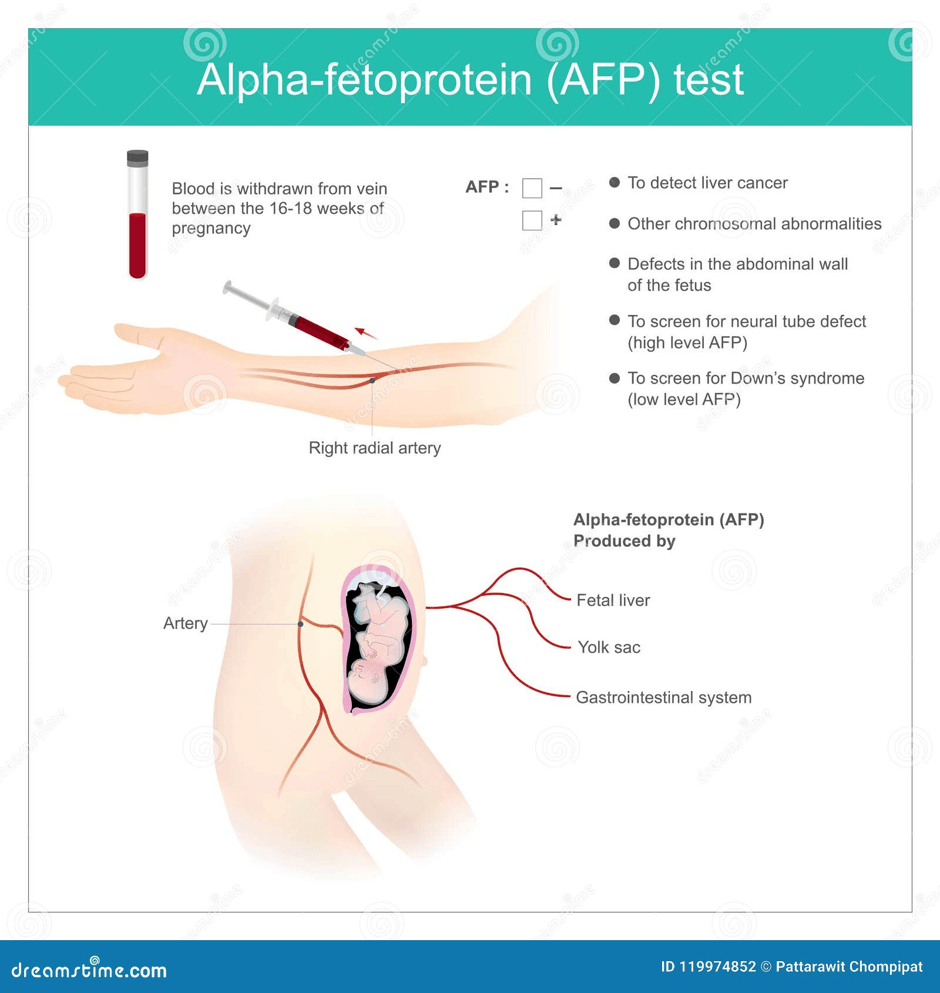
AFP is a protein formed in the liver and yolk sac of the fetus. It is present in the fetal serum and secondarily in maternal blood.
It is vital during the reproductive system as it is used for prenatal screening between the 15th and 20th week of pregnancy. High levels are associated with neural tube defects like spina bifida.
Open abdominal wall defects like gastroschisis. Fetal death and multiple pregnancies. Low levels are linked with Down syndrome. Ultrasound is performed if there are unexpectedly high or low levels.
Levels of AFP are linked to length of gestation and mother’s weight.
AFP is linked to liver tumours and teratomas.
Diagnostic hysteroscopy with Vaginoscopy ‘NO TOUCH’ technique By Dr. Pat
What if there is no complete response after chemotherapy and organ-sparing surgery?
Radiotherapy
Vaginal Melanoma
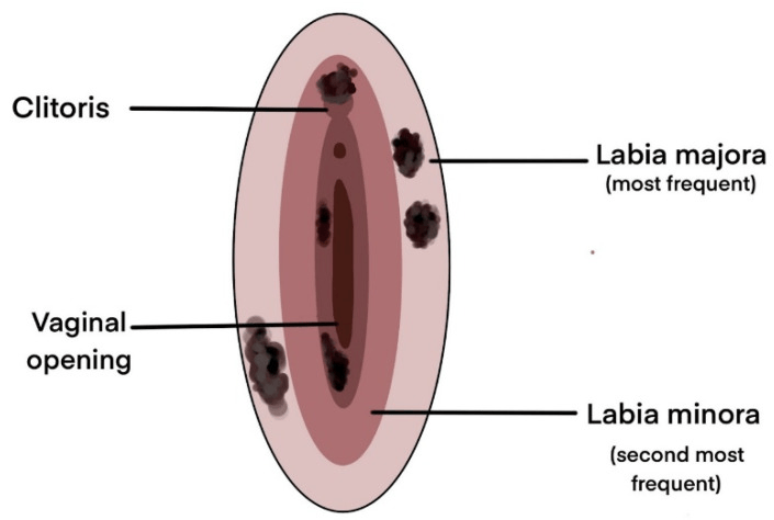
Rare cancer, diagnosed in elderly women.
No standard treatment because it’s rare.
Surgical excision (wide local excision) or colpectomy (removal of vagina and is another word of vaginectomy) with or without pelvic exenteration.
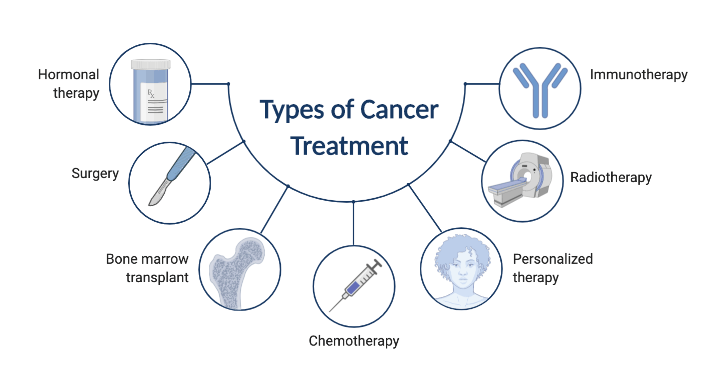
Adjuvant therapies (after surgery) – radiotherapy, immunotherapy (interferon alpha but limited evidence.
Palliative chemotherapy and radiotherapy – advanced cases.
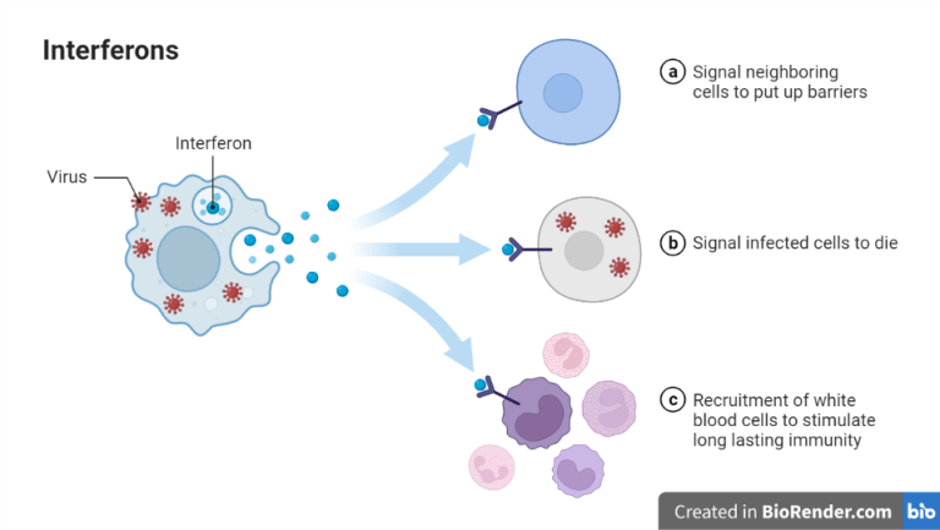
Interferons: INF-α, INF-β and INF-γ (FL-Immuno/07) By Frank Lectures
References
Hafeez Azeez (n.d.) Basic Surgical Procedures. Available at: https://www.slideshare.net/slideshow/basic-surgical-procedures/198359135 (Accessed: 5th November 2026)
International Society of Ultrasound in Obstetrics and Gynecology (ISUOG) (2026) Ovarian torsion. Available at:https://healthjade.net/ovarian-torsion/ (Accessed: 5th November 2026)
Lee, J.H., Jeong, Y.K., Park, J.K. and Hwang, J.C. (2003). “Ovarian Vascular Pedicle” Sign Revealing Organ of Origin of a Pelvic Mass Lesion on Helical CT. American Journal of Roentgenology, 181(1), pp.131–137. doi:10.2214/ajr.181.1.1810131.
National Cancer Institute (2026) Vaginal Cancer Treatment (PDQ®)–Health Professional Version. Available at: https://www.cancer.gov/types/vaginal/hp/vaginal-treatment-pdq (Accessed: 17th August 2026)
NHS England (n.d.) Proton beam therapy. Available at: https://www.england.nhs.uk/commissioning/spec-services/highly-spec-services/pbt/ (Accessed: 5th November 2026)
Nout, R.A., Calaminus, G., Planchamp, F., Chargari, C., Lax, S., Martelli, H., McCluggage, W.G., Morice, P., Pakiz, M., Schmid, M.P., Stunt, J., Timmermann, B., Vokuhl, C., Orbach, D. and Fotopoulou, C. (2023). ESTRO/ESGO/SIOPe Guidelines for the management of patients with vaginal cancer. International Journal of Gynecologic Cancer, [online] 33(8). doi:10.1136/ijgc-2023-004695.
Prajesh, P. (n.d.) Anatomy of female genital tract. Availablr at; https://www.slideshare.net/slideshow/anatomy-of-female-genital-tract-116217917/116217917#google_vignette (Accessed: 5th November 2026)
Tulandi, T. (2026) Ovarian transposition before pelvic radiation. Available at: https://www.uptodate.com/contents/ovarian-transposition-before-pelvic-radiation#H30824440 (Accessed: 8th October 2026)


Leave a Reply