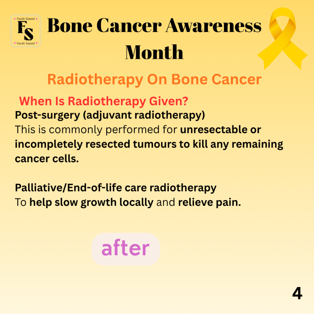
Proton Beam Therapy.
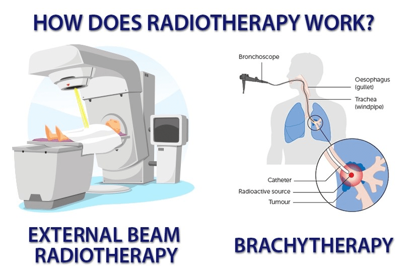
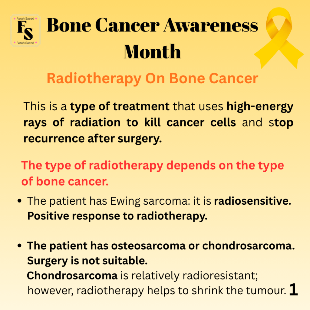
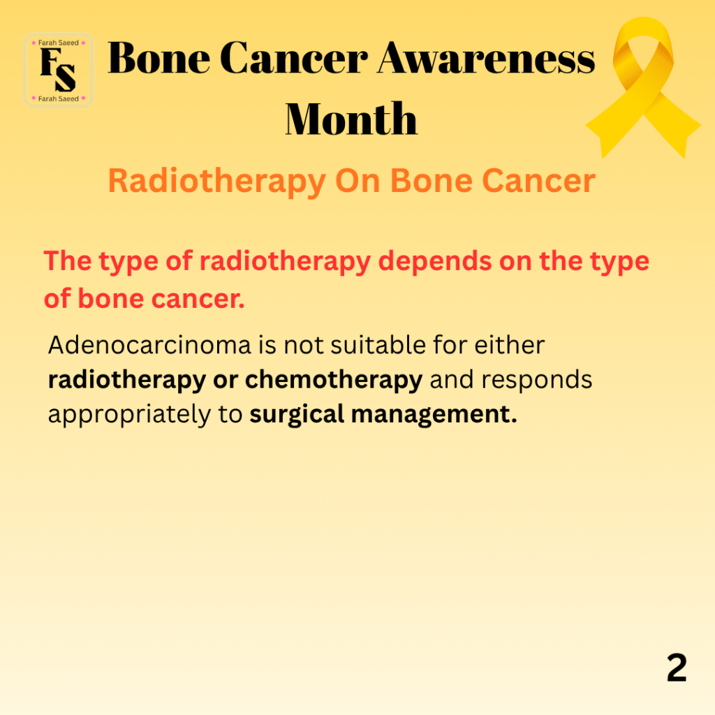
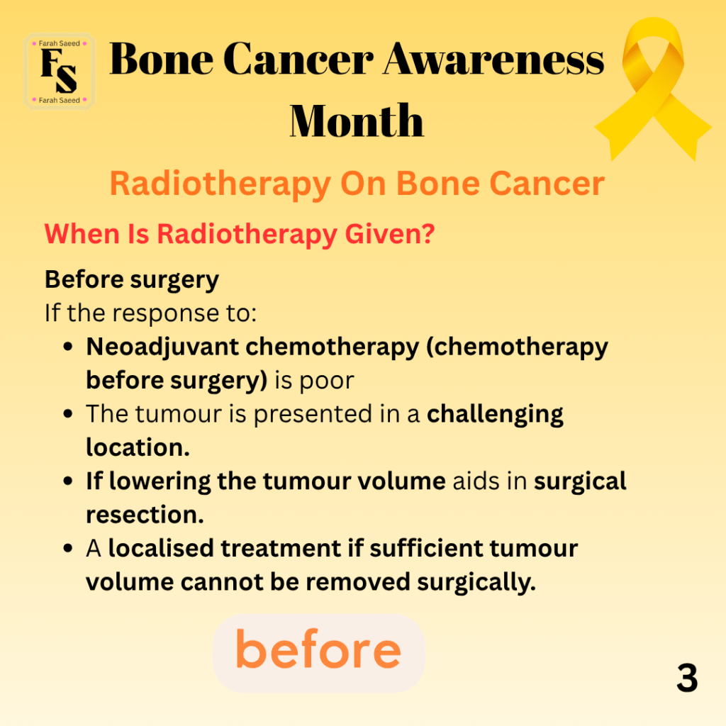
This is a common radiotherapy used to treat Ewing sarcoma, Chordoma and chondrosarcoma types of bone cancer with the aim having a more precise way of reaching the target without affecting any of the critical organs in the human body.
What Is Proton Beam Therapy and How Does it Work> By IOE Programme Dr Brian Lawenda
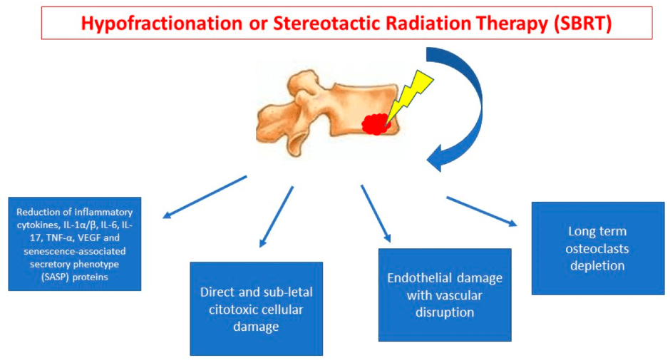
Stereotactic Body Radiotherapy (SBRT)
Radiotherapy is given at different angles where the tumour receives a high dose of radiation and tissues receive a lower dose.
It is a form of ablative therapy to ensure lower risk of recurrences.
This treatment is for bone cancers that may have spread to the lung.
What is Stereotactic Body Radiation Therapy? – By SLUCare Radiation Oncology
3D conformal radiotherapy or 3DCRT
Radiation beam is concentrated and fits the size of the tumour, avoiding healthy tissue. This is especially important in sensitive areas.
What is 3DCRT? By Manipal Hospitals
Radiofrequency Ablation
It is considered reliable, safe, minimally invasive, and effective in controlling bone cancer and relieving pain.
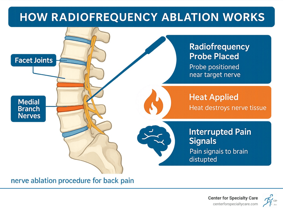
How does radiofrequency ablation work?
A probe enters the tumour and destroys tissue via frictional heating caused by ions targeting tissue using high-frequency electrical current.
This is guided using CT (computed tomography) or other types of scans.
Complications of radiofrequency ablation
Dehydration may occur, which leads to tissue resistance during ablation.
Low rate of major complications linked to secondary bone fractures.
Osteocool RF Ablation Animation By Medgadget
This video presents a device consisting of two probes, one at each end of the tumour. The probes have different coloured markers that determine to what extent the probes can enter and drill through: yellow (least depth), blue and green (very deep). Ablation can then be performed with subsequent cementoplasty, kyphoplasty and vertebroplasty.
Cementoplasty is a surgical procedure where poly-methyl-methacrylate cement is injected into the bone, especially vertebral bodies, where it is referred to as vertebroplasty. The aim is to treat or prevent fractures in weakened bones that may occur after thermal ablation or radiotherapy. Cementoplasty can provide pain relief and be secondary to microfracture stabilisation of the bone. However, one common complication is non-target cement instillation or “leaks” which may appear in the cortical bone or venous channels. The “leaked” cement is then hardened to prevent progression. If cement is added on adjacent nerve, it can cause neuropathic pain which is treated with medication or perineural steroid injection.
Kyphoplasty is a minimally invasive surgical procedure to treat compression fractures that may be caused by spinal tumours and multiple myeloma. It can also treat musculoskeletal inflammatory conditions called osteoporosis. This prevents conditions, for instance, kyphosis, where bones in the spine collapse. A small cut (incision) is made through the skin, and a hollow needle called a trocar enters the affected bone in the spine. An imaging technique known as fluoroscopy guides the trocar into position, and an inflatable balloon-like device enters the vertebrae. As the balloon grows/inflates, the vertebral height is achieved, and bone cement can then be added there. This prevents bones from further collapsing. It is estimated that 92% of people who underwent kyphoplasty have better pain relief. However, it is important to state that it does not treat the underlying bone loss that leads to the fracture.
Vertebroplasty is where liquid bone cement is injected via a needle into the vertebrae and acts like glue, holding the vertebrae together. This can be used in tumours that are either benign or malignant and do not heal and have severe back pain, and also conditions like osteoporosis where compression fractures are common. It is not used to treat chronic back pain caused by herniated discs or arthritis. Though vertebroplasty is a safe procedure with minimal to no complications reported. Most successfully is done within a few weeks to several months after the fracture occurs.
Laser ablation
Patients with bone metastases do not respond to standard treatment and require pain relief through this method.
What Is Laser Ablation By Henry Ford Health
How does laser ablation work?
Optical fibres transmit a type of energy called infrared light into the tumour to induce rapid heating of the target area. There are no neutral electrodes or medical devices. It normally penetrates half a centimetre, thus it is a minimally invasive procedure.
This causes proteins to break down (denature) and coagulation necrosis.
What is coagulation necrosis?
Coagulation necrosis is a type of tissue death that is distinctive from alternative cellular death. It is severe, irreversible, and commonly caused by lack of blood supply (ischaemia), lack of oxygen (hypoxia), and nutrients. Ischaemia results from blockage of arteries and hypoperfusion (decreased blood flow). Other causes of coagulation necrosis are exposure to toxic chemicals (cyanide) and metals (mercury). Burns and heat exposure beyond the optimum temperature. Bacterial infections can also result in coagulation necrosis.
This leads to depletion of adenosine triphosphate (ATP), which impairs ion pumps (Na⁺/K⁺-ATPase), resulting in an influx of sodium and water, causing swelling (oncosis). It affects the enzymes (proteases, endonucleases, and phospholipases) that degrade cellular components and structural proteins, begins to break down because of high levels of calcium entering the cell (calcium influx) and causing oxidative stress.
Other enzymes also break down, found in the lysosomes; a type of enzyme found in the cell that breaks down pathogens and large molecules. The dead cells release damage-associated molecular patterns (DAMPs) that attract immune cells involved in inflammation (e.g., neutrophils) that remove the debris of necrosis.
Amongst the organs that are affected by coagulation necrosis is the heart, where there is an increased risk of heart attack (myocardial infarction). Coagulative necrosis in the heart occurs within 12–24 hours of ischemic injury. Other organs are also affected by coagulative necrosis, like the kidneys, spleen, and adrenal glands (chestnut-shaped organs on top of the kidneys). Kidneys present distinctive areas of necrotic tubules. The spleen and adrenal glands have obstruction, e.g., a blood clot (thrombus), or when there is a blood clot, air, or fat that causes a lodge from one area to another (embolus). Other reports are damage to the testicles as a result of the twisting of the seminal cords affecting the blood supply (testicular torsion).
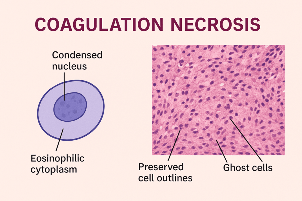
What are the microscopic features of coagulation necrosis?
The necrosis is characterised by a breakdown of proteins and enzymes; hence the term ‘coagulation’ that creates a solidifying effect despite cellular death. The tissue can be stained with a coloured dye called Haematoxylin and Eosin (H and E), giving a red stain feature.
Under the microscope, there is loss of nuclei; this is where the nuclear material disappears, where they fade (karyolysis), shrink (pyknosis), and fragment (karyorrhexis), causing irreversible cell death.
A ghost-like appearance where the outlines become visible for several days despite nuclear loss and maintain shape due to early inactivation of hydrolytic enzymes that prevent immediate cell lysis and the preservation of structural proteins. There is no tissue softening (liquefaction) to maintain firm consistency.
Initially, there is an absence of inflammatory cells, but after some time, Immune cells then perform immediate effects and begin phagocytosis (macrophages) and neutrophils (clean up necrotic debris).
The cytoplasm of cells that necrotic cells becomes intensely pink (eosinophilic) because of breakdown of proteins and loss of RNA in basophils. Eosinophils and basophils are other types of polymononuclear granulocytes (type of immune cells) that are involved in hypersensitivity reactions and allergies.
What are the complications of laser ablation?
Small size of the ablation zone for single fibre, which makes it unsuitable for large tumours.
Argon Beam Coagulation
Its purpose is to control the surface during major surgeries.
How does it work?
Argon is an inert (unreactive) gas that can be used in surgery such as argon beam coagulation, where there is conduction of unipolar electrical current delivered through the fine beam, and it affects the superficial layers (top layers) without affecting deeper layers, causing coagulation, shrinkage of the main target tissue, and dessication.
Fast movement and low settings: superficial layers of the skin to prevent damage of underlying layers.
Slow movement and high settings: Deeper layers of the tissue and becomes more intensive. This can be used to ablate a lesion
APC stands for Argon Plasma Coagulation by Erbe Group, describing the technique.
Argon Beam Coagulation (ABC) for Total Joint Procedures By CONMED Product Video – This presents components of the argon beam coagulation.
How to use argon gas coagulator By Farid Gutierrez
Cryoablation
The aim is to use cold treatment to be effective against primary and secondary bone cancers that are painful.
How does cryoablation work?
The tumour tissue is cooled to -20 degrees Celsius and transfers energy from the inserted probe to surrounding tissue via conduction and convection.
This requires imaging for guidance: ultrasound and magnetic resonance imaging (MRI) to identify the tumour or the ablation zone, and which areas are susceptible to damage from heat injury and cell death.
Cell death occurs when immediately adjacent tissues have contact with the probes. The probe enters via intracellular ice formation and destroys cells.
Further away, there is a cooling effect that causes osmosis across the cell membrane. There is secondary dehydration and death.
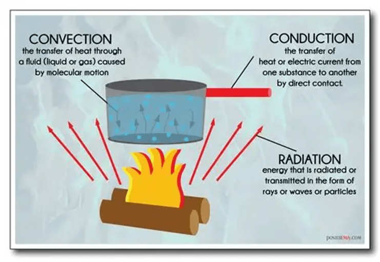
What are the complications of the cryoablation procedure?
Peripheral bone necrosis and cold injury to soft tissues in the surrounding target area. It is also complex and high-cost.
It has a recurrence rate of 11.2% for benign-aggressive and malignant bone tumours.
Cryoablation: How it works by Boston Scientific IO and Embolization
Phenol-local adjuvant
It is a type of chemical that induces tumour cell death, kills bacteria (bactericidal), stops bacterial growth (bacteriostatic), local anaesthetic (sleepy drug), and can kill cells (cytotoxic).
How Does Phenol Work?
It induces cell death of tumour cells, minimising risk, and can destroy 1 to 1.5 mm of tumour tissue, resulting in the breakdown of cellular proteins and DNA damage, causing tumour cell necrosis.
What are the complications of the Phenol treatment?
It is a challenge to penetrate tissues, and this limits cell death only on the top layer of the bone tissue. This can be alleviated by using polymethyl methacrylate (PMMA), which causes mechanical tumoural cavities with force, stress, tension, and strain impacting tumour growth and progression and may be involved in degenerative changes.
Recommended reading
Click here for more information and reading material
https://www.cancerresearchuk.org/about-cancer/bone-cancer/treatment/radiotherapy-treatment
https://oncodaily.com/oncolibrary/radiotherapy/radiotherapy-for-bone-cancer
https://www.frontiersin.org/journals/oncology/articles/10.3389/fonc.2025.1648849/full
References
Bădilă, A.E., Rădulescu, D.M., Niculescu, A.-G., Grumezescu, A.M., Rădulescu, M. and Rădulescu, A.R. (2021). Recent Advances in the Treatment of Bone Metastases and Primary Bone Tumors: An Up-to-Date Review. Cancers, [online] 13(16), p.4229. doi:https://doi.org/10.3390/cancers13164229.
Bone Cancer Research Trust (2026) Giant Cell Tumour of the Bone Available at:https://www.bcrt.org.uk/information/information-by-type/giant-cell-tumor/ (Accessed: 4th April 2026)
Cancer Research UK (n.d.) Radiotherapy for bone cancer Available at: https://www.cancerresearchuk.org/about-cancer/bone-cancer/treatment/radiotherapy-treatment (Accessed: 5th June 2026)
Cleveland Clinic (2023) Kyphoplasty. Available at: https://my.clevelandclinic.org/health/procedures/kyphoplasty (Acccessed: 2nd August 2026)
Najid, M. (2025) Coagulation Necrosis: Definition, Histology, and Clinical Significance. Available at: https://cancerbiologyresearch.com/coagulation-necrosis-definition-histology-and-clinical-significance/ (Accessed: 9th June 2026)
National Health Service (2025) Treatment for bone cancer. Available at: https://www.nhs.uk/conditions/bone-cancer/treatment/ (Accessed: 4th April 2026)
Pietrangelo, A. (2019) What is fibrosarcoma and how is it treated? Available at: https://www.healthline.com/health/fibrosarcoma (Accessed 3rd April 2026)
Pullan, J. and Lotfollahzadeh, S. (2024). Primary Bone Cancer. Available at: https://www.ncbi.nlm.nih.gov/books/NBK560830/ (Accessed: 9th April 2026)
Stanborough, R.O., Long, J.R. and Garner, H.W. (2022). Bone and Soft Tissue Tumors. Radiologic Clinics of North America, 60(2), pp.311–326. doi:10.1016/j.rcl.2021.11.009.
The Royal Orthopaedic Hospital (2026) Vertebroplasty. Available at: https://roh.nhs.uk/services-information/oncology/vertebroplasty (Accessed: 2nd August 2026)
Updated July 2026 Next Review July 2028


Leave a Reply