
FST is an educational resource, if you would like further information on chemotherapies used in cancer treatment, please visit the British National Formulary (BNF)https://bnf.nice.org.uk/
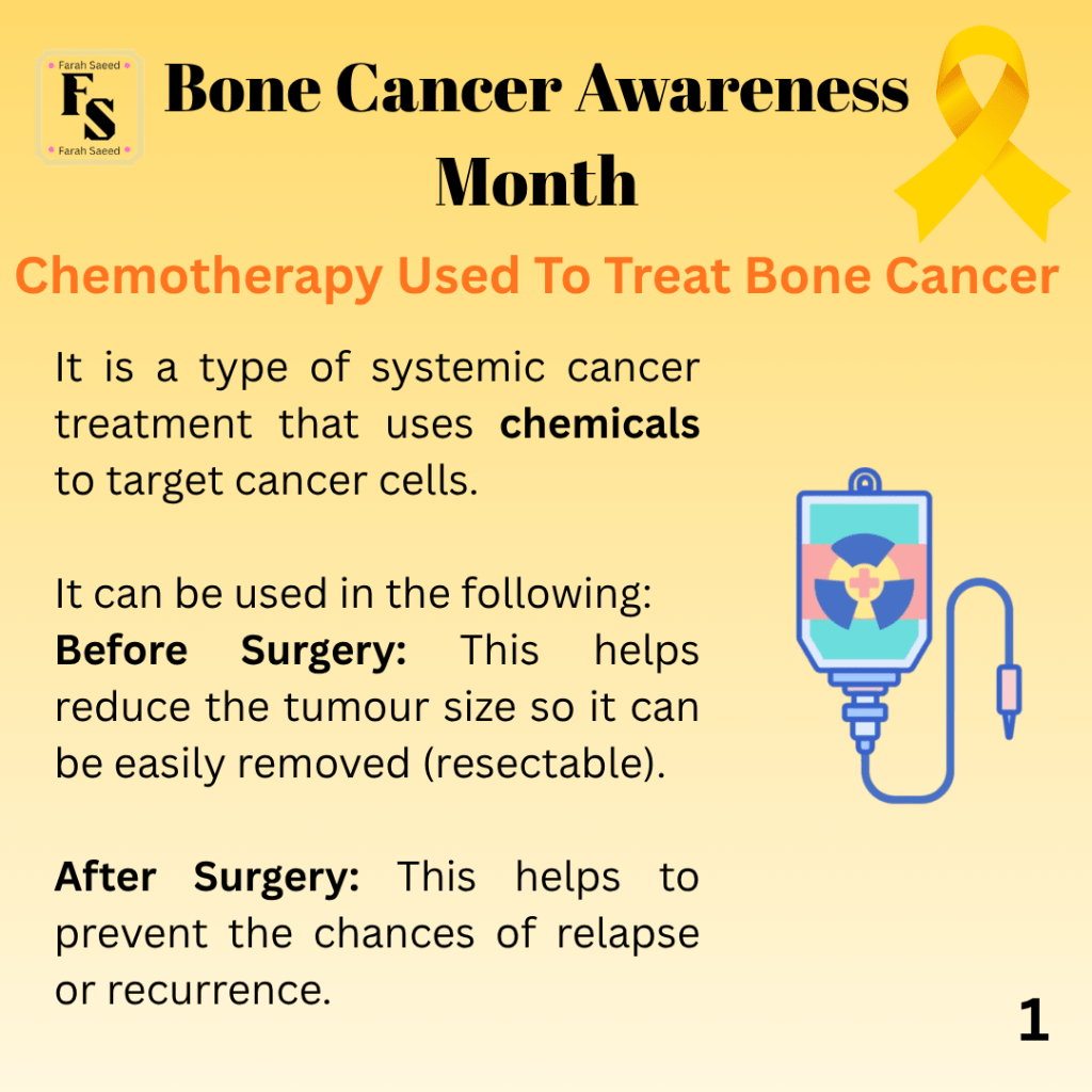
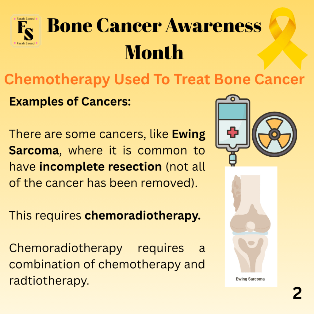
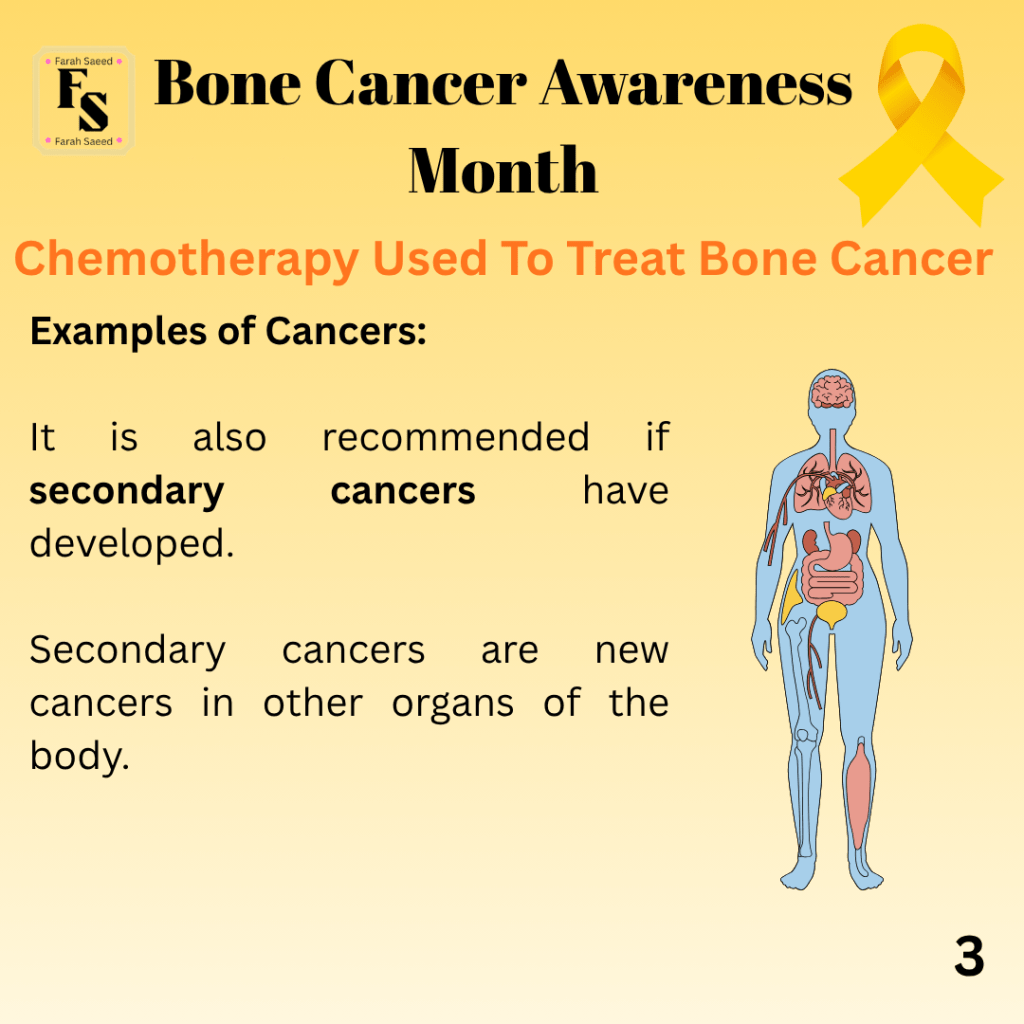
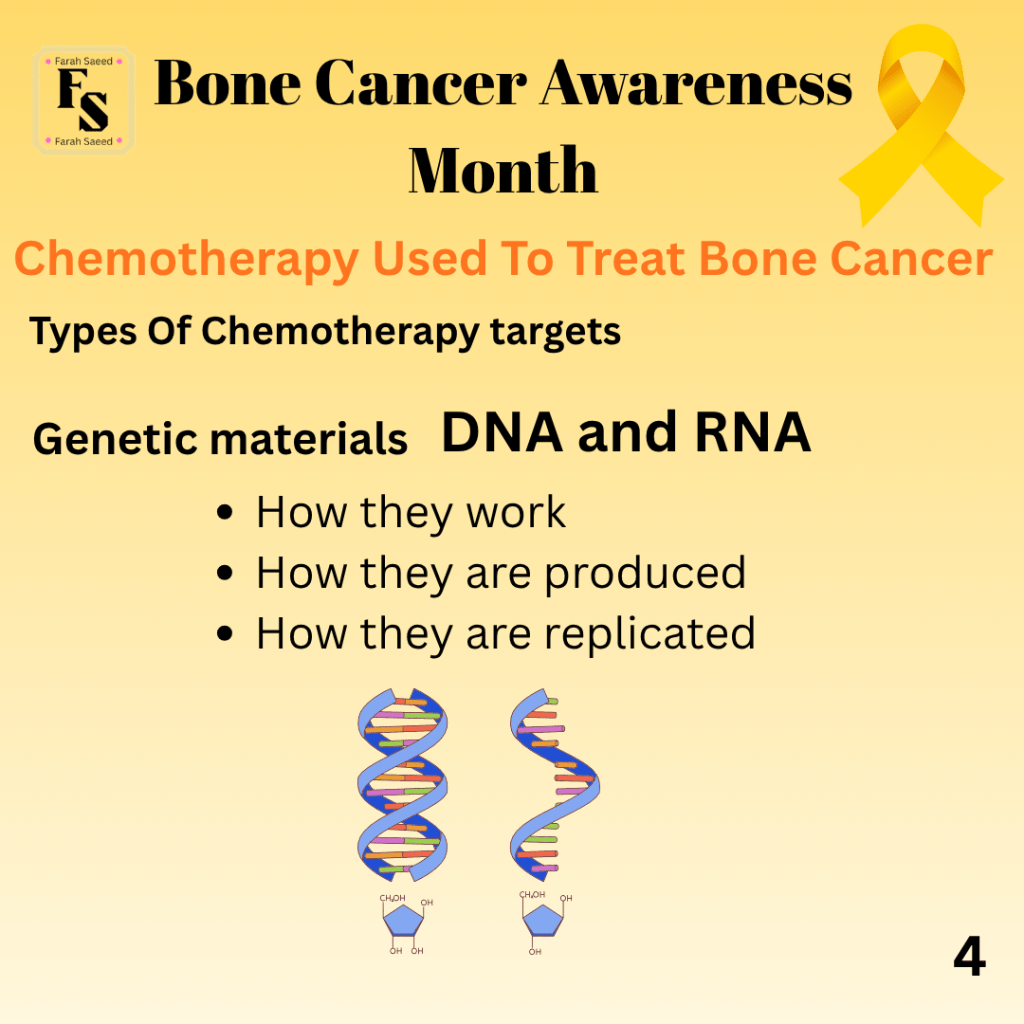
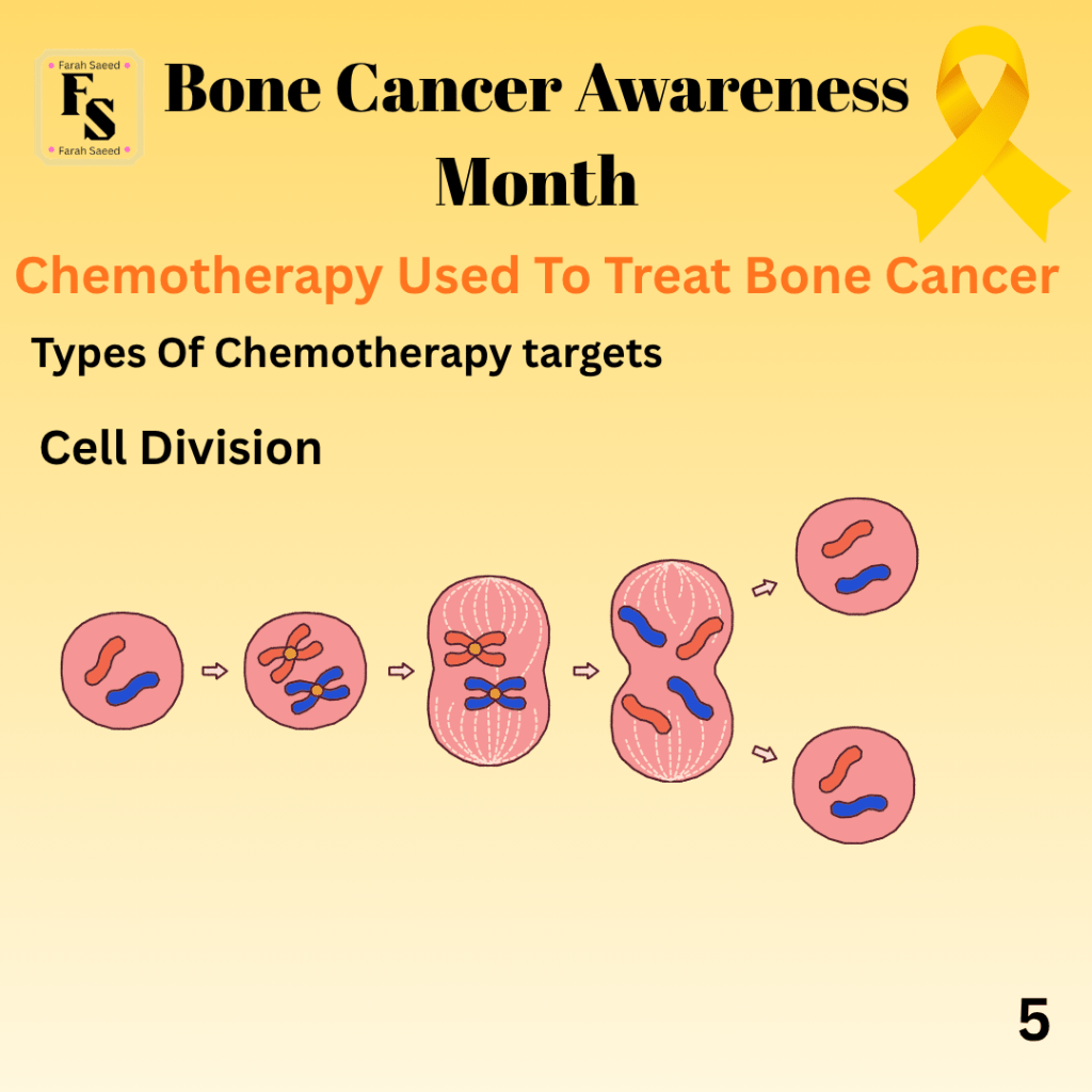
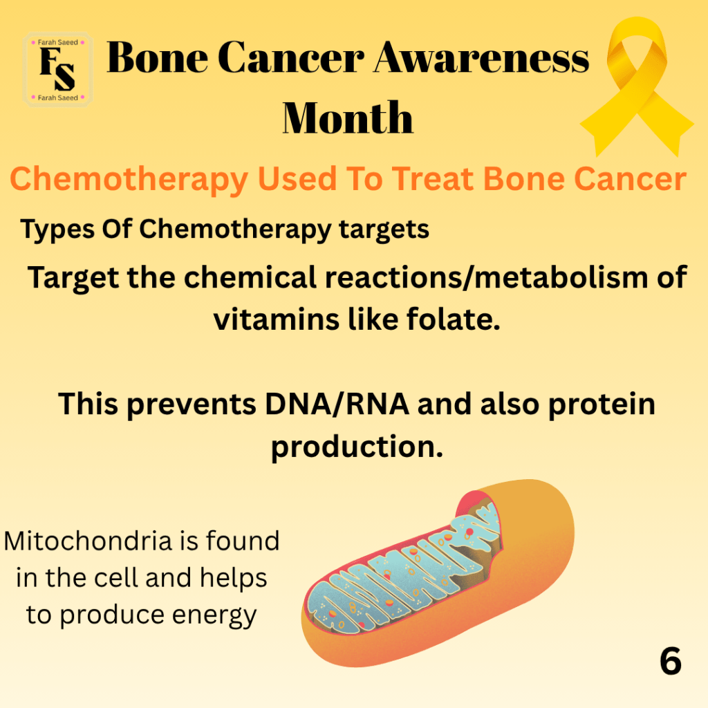
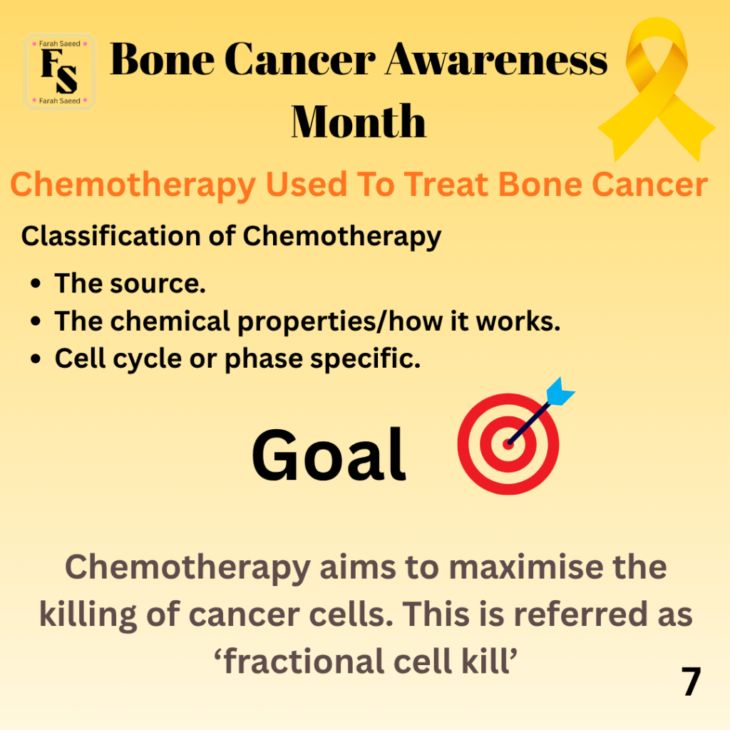
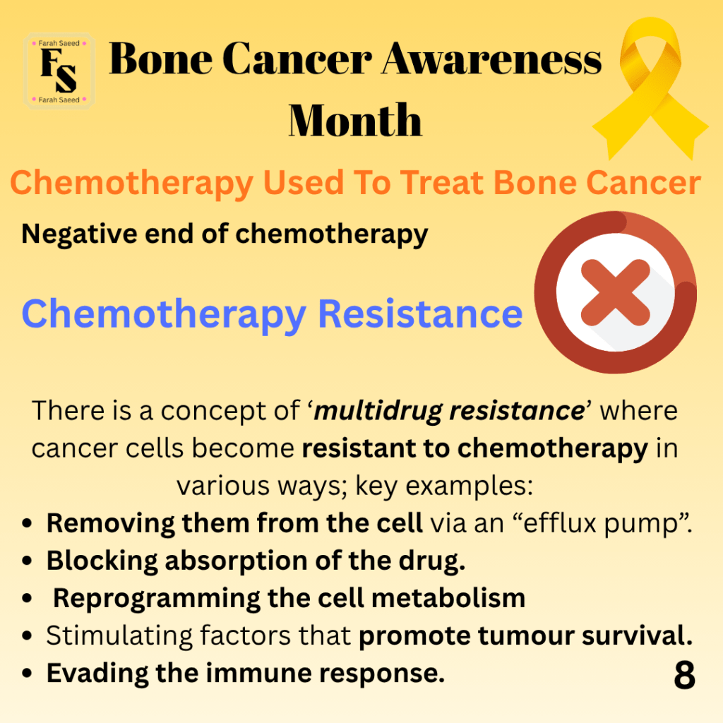
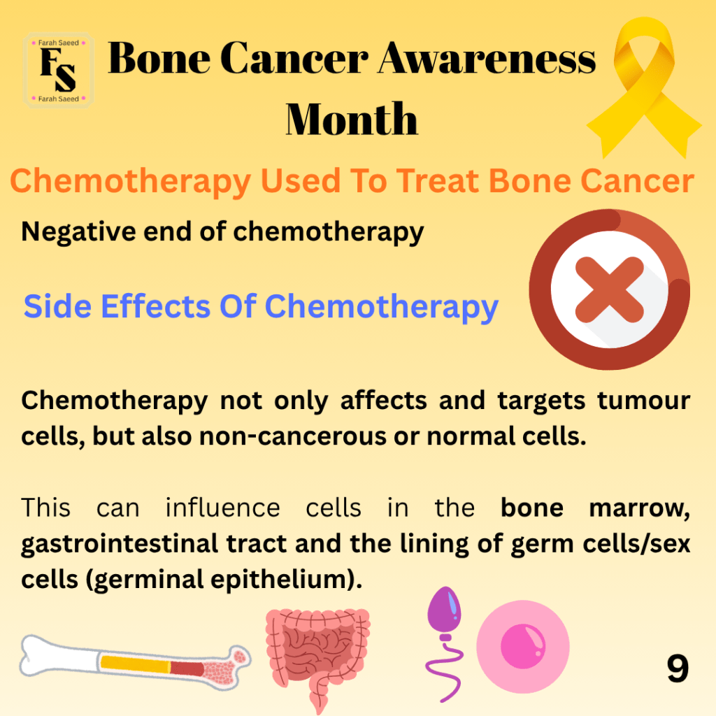
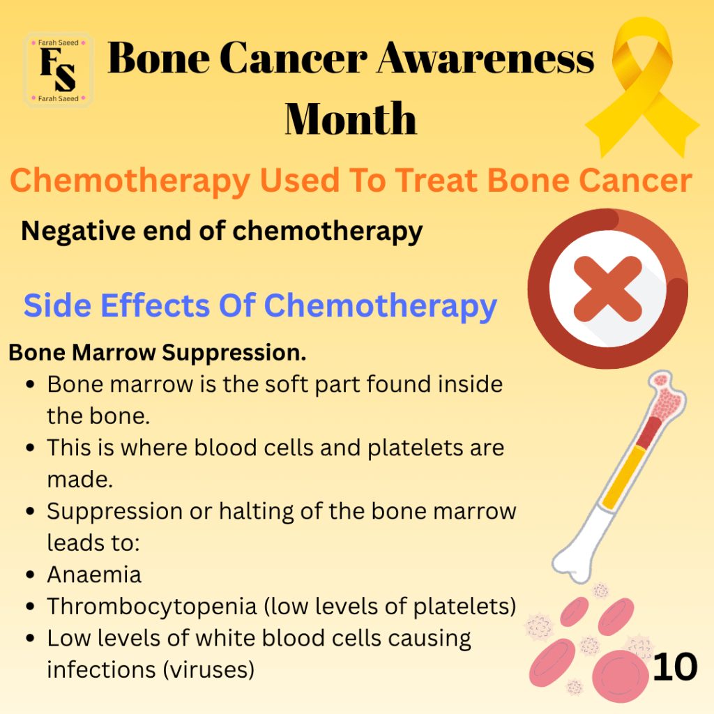
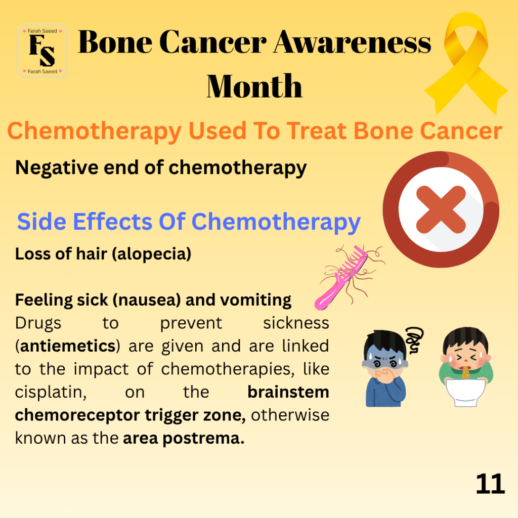
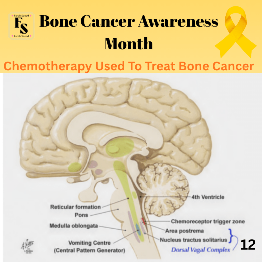
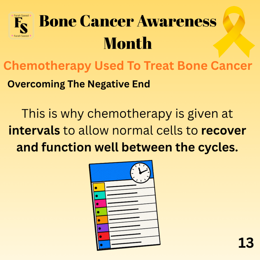
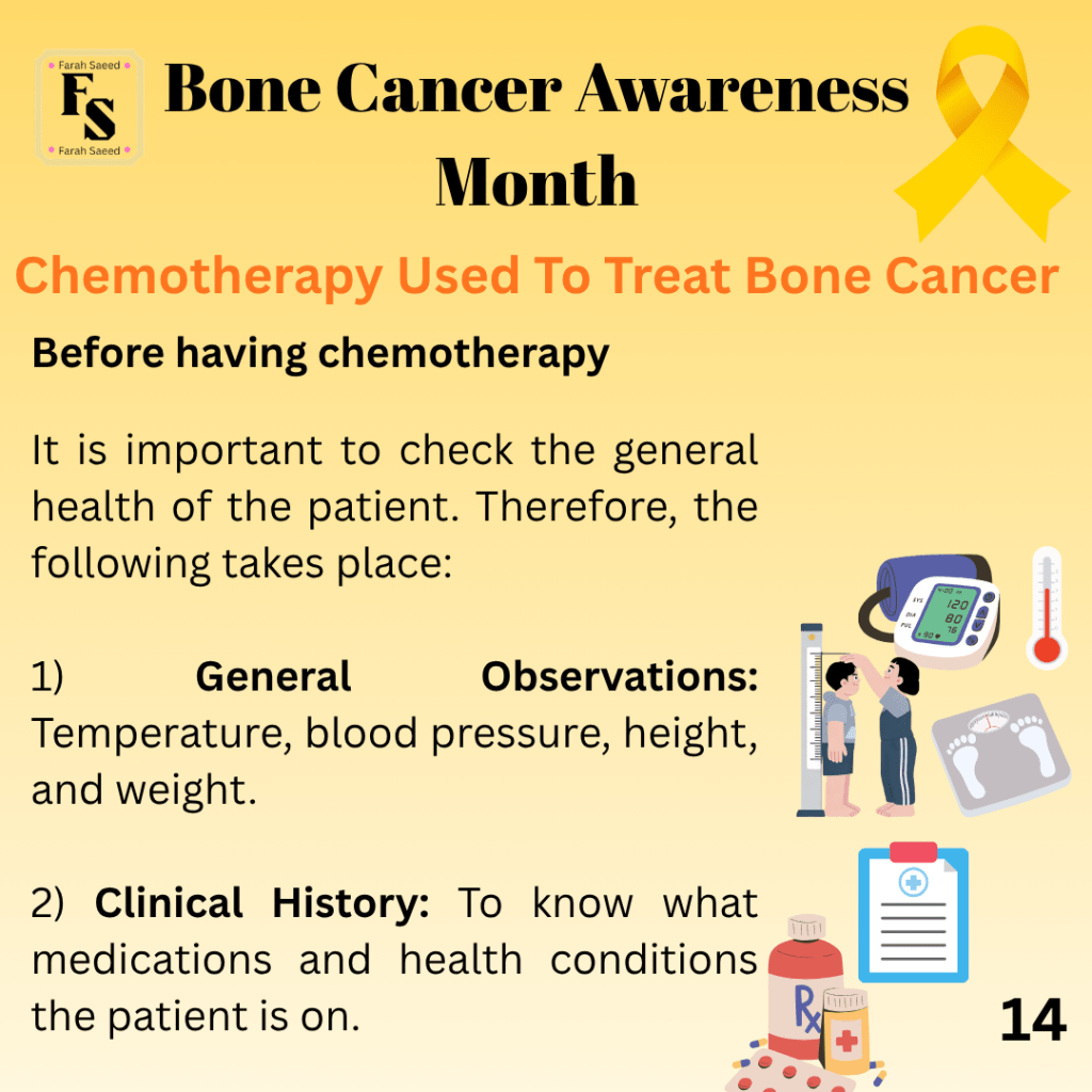
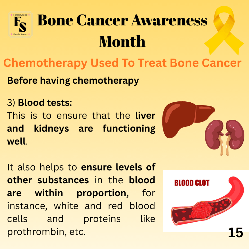
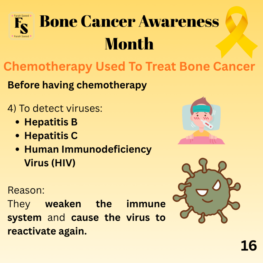
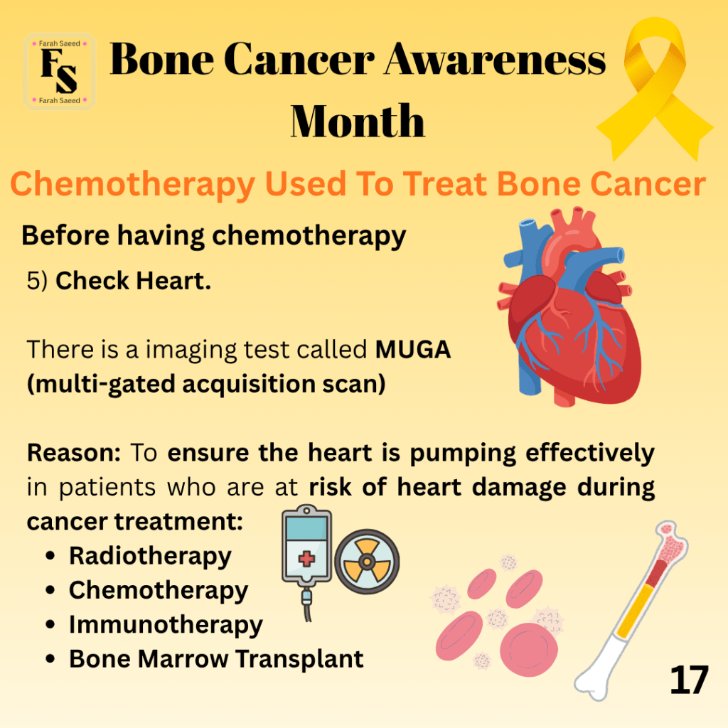
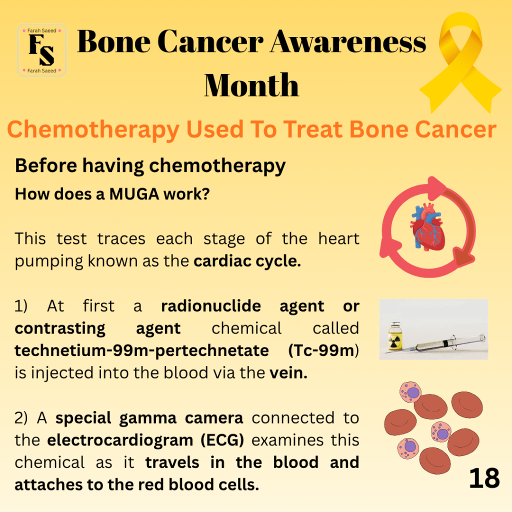
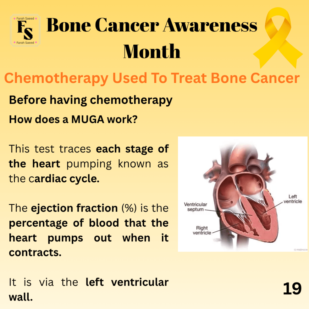
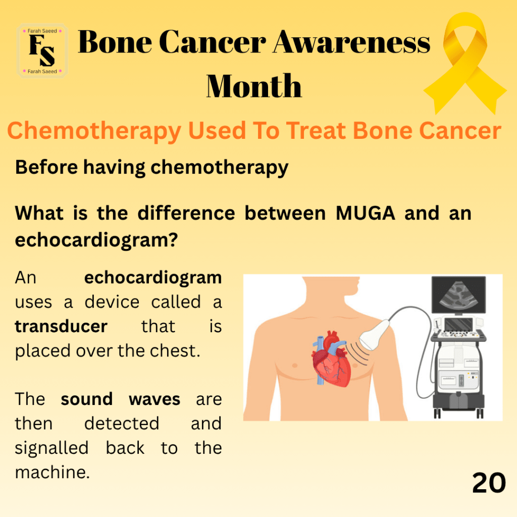
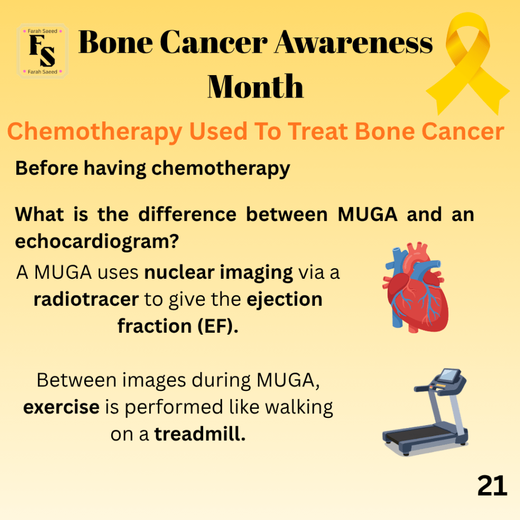
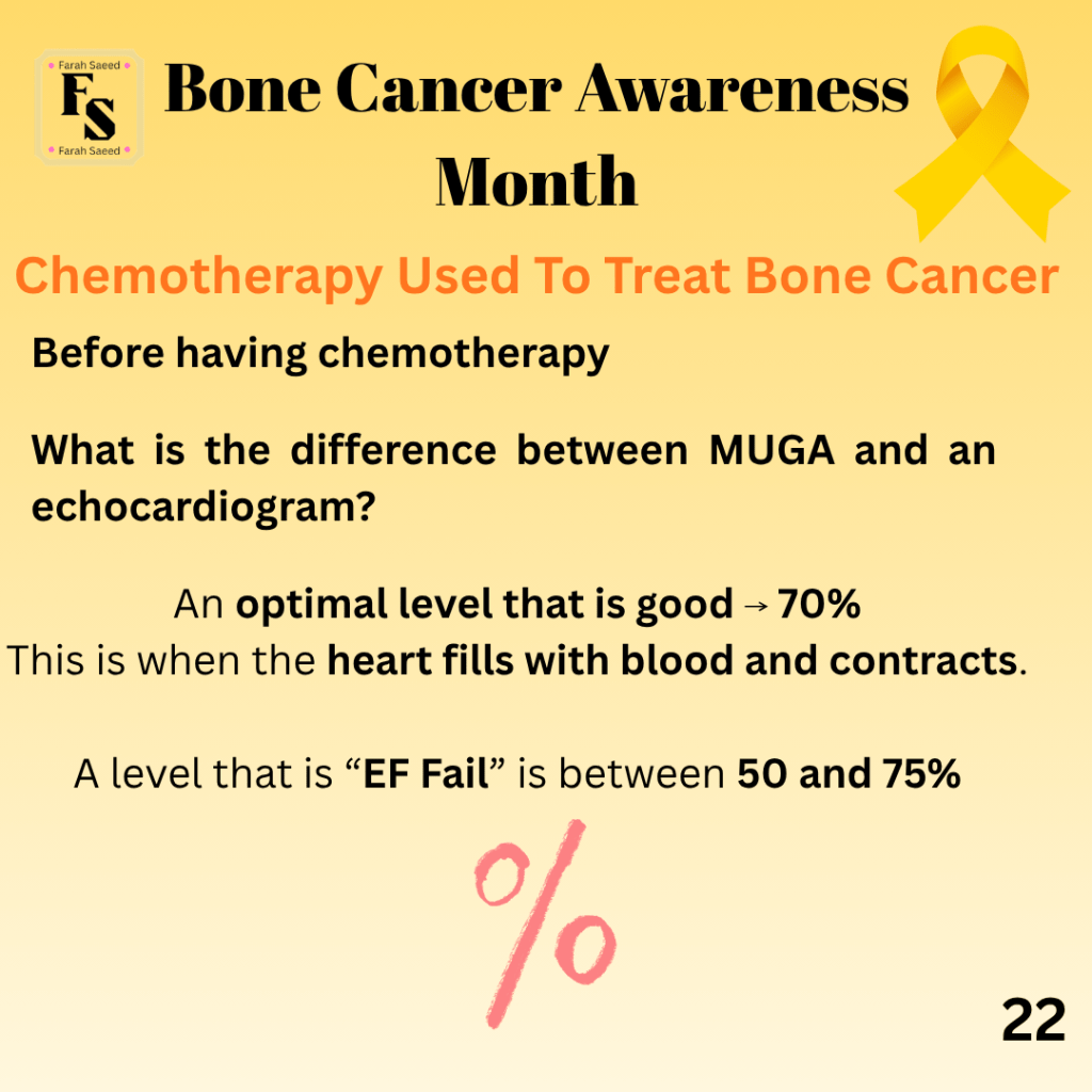
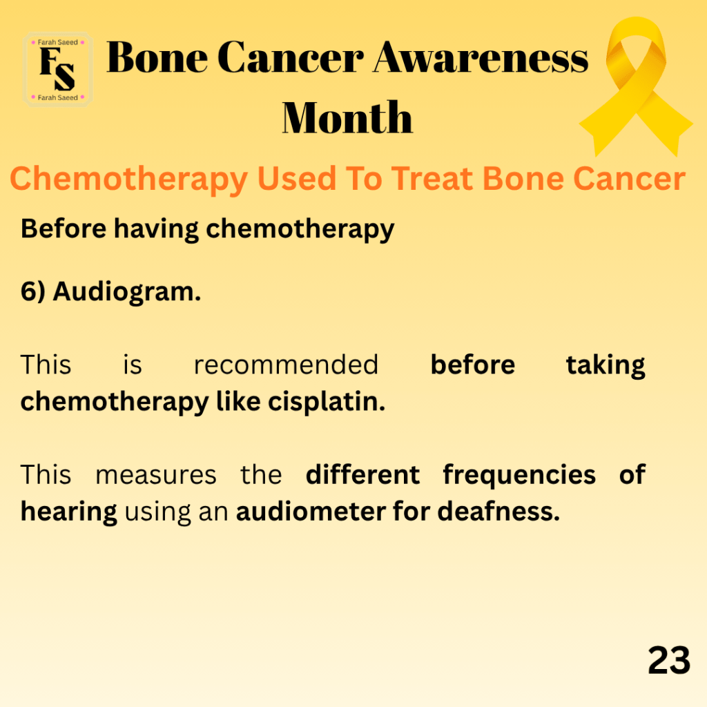
Supporting Images of Chemotherapy
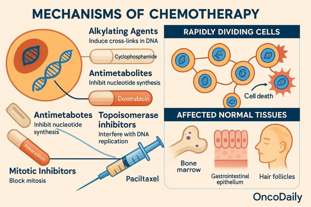
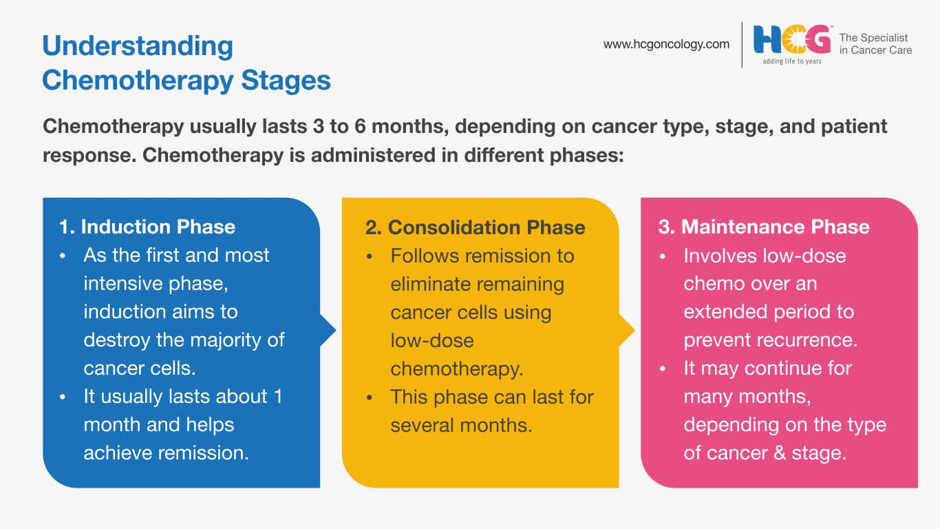
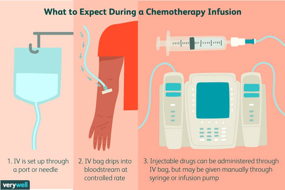
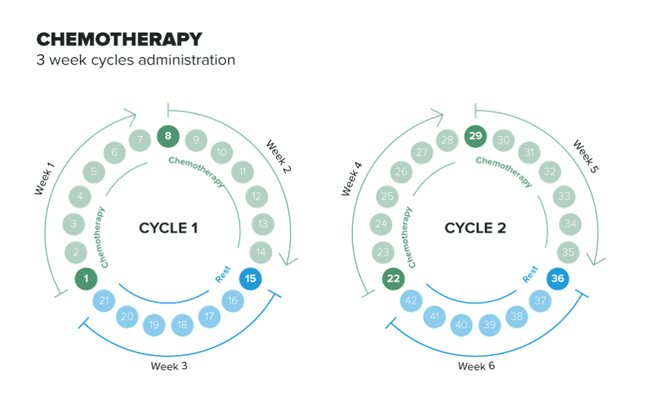
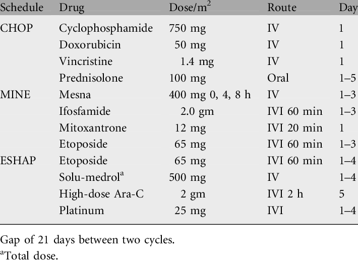
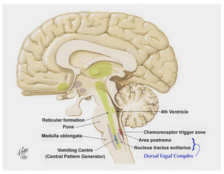
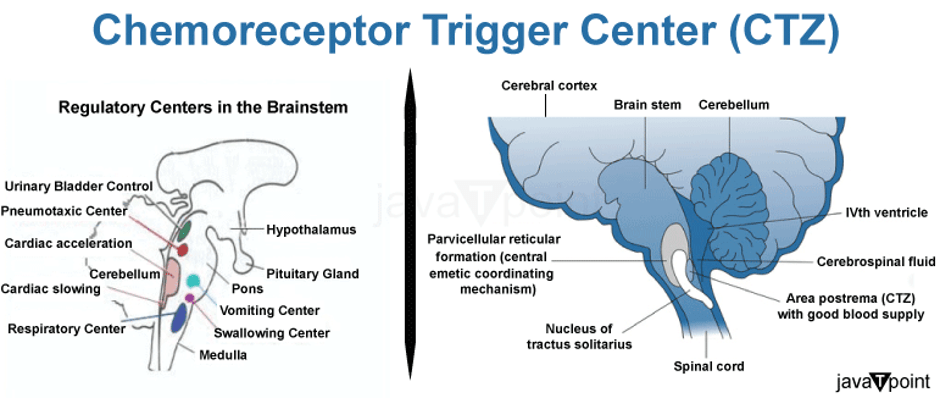
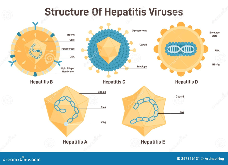
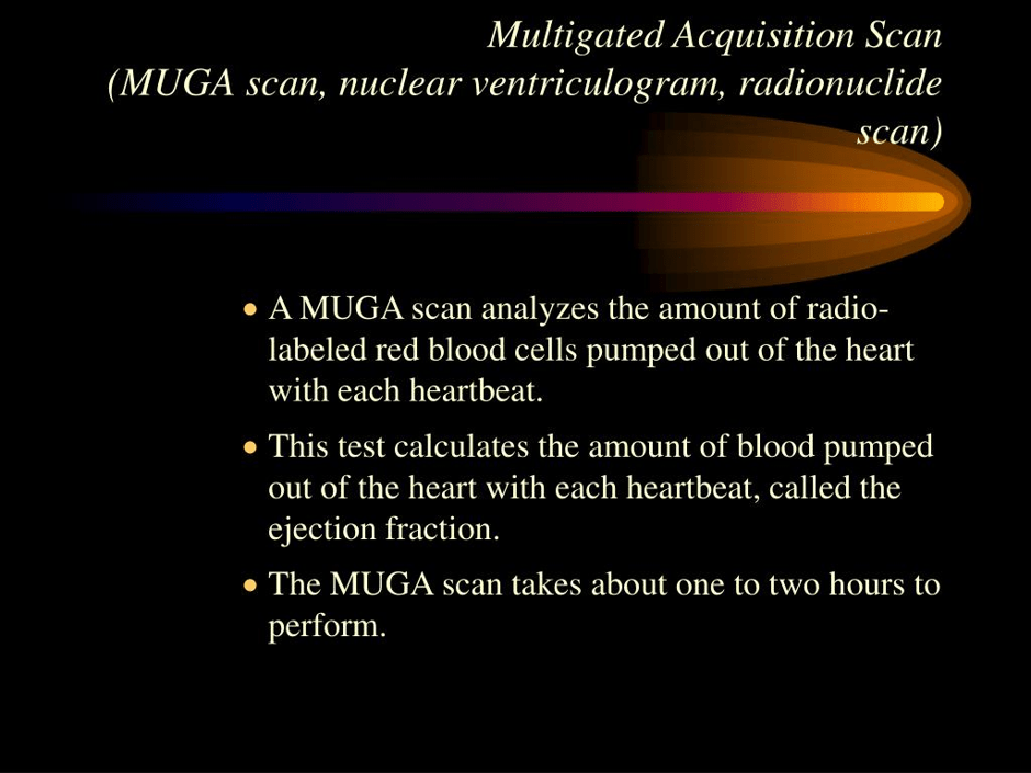
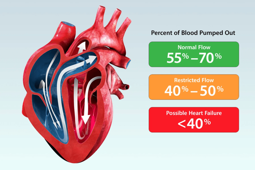
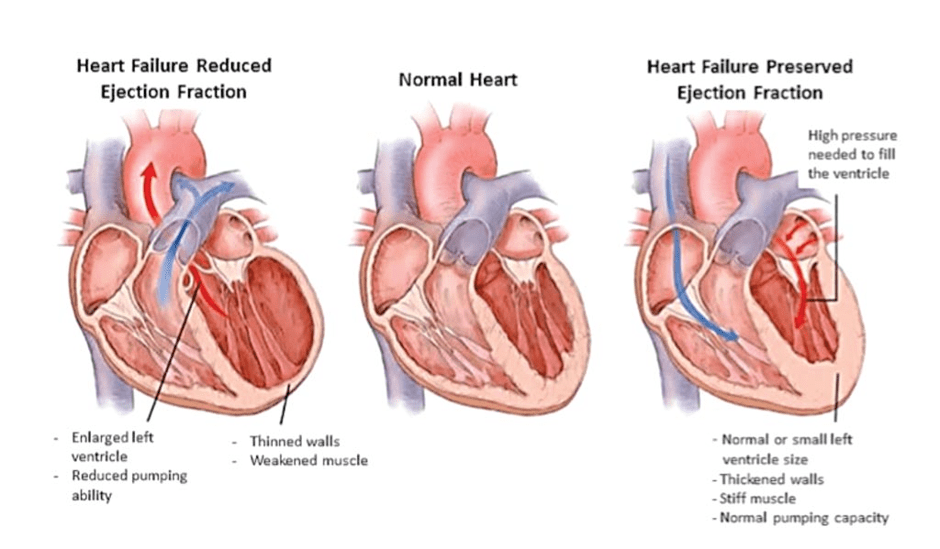
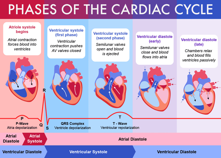
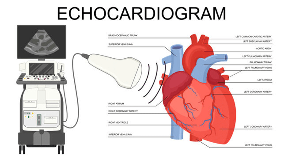
Main Types Of Chemotherapy
Supporting Images Of Chemotherapy
About Mifamurtide
It is a type of targeted therapy that is prescribed for patients between 2 and 30 years old.
It is commonly given with chemotherapy after surgery.
The aim is to remove residual micrometastases that were not taken by chemotherapy after surgery (adjuvant chemotherapy).
The purpose of Adjuvant Therapy is to decrease the rate of local recurrence after having a curettage. This prevents the need for additional resectional and reconstruction surgeries.
How does it work?
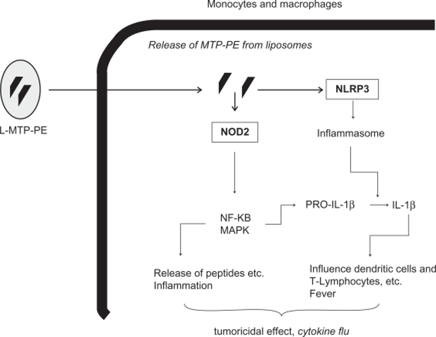
It aims to boost the immune system, specifically macrophages and monocytes.
It is involved in the signalling pathways that are involved in cellular growth, proliferation, and other cellular functions.
The activation of NFKB and MAPK promotes inflammation and the release of small proteins called cytokines.
What examples of receptors are involved in how Mifamurtide works?
There are pattern-recognition molecules that are detectable by Toll-like receptors and oligomerization domain (NOD)-like receptors (NOD1, NOD2 and NLRP3).
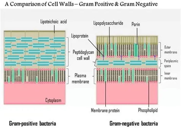
They are sensitive and can recognise particular aspects in the peptidoglycan polymer found in the wall of the bacteria.
The NOD2 receptor and MDP
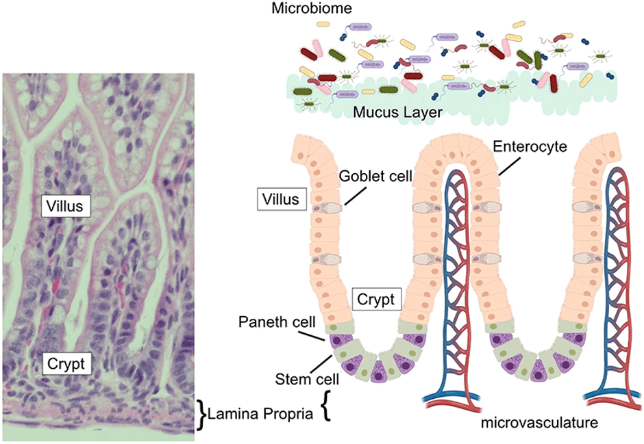
NOD2 is tissue-specific and can be found in monocytes, macrophages, dendritic cells, and in the wall of the cells of the intestines called Paneth cells.
A key example of a pattern-recognition molecule is muramyl dipeptide (MDP) found in the wall. This links to the source of Mifamurtide (L-MTP-PE) is a synthetic drug and a derivative of muramyl dipeptide (MDP) found in the wall of bacteria.
There are two types of intestines: large and small.
The Paneth cells are found throughout the small intestine (duodenum, jejunum and ileum) in small pouches called crypts of Lieberkühn.
The function of the small intestine is to break down and absorb food into the blood to target cells.
Paneth cells help make small proteins called defensins which has a antimicrobial effect on bacteria and other microorganisms that cause disease (pathogenic) and do not cause disease (non-pathogenic, commensal flora).
Proinflammatory cytokines released are interleukin-1 beta (IL-1β), interleukin-6 (IL-6), and tumour necrosis factor (TNF-α), chemokines, antimicrobial peptides, and adhesion molecules.
NOD-like receptors also recognise that NFKB signalling can have an amino-terminal caspase recruitment domain.
Consequently, it increases the levels of C-reactive protein and neopterine in the blood, indicating inflammation.
When is Mifamurtide given?
It is given intravenously via a vein in the blood, with breaks between cycles, at a dose of 2 mg/m2.
Each cycle lasts a week, and this treatment is given for 9 months (36 cycles) using the method stated below for the first 12 weeks.

After 12 weeks, it transfers to once a week for the next 24 weeks.
Please click here for more info via Cancer Research UK https://www.cancerresearchuk.org/about-cancer/treatment/drugs/mifamurtide.
Supporting Images Of Mifamurtide
Examples of chemotherapy for Ewing Sarcoma according to NCCN guidelines
Example one:
VDC/IE is recommended in accordance to the NCCN guidelines.
Primary chemotherapy treatment
VDC/IE is a multiagent chemotherapy consisting of Vincristine, Doxorubicin, Cyclophosphamide, and then alternating with Ifosfamide and Etoposide.
This can be taken for at least 9 weeks iA
Purpose: This is to decrease the tumour and achieve complete resection
Options for local control depend on age, tumour location, tumour size, response to chemotherapy, patient preference and anticipated morbidity.
1)Radiotherapy with chemotherapy can be used for local control.
2) Examples of surgery: wide excision, amputation in selected cases; adjuvant treatment can be given.
3) Chemotherapy post-surgery (adjuvant). This helps to minimise the risk of local recurrence after having curettage. This prevents the need for further surgery for resection and reconstruction.
Please visit the surgery on bone cancer section for further detail.
The duration or the length of time needed for chemotherapy after surgery like wide excision or amputation is between 28 and 49 weeks. This depends on the type of regimen and dosing schedule.
Examples of chemotherapy for relapsed cancers (returning cancers) according to NCCN guidelines
Clinical trials for new treatment
Example one
Vincristine, irinotecan, and temozolomide for relapsed or refractory Ewing sarcoma.
The overall response rate is 68%.
Example two
Irinotecan and topotecan with cyclophosphamide and temozolomide are linked with favourable response.
Example three
The CABONE trial, a multicenter, single-arm phase II trial, evaluated the activity of cabozantinib in patients with advanced Ewing sarcoma and osteosarcoma.
What is Cabozantinib?
This is a second-line treatment for patients with relapsed, refractory, or metastatic osteosarcoma.
It is an inhibitor of two main receptors: vascular endothelial growth factor receptor (VEGFR2) and the MET receptor.
The MET receptor signalling in non-cancerous conditions.
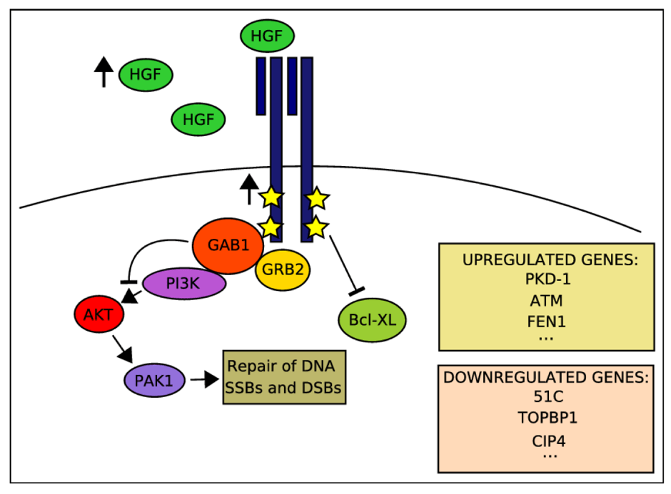
The tyrosine kinase receptor (MET) and its target substrate, Hepatocyte growth factor (HGF) are needed for growth and survival of cells in non-cancerous conditions.
The cells are found on the epithelial lining and migration of muscle progenitors (starting cells of muscles).
Other functions including to heal tissue damage and regenerating or making sites after healing in several organs.
MET receptor signalling and cancerous conditions
In cancerous conditions, MET stimulates the survival of cancer cells through production of new blood vessels that provide nutrients (angiogenesis), invasion, and facilitate the production of secondary cancers. They can also have mutated genetic lesions that maintain their gain of function in some primary tumours.
How does the MET signalling work?
The MET receptor binds to its target molecule and becomes activated via phosphorylation (the addition of phosphate groups).
They then interact with the GRB2-associated binding protein 1 (GAB1) Mult adaptor protein, to transduce the signal and recruit multiple proteins.
This causes the transcription of target genes for repair, growth and other cellular events.
The MET receptor also interacts with other molecules, e.g.
α6β4 integrin – to help with the downstream events in the signalling pathway
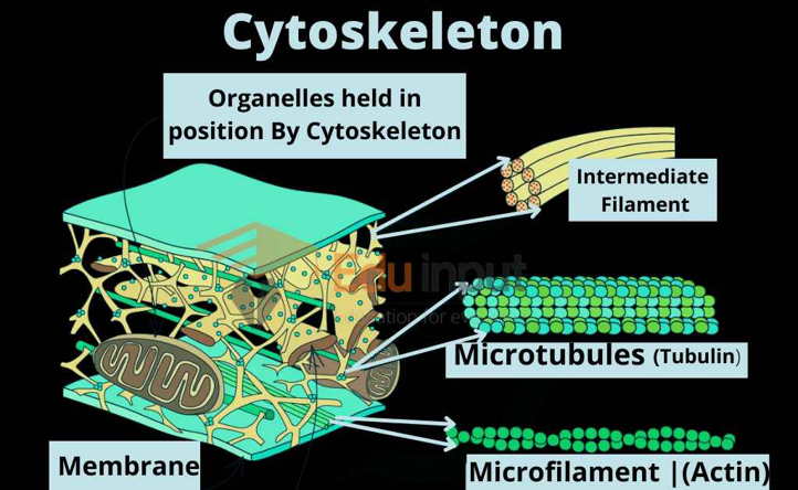
CD44 adhesion molecule – to help with the organisation of the cytoskeleton (the outer part of the cell)
The MET receptor also helps to support other signalling pathways, for instance, extracellular signal-regulated kinases (ERKs) and signal transducer and activator of transcription 3 (STAT3).
Supporting Images of Cabozantinib
What is the overall survival?
The overall survival (OS) was 84% at 6 months, 48% at 12 months and 14% at 24 months.
The median OS was 10.2 months and the median progression-free survival (PFS) of 4.4 months.
42% of patients had a metabolic tumour response, and it was well-tolerated in patients with advanced cancers. There were several side effects, e.g.
hypophosphatemia (low levels of phosphate in the blood)
high levels of the enzyme aspartate aminotransferase
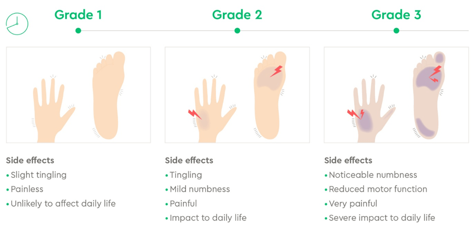
palmar-plantar syndrome (hand-foot condition where skin is affected).
About Palmar-plantar syndrome
If one has a lighter skin tone, it becomes red. If there is a darker skin tone, areas become darker. The skin may also appear sore or painful or tingle or swell or peel or crack or blister.
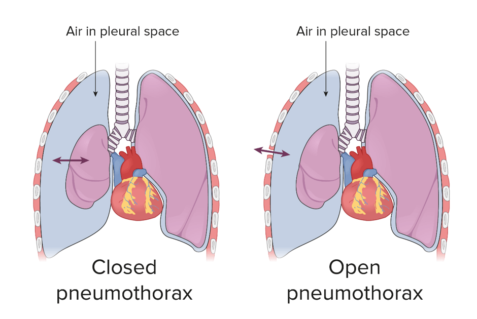
Pneumothorax – air space in the pleural cavity near the lungs.
Neutropenia – low levels of neutrophils (type of white blood cells)
Chemotherapy For Osteosarcoma
First line of treatment
Cisplatin, Doxorubicin and High-dose methotrexate (MAP)
MAP is preferred in patients aged 40 years with excellent performance status.
Delayed elimination due to duct or renal impairment – a chemical called glucarpidase is strongly recommended in patients receiving high-dose methotrexate.
What is Glucarpidase?
Glucarpidase is a bacterial recombinant enzyme that prevents the kidneys from clearing methotrexate to maximise efficiency but lower toxicity level by hydrolysing it to inactive metabolites:
4-deoxy-4-amino-N10-methylpteroic acid (DAMPA) and glutamate.
The liver undergoes detoxification
What other chemotherapeutic agents can be used to treat osteosarcoma for patients with non-metastatic resectable osteosarcoma?
Suggestion One
Cisplatin, doxorubicin, and methotrexate
Suggestion Two
Cisplatin, doxorubicin, methotrexate, and ifosfamide
Commentary on Suggestion One and Two:
Studies revealed there was no difference in the 6-year Event-Free Trial (EFS) which was 63 for Suggestion One and 64 for Suggestion Two.
However, their overall survival was different, 74 for Suggestion One and 70 for Suggestion Two.
Overall Survival (OS) is the time from diagnosis or the start of treatment until death from any cause.
Progression-Free Survival (PFS) is the time from the start of the treatment or randomization until disease progression or death from any cause, whichever occurs first.
Event-Free Survival is defined as the time from the start of treatment until the first presence of the predefined, negative clinical outcome (Event).
Suggestion Three: Gemcitabine
It can occur as a single agent: Gemcitabine (single agent)
OR
a combined regimen either:
A) Docetaxel and Gemcitabine.
B) Cyclophosphamide and topotecan.
C) High dose methotrexate with or without etoposide and ifosfamide.
D) Ifosfamide, carboplatin and etoposide – This has been effective in treating replaced or refractory bone sarcoma.
What can be used for second line therapy for osteosarcoma?
A) Sofarenib
This is a type of targeted therapy, not chemotherapy.
The Progression Free Survival (PFS) at 4 months (primary endpoint) was 46%.
The Median Progression Free Survival was 4 months.
The Overall Survival was 7 months.
Chemotherapy For Adamantinoma
Surgery is best option and tend to nor respond to chemotherapy.
There is a change of being fully cured if all tumour cells are removed.
References
Bădilă, A.E., Rădulescu, D.M., Niculescu, A.-G., Grumezescu, A.M., Rădulescu, M. and Rădulescu, A.R. (2021). Recent Advances in the Treatment of Bone Metastases and Primary Bone Tumors: An Up-to-Date Review. Cancers, [online] 13(16), p.4229. doi:https://doi.org/10.3390/cancers13164229.
Basic Medical Key (2016) Lower Digestive Tract. Available at: https://basicmedicalkey.com/lower-digestive-segment/ (Accessed: 25th June 2025)
Budhu, J.A., Wen, P.Y. and Lee, E.Q. (2022). Neurologic complications of oncologic therapy. Handbook of Neuro-Oncology Neuroimaging, pp.131–155. doi:https://doi.org/10.1016/b978-0-12-822835-7.00013-5.
Cancer Research UK (2026) Mifamurtide (Mepact) Available at: https://www.cancerresearchuk.org/about-cancer/treatment/drugs/mifamurtide (Accessed: 11th June 2026)
Cleveland Clinic (2022) MUGA Scan. Available at: https://my.clevelandclinic.org/health/diagnostics/17247-multigated-acquisition-scan-muga (Accessed: 4th June 2026)
Gemcitabine (n.d.) Gemcitabine. Available at: https://www.cancerresearchuk.org/about-cancer/treatment/drugs/gemcitabine (Accessed: 9th June 2026)
Kager, L., Pötschger and Bielack (2010). Review of mifamurtide in the treatment of patients with osteosarcoma. Therapeutics and Clinical Risk Management, p.279. doi:https://doi.org/10.2147/tcrm.s5688.
Marshall, E. (2026) Actions of Protein Kinases (PKs) Downstream of Akt. Available at: https://www.biorender.com/template/actions-of-protein-kinases-pks-downstream-of-akt (Accessed: 9th June 2026)
Najid, M. (2025) Coagulation Necrosis: Definition, Histology, and Clinical Significance. Available at: https://cancerbiologyresearch.com/coagulation-necrosis-definition-histology-and-clinical-significance/ (Accessed: 9th June 2026)
National Cancer Institute (2019) Adamantinoma https://www.cancer.gov/pediatric-adult-rare-tumor/rare-tumors/rare-bone-tumors/adamantinoma (Accessed: 4th April 2026)
National Health Service (2025) Treatment for bone cancer. Available at: https://www.nhs.uk/conditions/bone-cancer/treatment/ (Accessed: 4th April 2026)
National Institute for Health and Care Excellence (2026) Glucarpidase. Available at: https://bnf.nice.org.uk/drugs/glucarpidase/ (Accessed: 11th June 2026)
National Institute For Health And Care Excellence (2026) How dose carbozantinib work? Available at: https://www.cancerresearchuk.org/about-cancer/treatment/drugs/cabozantinib-tablets#how-does-cabozantinib-work (Accessed: 9th June 2026)
Nair, R., Prabhash, K., Sengar, M., Bakshi, A., Gujral, S., Gupta, S. and Parikh, P. (2007). The effect of short-term intensive chemotherapy on reactivation of tuberculosis. Annals of Oncology, 18(7), pp.1243–1245. doi:10.1093/annonc/mdm107.
Pietrangelo, A. (2019) What is fibrosarcoma and how is it treated? Available at: https://www.healthline.com/health/fibrosarcoma (Accessed 3rd April 2026)
Pullan, J. and Lotfollahzadeh, S. (2024). Primary Bone Cancer. Available at: https://www.ncbi.nlm.nih.gov/books/NBK560830/ (Accessed: 9th April 2026)
Schnur, M.B. (2023) Alkylating Agents How Do They Work. Available at: https://www.nursingcenter.com/blogs-plus/blogs/blogs-post/Alkylating-Agents (Accessed: 1st August 2026)
Science Insights (2025) Event-Free Survival vs. Progression-Free Survival. Available at: https://scienceinsights.org/event-free-survival-vs-progression-free-survival/ (Accessed: 1st August 2026)
Thomas, A., Bates, S.M Figg, W. and Pommier, Y. (2017) DNA Topoisomerase-targeting drugs Available at: https://oncohemakey.com/dna-topoisomerase-targeting-drugs/ (Accessed: 9th June 2026)
Trusolino, L., Bertotti, A. and Comoglio, P.M. (2010). MET signalling: principles and functions in development, organ regeneration and cancer. Nature Reviews Molecular Cell Biology, 11(12), pp.834–848. doi:https://doi.org/10.1038/nrm3012.
Tumor Surgery (2026) Adamantinoma: A Rare Bone Tumor of the Tibia Explained. Available at: https://www.tumorsurgery.org/2025/12/16/adamantinoma-rare-bone-tumor-tibia/ (Accessed: 4th April 2026)
Your Genome (n.d.) What is mitosis? Available at: https://www.yourgenome.org/theme/what-is-mitosis/ (Accessed: 4th June 2026)
Updated July 2026 Next Review July 2028


Leave a Reply