Aims And Objectives
This article aims to provide an overview for the general public on the following:
To understand the difference between the key terms: menstrual cycle, menstruation, menarche and menopause.
The different types of menstruation.
The process of Oogenesis (how the egg cell is formed).
The process of folliculogenesis (how the follicles are formed).
The process of the menstrual cycle and how the hormones and womb change throughout the menstrual cycle.
The colours that may appear during menstruation.
What Is The Difference Between The Menstrual Cycle, Menstruation, Menarche, and Menopause?
It is important to understand menstruation because both menstruation, menopause, and pathological conditions associated with menstruation increase the risk of gynaecological cancers, especially endometrium (womb), ovaries, vagina and vulva.
The Menstrual Cycle
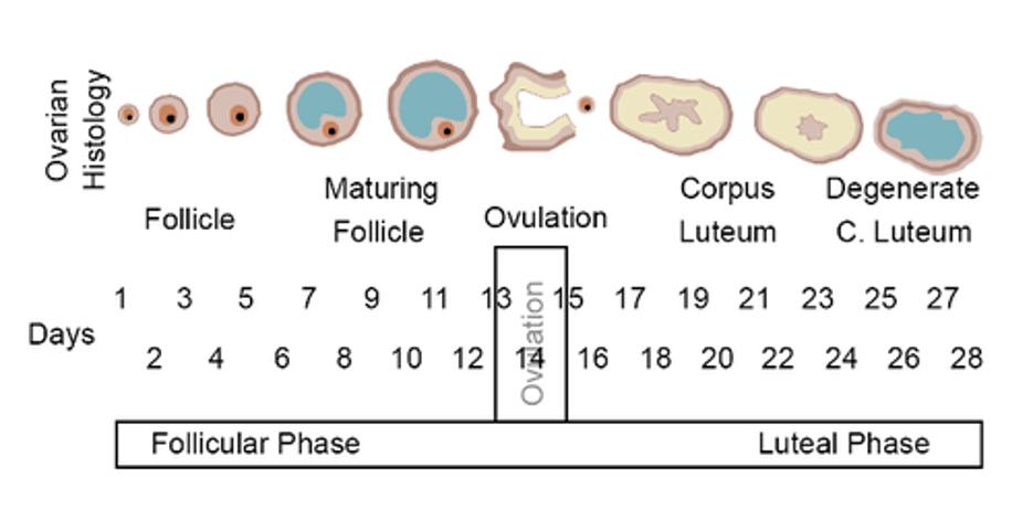
The menstrual cycle is a period of 28 days in which an egg cell, or ovum, aims to be fertilised, and if it doesn’t, menstruation takes place. Fertilization is the joining of the egg cell and the sperm cell.
Menstruation
Menstruation is the release of blood from the endometrium, the lining of the womb, if fertilization does not take place. There are three main types of menstruation: anovular, vicarious, and retrograde.
Anovular Menstruation
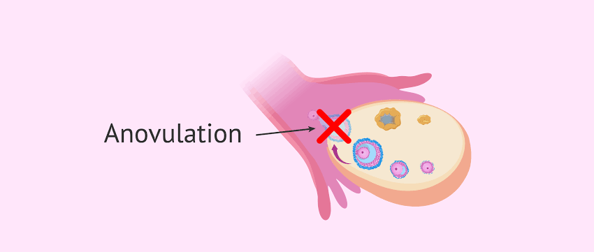
Anovular menstruation is the discharge of blood without the previous release of an egg from the ovary.
Vicarious Menstruation
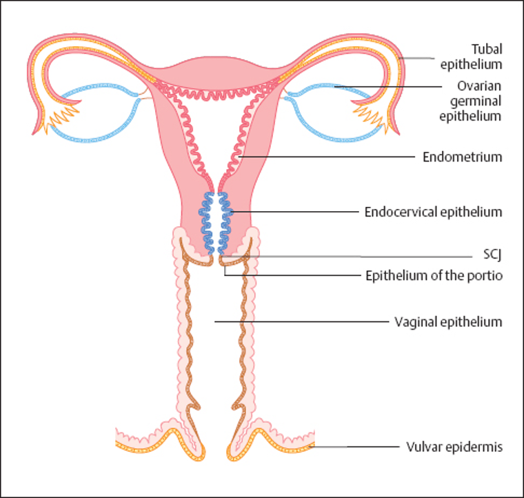
Vicarious menstruation is the bleeding from the mucous membrane in other parts of the female reproductive tract other than the endometrium. The mucous membrane is found on the outermost epithelial layer.
Retrograde Menstruation
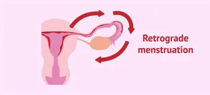
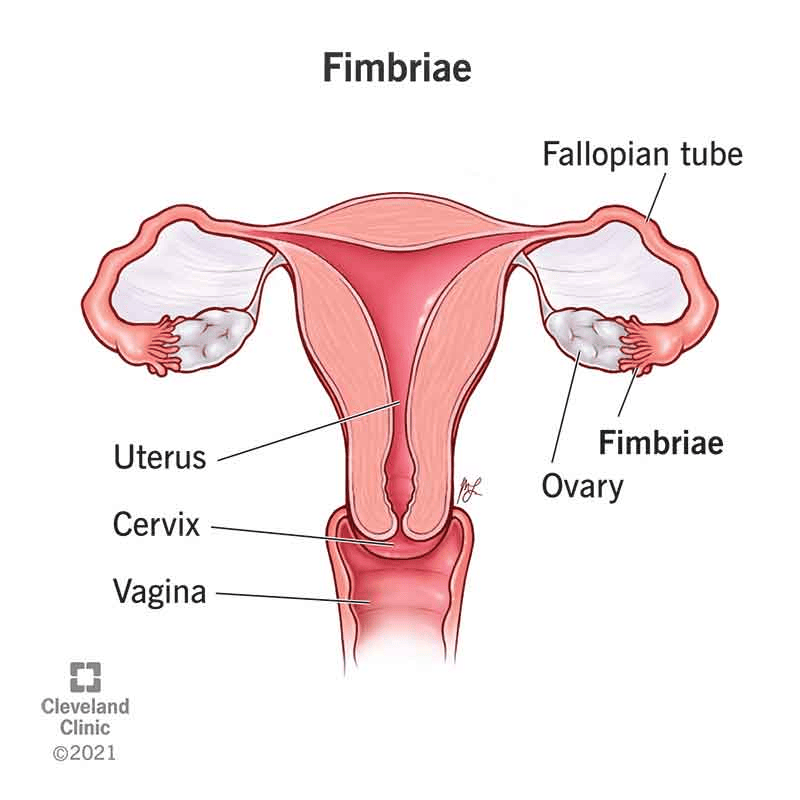
Retrograde menstruation is the backflow of blood and endometrial cells through the fallopian tubes, or oviducts. The fallopian tubes are what connect the ovaries to the womb.
Menarche
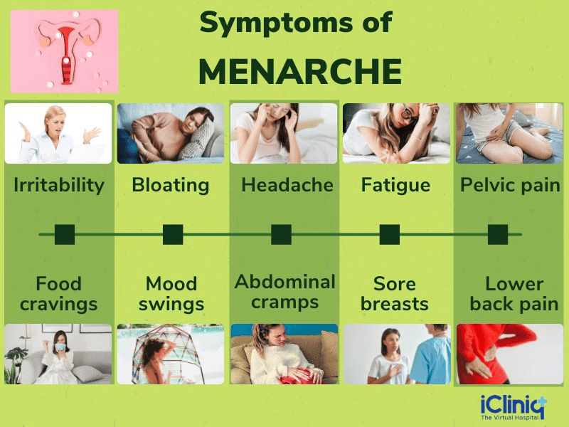
Menarche is the first menstruation that commonly occurs during puberty as part of growth and development. Other changes that occur during puberty are the growth of the chest (breasts), hair in the private parts (pubic hair), and the armpit (axillary).
Menopause
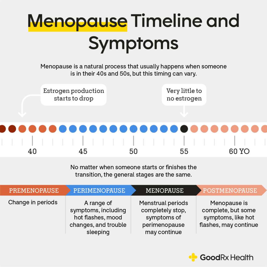
Menopause is the process in which the ovaries stop producing egg cells every four weeks, the menstrual cycle becomes less regular, and menstruation stops. This commonly occurs between 45 and 55 years. The cause of this is that the ovaries and eggs become less responsive to the gonadotropins (follicle-stimulating hormone and luteinizing hormone), especially with a marked decrease in plasma oestrogen (female hormone) concentration.
What symptoms are associated with menstruation but increase the risk of gynaecological cancers?
Oligomenorrhoea
Oligomenorrhoea is when menstruation is irregular.
Amenorrhoea
Amenorrhoea is the absence of menstruation.
Metrorrhagia
Metrorrhagia is the bleeding between menstrual cycles.
It may be heavy, long-term, temporary, or throughout life. It is commonly caused by:
- Stress
- Hormonal changes
- Contraceptive/birth control (starting or stopping), which can cause abnormal bleeding.
- Blood spotting after two weeks following the period suggests that the mature egg has been released (ovulation). Upon ovulation, the oocyte is captured by the fimbriae.
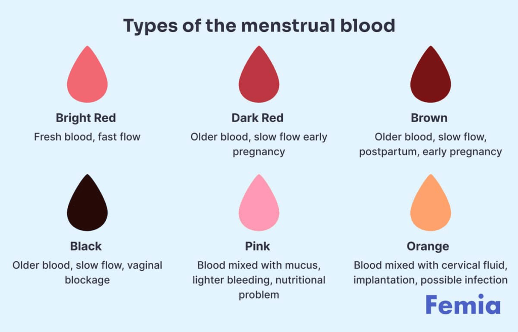
There may appear light bleeding or spotting at different moments of the menstrual cycle that vary from dark brown to light pink. There may also be mucus or clots. The cause of this is suggestive of the following:
- How heavy the blood is
- How often it appears – bleeding after going through menopause.
- Bleeding between periods
20% of women are asymptomatic, meaning they do not experience any symptoms.
What conditions are associated with menstruation but increase the risk of gynaecological cancers?
Endometriosis
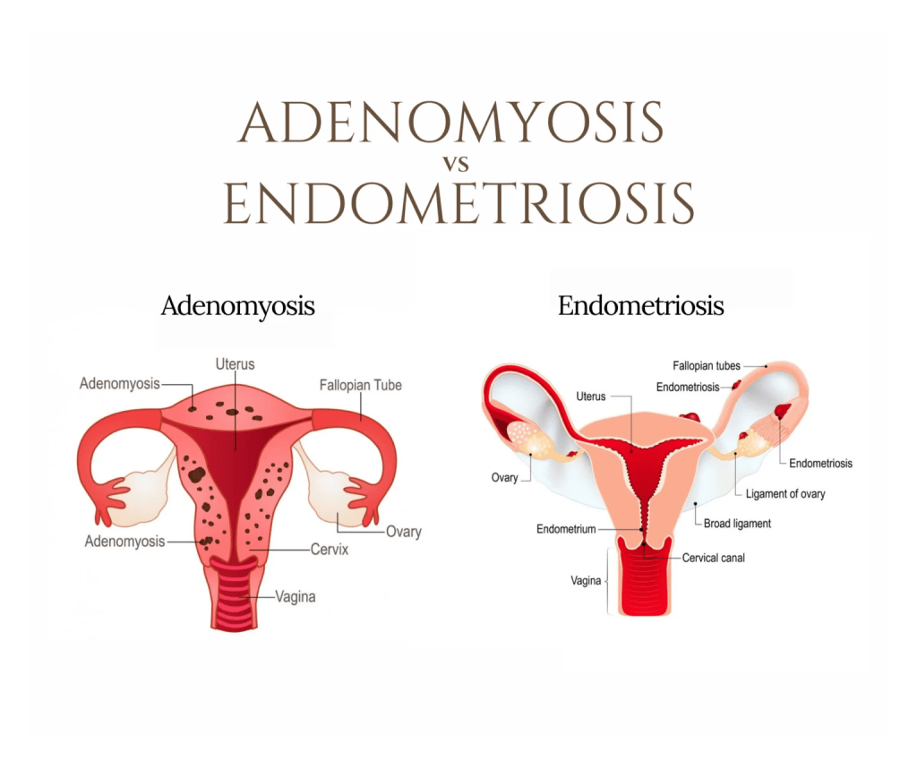
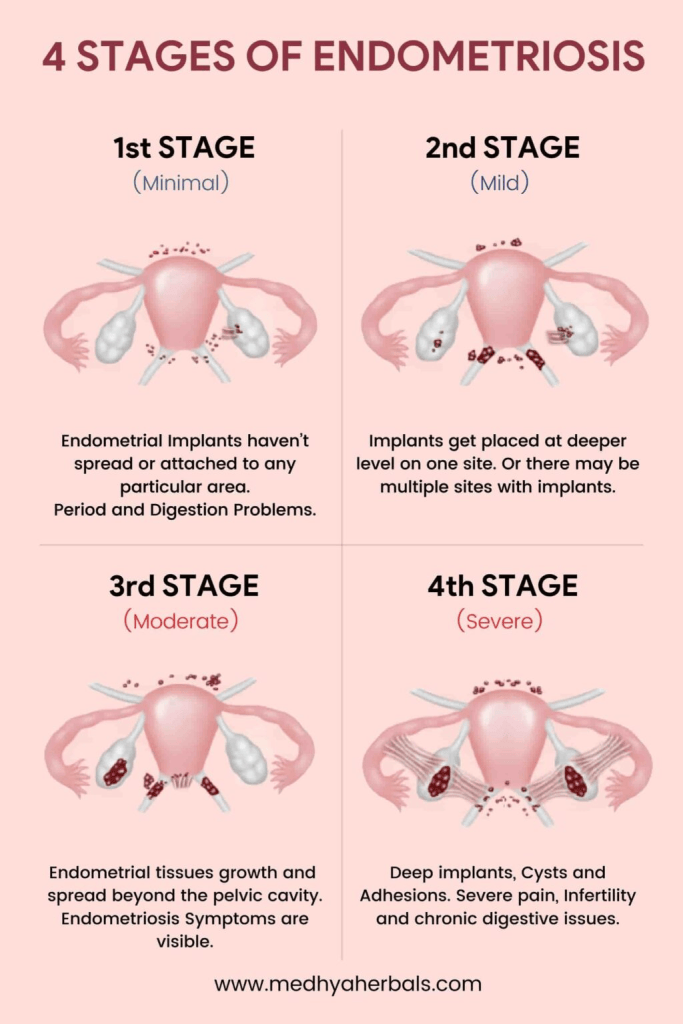
This is primarily caused by retrograde menstruation, where there is backflow into the fallopian tube. This may appear with pelvic pain, dyspareunia, and severe dysmenorrhoea. If the tissues have affected all of the uterine wall, then it is referred to as adenomyosis.
Dysmenorrhoea is when there is pain in the belly and cramps that can affect the lower back and thighs when menstruation occurs. There are two forms:
– Primary dysmenorrhoea, where there are painful periods after menarche (first period). This is associated with the overproduction of fatty acids (eicosanoids) called prostaglandin F2-alpha by the endometrium.
– Secondary dysmenorrhoea is when there are fibroids or endometriosis.
Dyspareunia: This is when there is painful sexual intercourse experienced by a woman, and it may be linked to the vagina (vaginismus), endometriosis, or inflammation in the pelvis (pelvic inflammatory disease).
Pelvic Inflammatory Disease (PID)
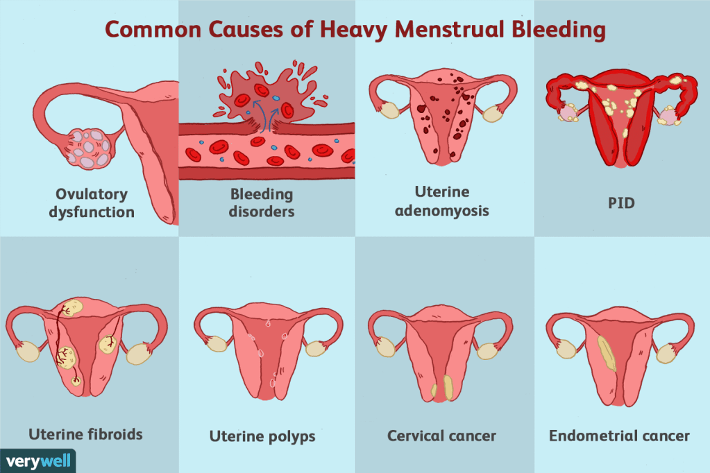
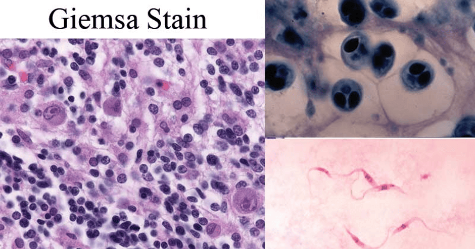
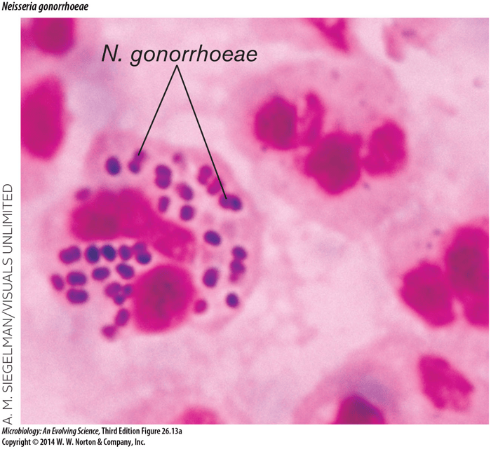
This is commonly associated with infection coming from the vagina. For instance, Chlamydia trachomatis and Neisseria gonorrhoeae. Both can cause either an acute (sudden) or long-term (chronic) infection that affects the uterus, fallopian tubes and ovaries.
It is commonly characterised by:
- Lower abdominal pain
- Abnormal vaginal bleeding
- Vaginal discharge.
- A blockage of the fallopian tubes can also lead to PID, ectopic pregnancy (pregnancy outside the womb), or possibly infertility. Infertility is when there are minimal ovulatory menstrual cycles in which the egg is released.
Polycystic Ovary Syndrome
This is also known as Stein-Leventhal syndrome.
This is commonly associated with the following:
Amenorrhoea (absent periods)
Oligomenorrhoea (infrequent periods)
High levels of androgens (male hormones)
Hirsutism (pigmented hair on face, chest, belly and upper back. It is caused by high levels of androgens.
Large-sized ovaries with fluid-filled cysts due to an imbalance between gonadotropin hormones: follicle-stimulating hormone (FSH) and Luteinizing hormone (LH).
It can also lead to infertility, overweight, diabetes mellitus (type 2), and heart disease.
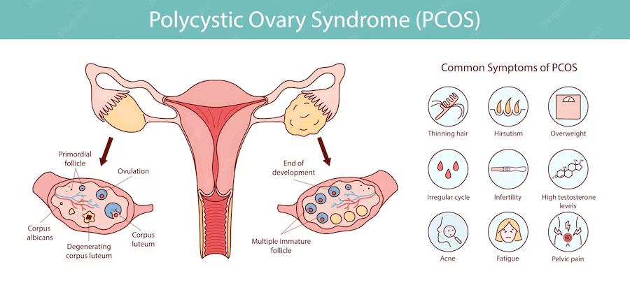
Collectively, these are all possible risk factors for gynaecological cancers.
Oogenesis
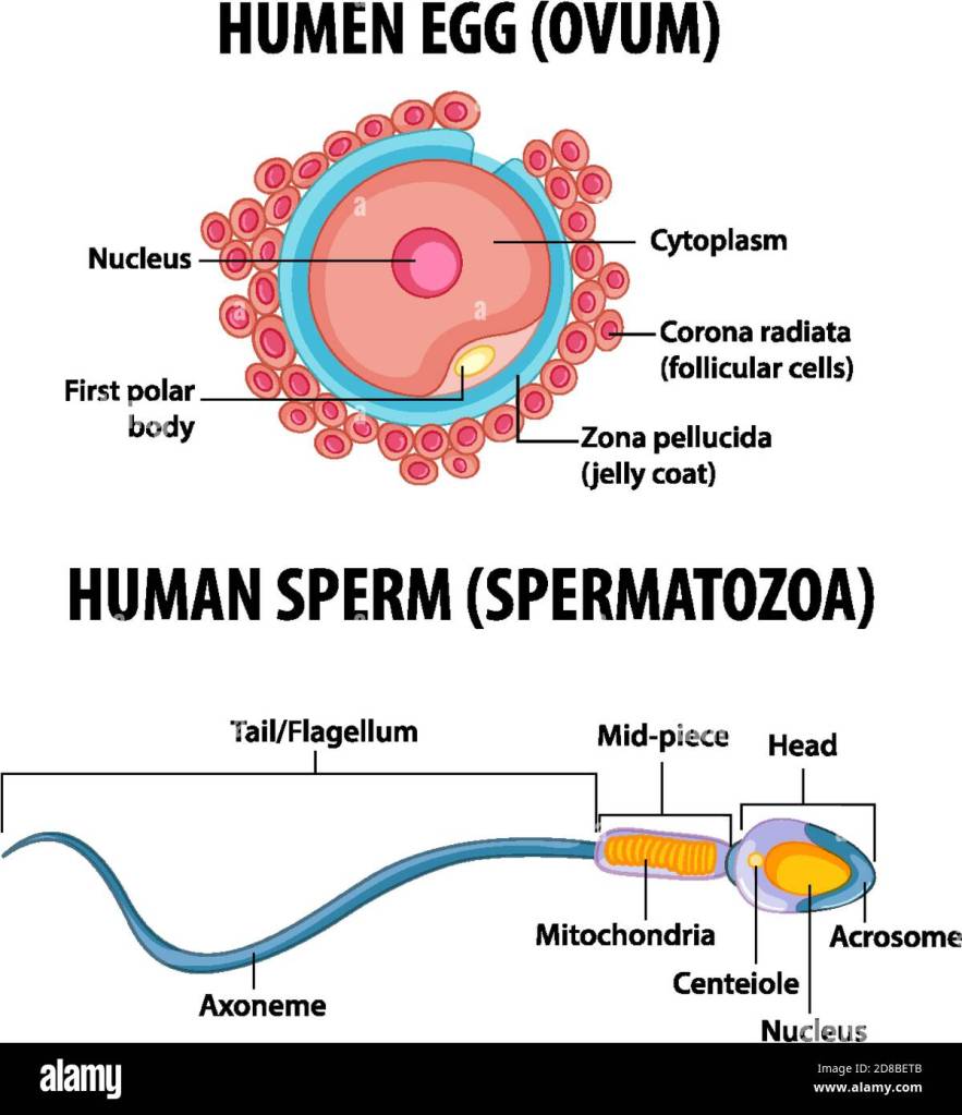
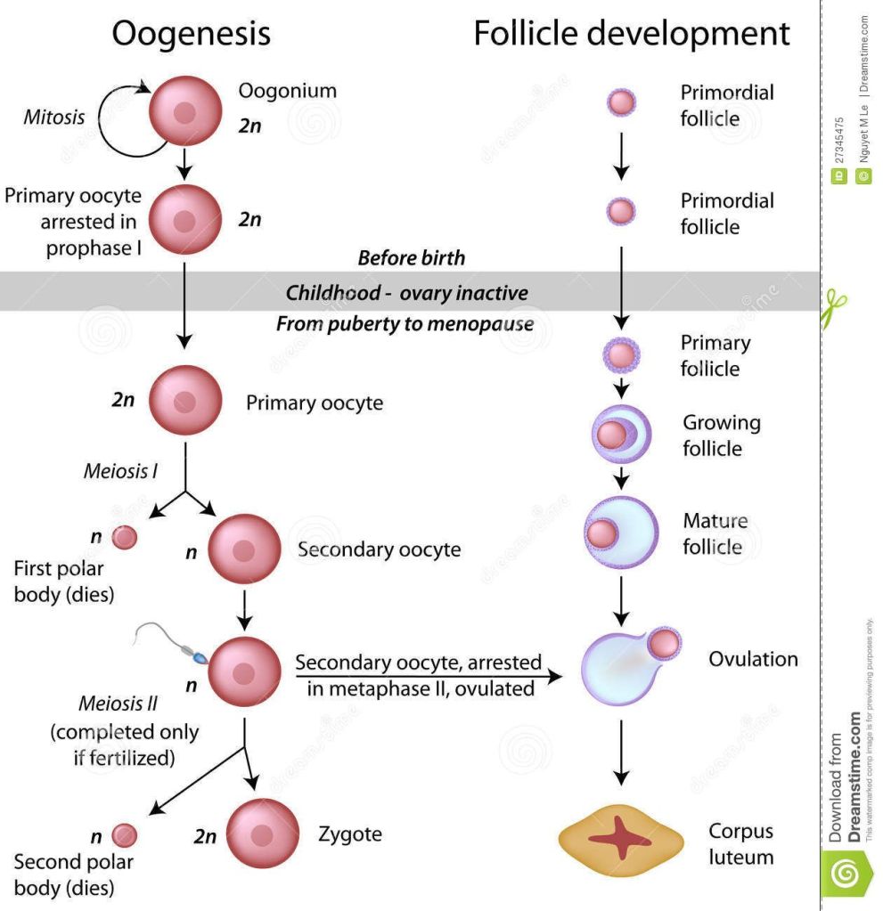
Oogenesis is defined as the production of the female gametes/sex cells called the egg or ovum (ova – plural)
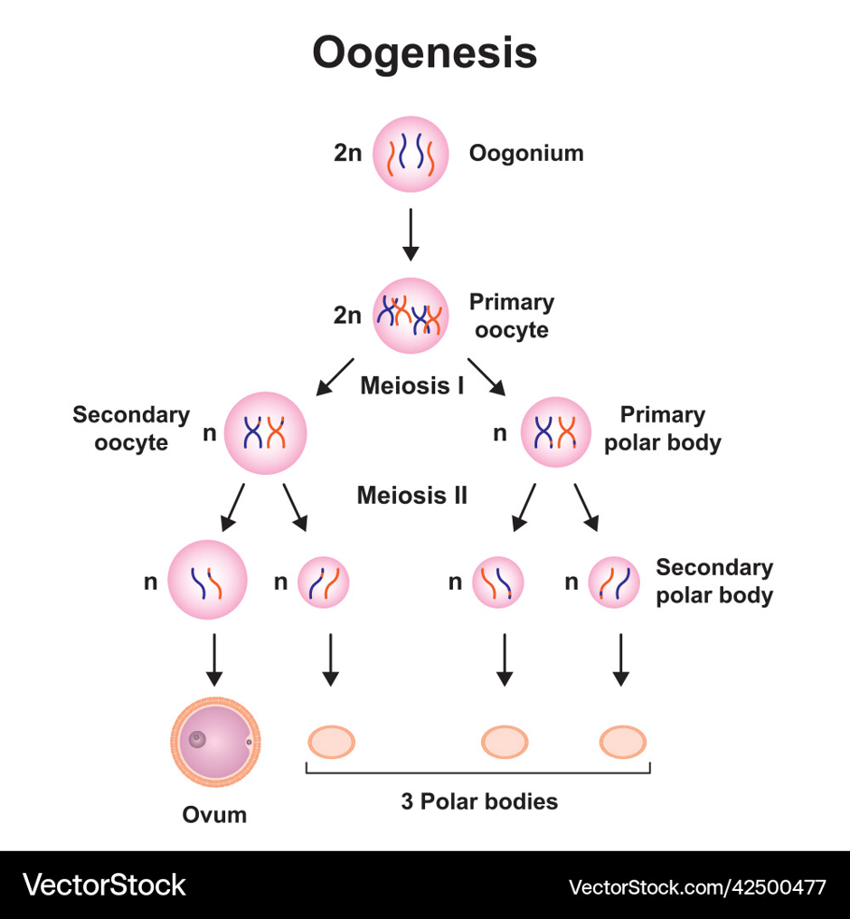
First Step Of Oogenesis: The Production Of Oogenia
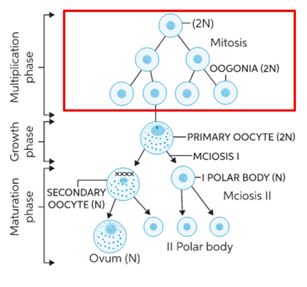
At first, the primitive germ cells/oogonia (single term: oogonium) undergo mitosis.
Germ cells, or gonocytes, are cells of the embryo that have the ability to develop into a gamete, in this case an ovum.
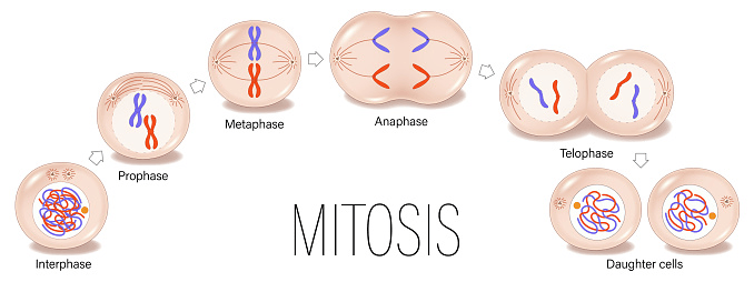
Mitosis is a type of cell division that facilitates growth and repair. It is a form of asexual reproduction. A single cell can produce two genetically identical cells called daughter cells. A normal cell has 23 pairs of chromosomes (2n; n = 23).
Chromosomes are threadlike structures that contain the genetic material.
Oogonia rapidly divide in the utero by about 7 million germ cells by the 7th month of gestation/pregnancy.
Most of the oogonia decrease while the remainder go on to make primary oocytes.
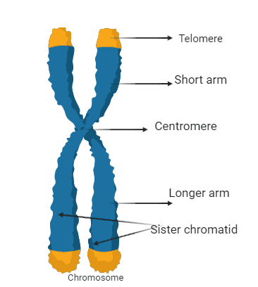
The structure of the chromosome. There are two sister chromatids that are joined together to form a chromosome. Each sister chromatid has a short arm (p) and a long arm (q). The tip end of each sister chromatid is a telomere. The centre of a chromosome is the centromere.
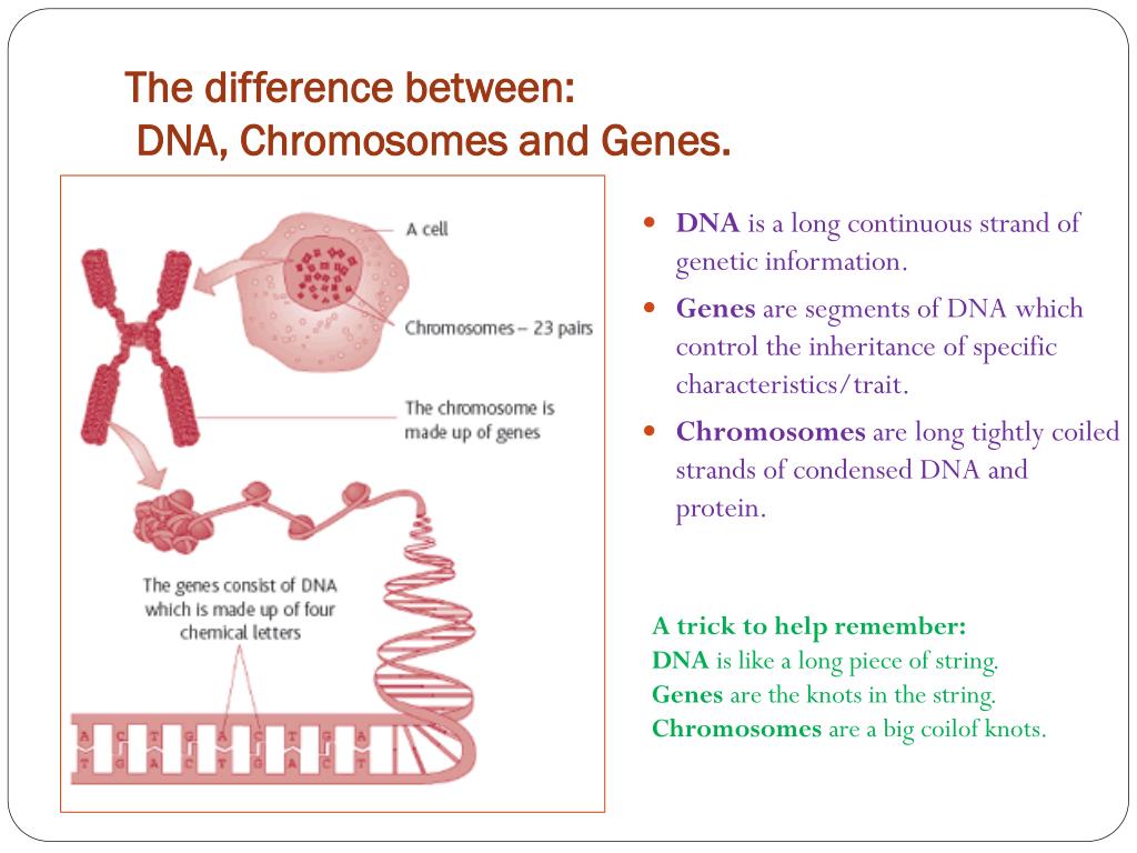
The nucleus is an organelle that controls the cell. It contains genetic information that is tightly coiled with DNA and protein (histones) to maintain stability. Genes are short sections of DNA that control a particular trait. Deoxyribonucleic acid is a long strand of genetic information.
The Second Step of Oogenesis: The production of primary oocytes.
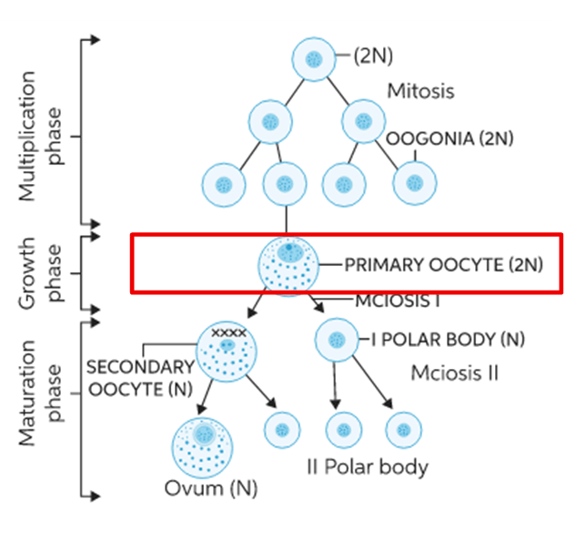
The oogonia develop into primary oocytes.
This occurs during the foetus (developing baby).
At birth, all the eggs remain as primary oocytes containing 46 chromosomes in each.
They then stop mitosis – this is referred to as meiotic arrest until puberty/menarche (first period).
This commonly occurs during prophase I.
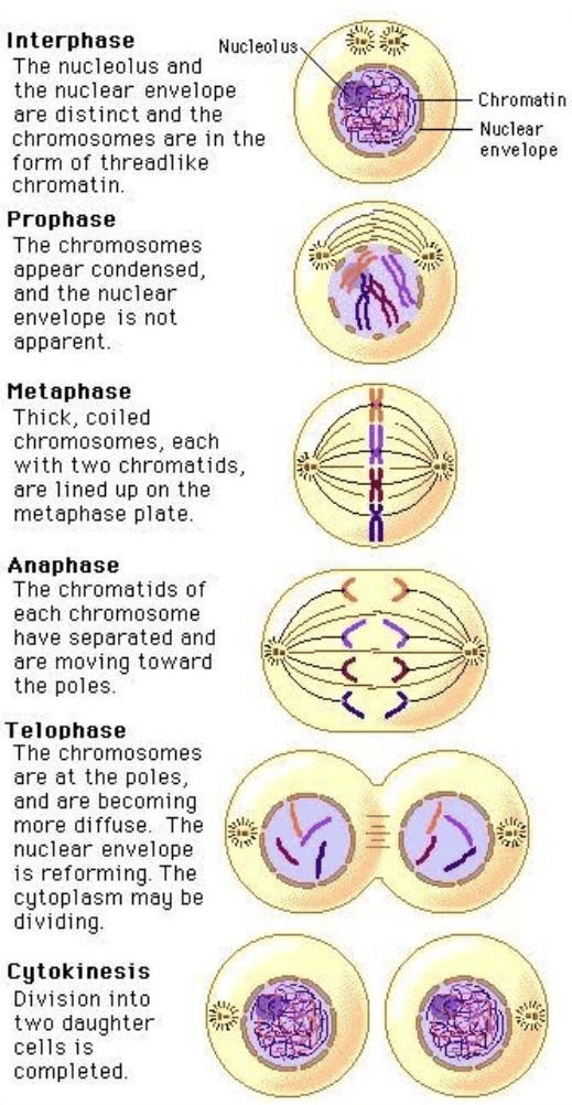
Mitosis has four main steps to divide the cell into two identical daughter cells, each with 23 pairs of chromosomes.
PMAT (Prophase, Metaphase, Anaphase, Telophase).
The Third Step Of Oogenesis: The Formation of the Secondary Oocyte.
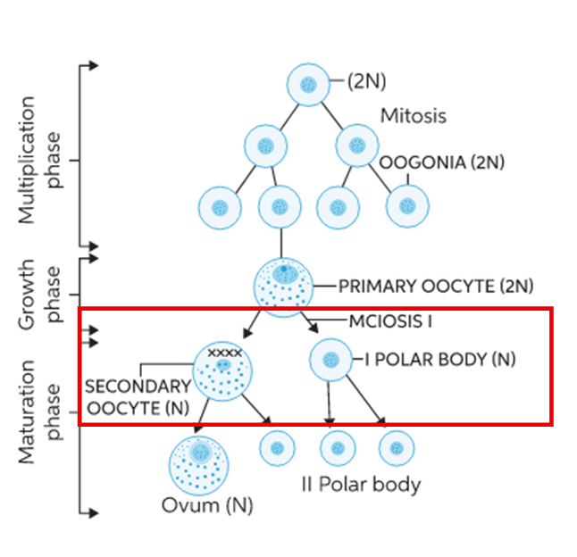
At puberty, the primary oocyte that is ready to be released from the ovary (ovulation) complete their first meiosis before it takes place to form the secondary oocyte.
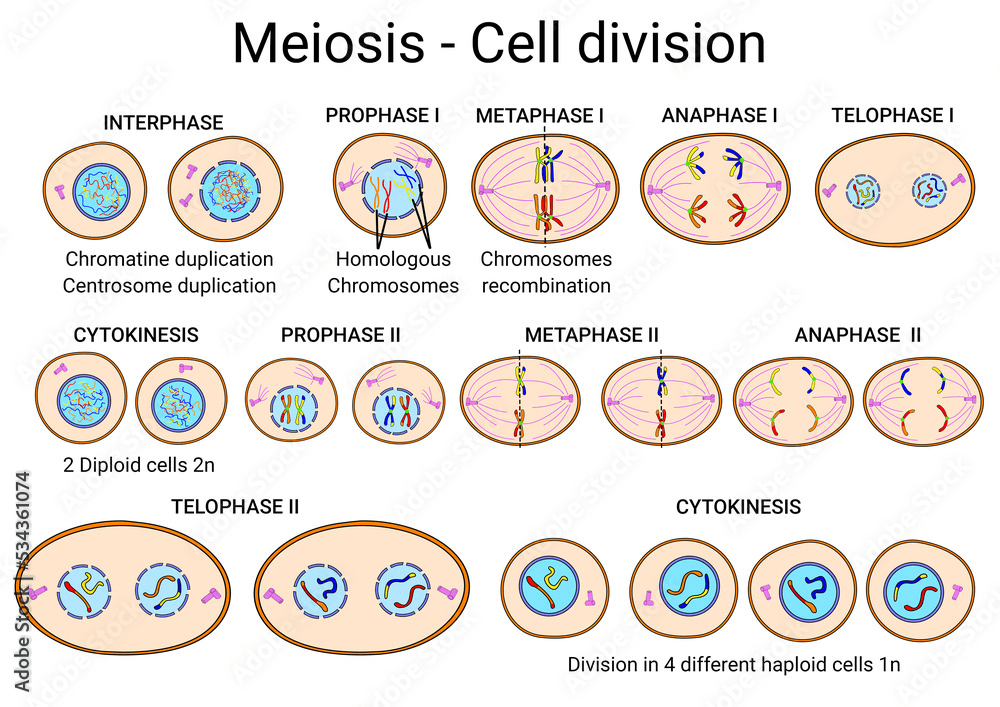
Meiosis is a type of cell division where the original cell divides into half the number, so each daughter cell has 23 chromosomes. It has two sets of cell division. It goes through the PMAT (Prophase, Metaphase, Anaphase, Telophase) twice.
However, during this stage, one daughter cell has cytoplasm where all the chemical reactions take place and contains 23 chromosomes.
The other daughter cell becomes a first polar body – small and non-functional.
The Fourth Step Of Oogenesis: The Second Meiosis
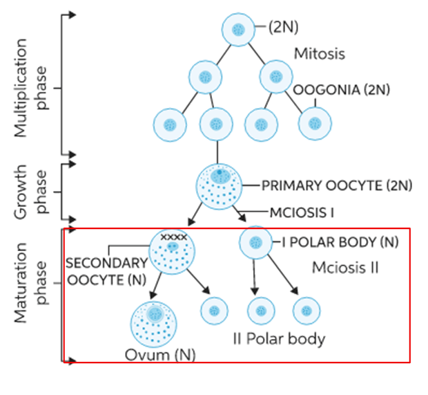
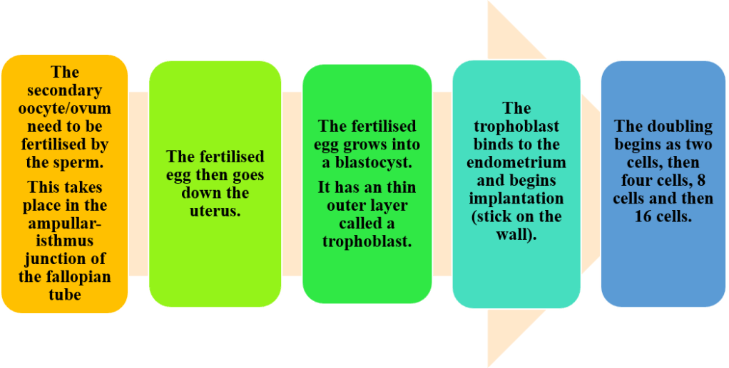
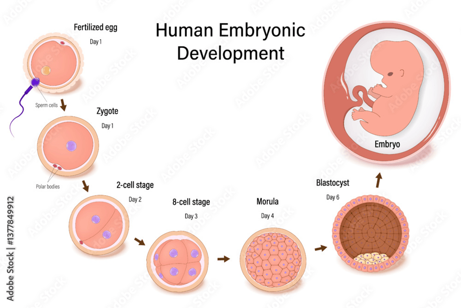
Once the egg has been released from the ovary, it enters the fallopian tube; however, the secondary oocyte can be fertilised/joined by the sperm (male sex cell)
This joining between the secondary oocyte and sperm leads to the formation of a daughter cell called an ovum containing 23 chromosomes and a second polar body.
The second polar body is small and non-functional.
The Menstrual Cycle
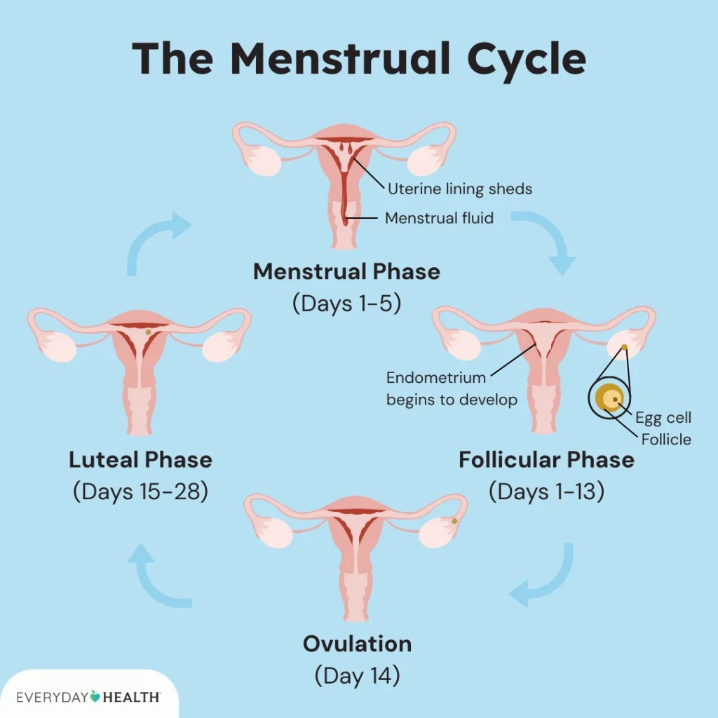
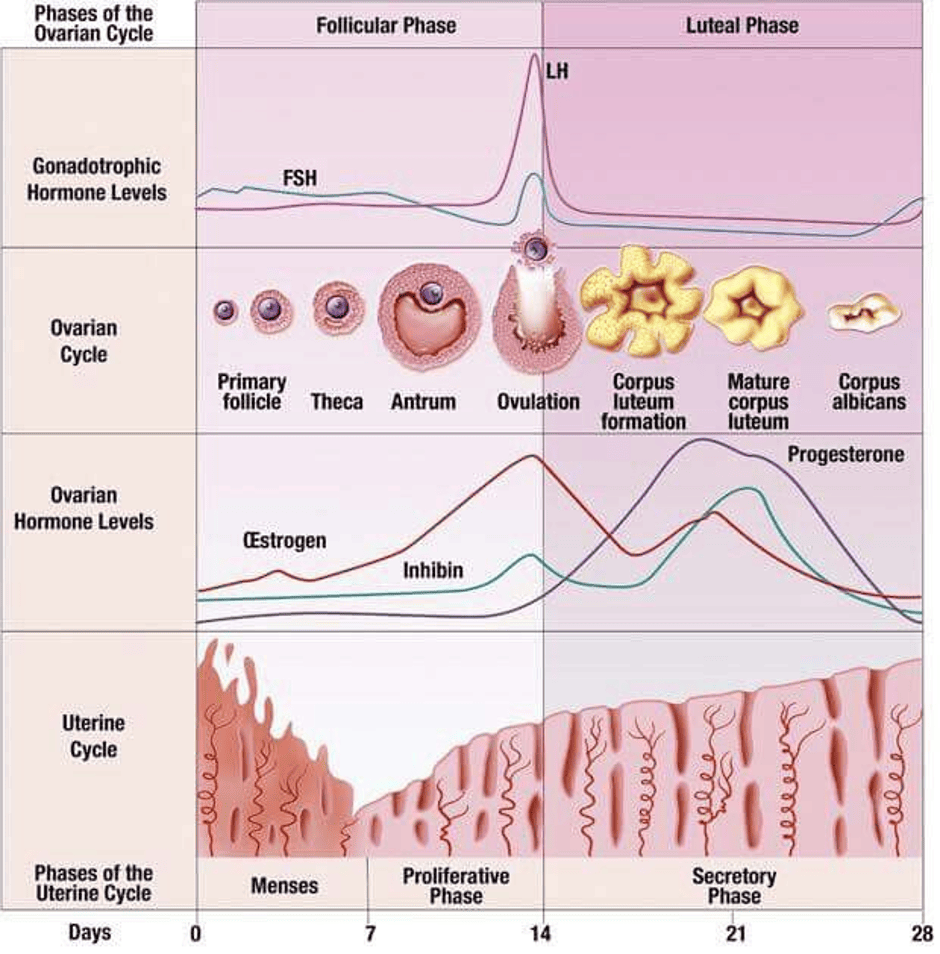
The menstrual cycle is divided into the follicular phase, ovulation, and luteal phase.
The follicular phase is the growth and maturation of the egg cell.
Ovulation is the release of the mature egg from the ovary. This commonly takes place on Day 13 or 14 of the menstrual cycle.
The Luteal phase lasts for 14 days; however, the follicular phase is more variable.
The Follicular Phase
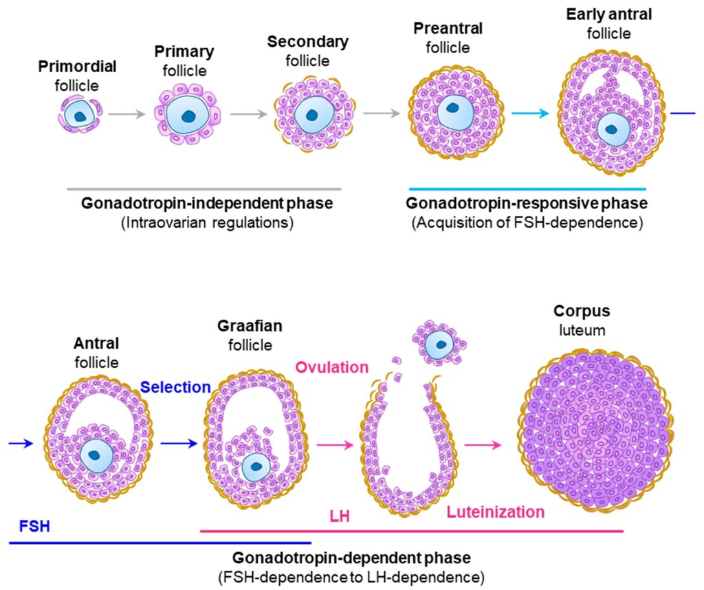
Folliculogenesis – The female egg exists in a structure called a follicle.
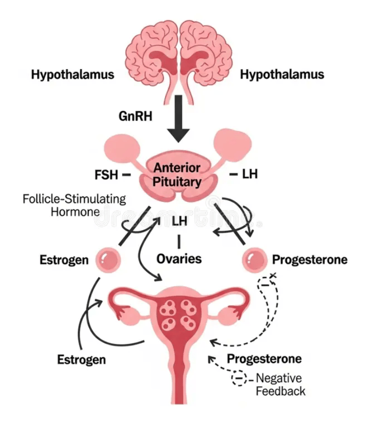
Key hormones involved are follicle-stimulating hormone (FSH) and Luteinizing hormone (LH). The anterior pituitary gland produces them. They are stimulated by GnRH (gonadotropin-releasing hormone) that is released via the hypothalamo-pituitary portal vessels. Gonadotropin-releasing hormone receptors are found on the anterior pituitary gland.
Their existence begins during the formation of the primary oocyte.
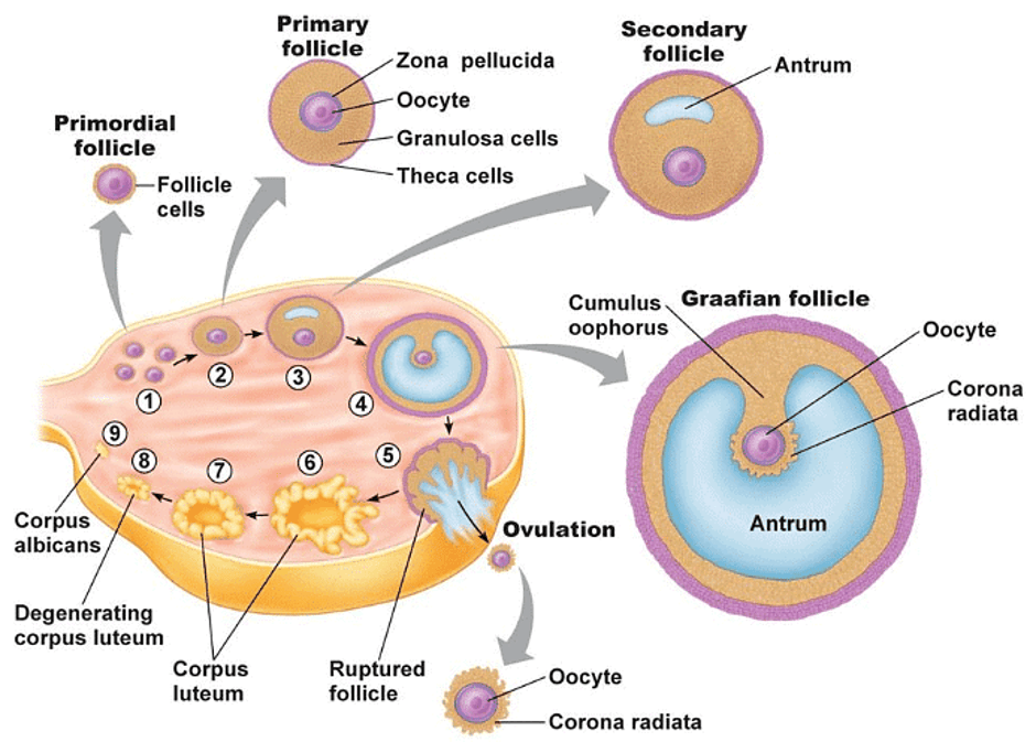
Step One Of Folliculogenesis: Primordial Follicles
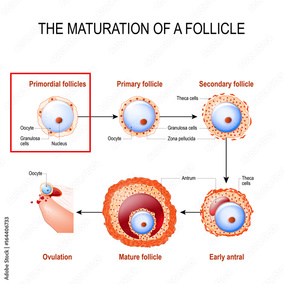
They are a single layer of cells called granulosa cells around the Diplotene oocyte.
The follicle-stimulating hormone (FSH) helps with the growth of the follicle.
Step Two Of Folliculogenesis: Primary Follicles
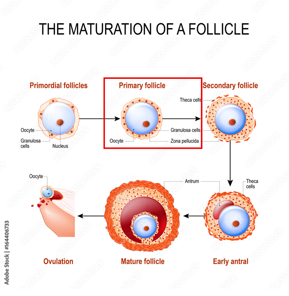
The oocyte grows and develops additional concentric layers of granulosa cells.
The granulosa cells secrete several hormones:
- Small amount of progesterone
- Inhibin
- Oestrogen.
The role of inhibin is to prevent the production of Follicle Stimulating Hormone (FSH).
The zona pellucida then develops and separates the oocyte from the inner granulosa cells.
Step Three of Folliculogenesis: Preantral follicle
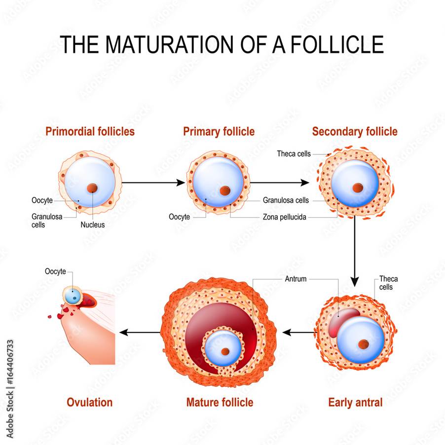
Continuous growth occurs via mitosis of granulosa cells that differentiate to form theca cells.
Theca cells – It helps to make oestrogen in granulosa cells.
The steroidogenic cells are the theca and granulosa cells.
One of the types of inhibin, Inhibin B, is the major circulating form produced in the antral granulosa cells. It increases FSH-induced oestrogen production in granulosa cells and luteinizing hormone (LH)-induced androgen production in theca cells.
Step Four Of Folliculogenesis: Early Antral Follicle
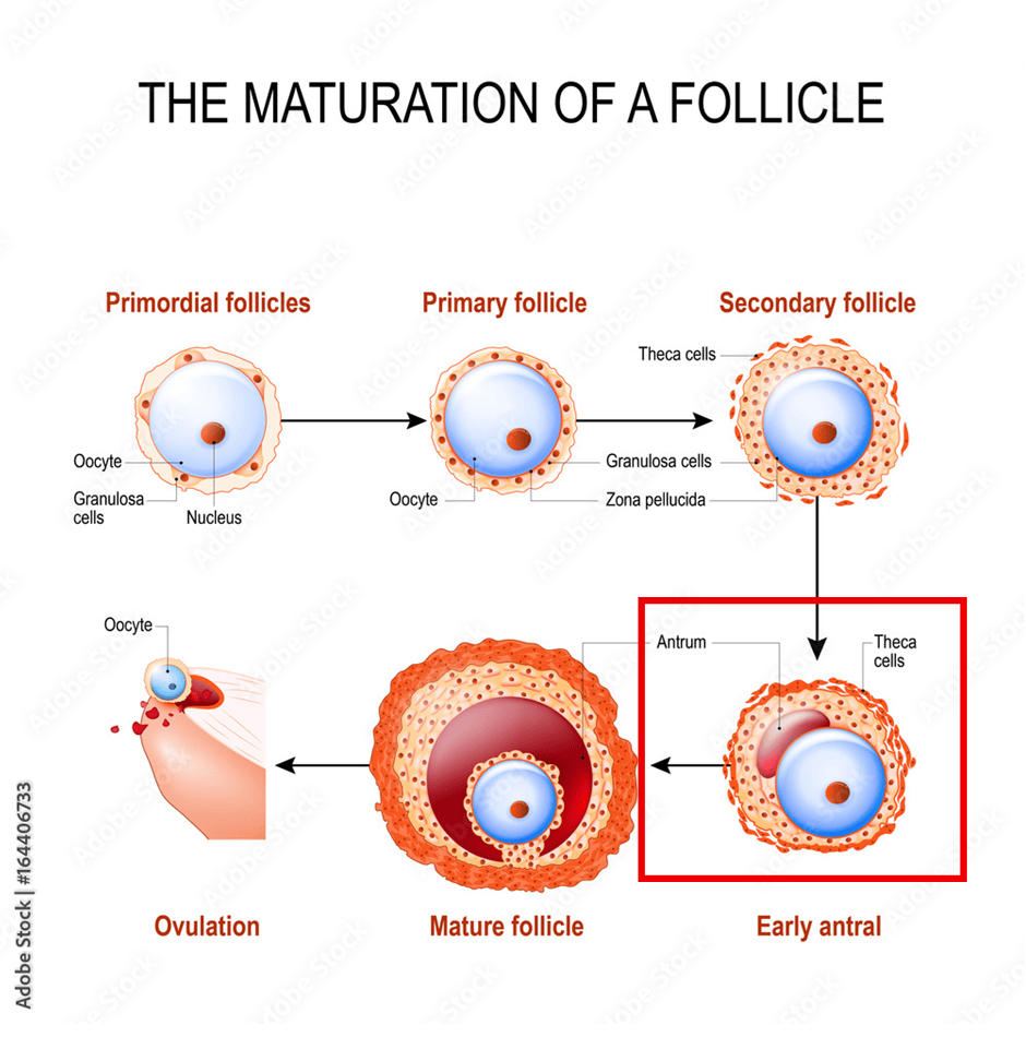
The primary oocyte reaches full size, and a fluid-filled space called the antrum begins to form.
The growth of preantral and early antral occur throughout infancy and childhood and during the menstrual cycle.
At the start of the menstrual cycle: 10 to 25 preantral and early antral follicles develop into larger antral follicles.
After a week (7 days), one of the antral follicles becomes a dominant follicle and is associated with the amount of oestrogen being produced.
Non-dominant/inactive follicles degenerate and undergo a cell death process of atresia.
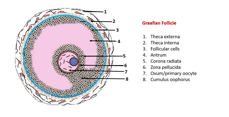
Step Five Of Folliculogenesis: The Graafian Follicle
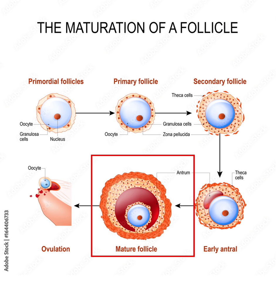
The dominant follicle increases in size due to fluid.
The size increases to about 20 to 25 mm.
The antrum size increases.
The theca and granulosa cells increase in size and project onto the antrum to form a cumulus oophorus.
Theca cells are luteinized and produce sex hormones.
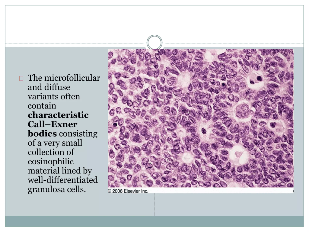
It contains Call-Exner bodies (rosette-like formation with central filament/tubes and lots of eosinophils.
Basal lamina: A thin membrane or layer of tissue.
There is no reticulum – network of tubules or blood vessels.
The different types of cells produce an environment that is supported by hormones for folliculogenesis and early stages of pregnancy.
Before ovulation (release of mature follicle from the ovary), the primary oocyte comes from meiotic arrest and completes first meiosis to form a secondary oocyte.
During this process, the cumulus separates from the follicle so the oocyte can float in the antrum.
The mature follicle (Graafian follicle) is then formed.
Ovulation
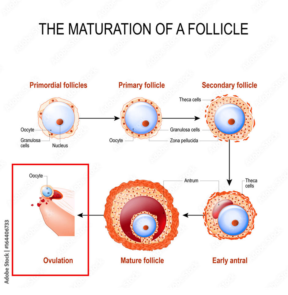
After approximately 36 hours following the LH surge, the release of the mature Graafian follicle from the ovarian surface epithelium (OSE) takes place.
The oocyte is captured by the fimbria.
It travels into the uterus via the fallopian tube.
The thin walls of the follicle and ovary rupture because of the enzymes that break through.
The secondary oocyte is surrounded by the zona pellucida, granulosa cells, and the cumulus oophorous and the antrum is released around day 13 or 14.
Multiple births may arise when two or more mature follicles are released, and more than one egg is released.
Luteal Phase
The Graafian mature follicle releases the antral fluid and secondary oocyte/egg.
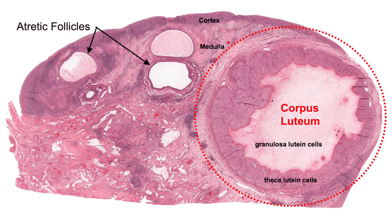
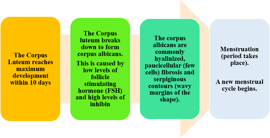
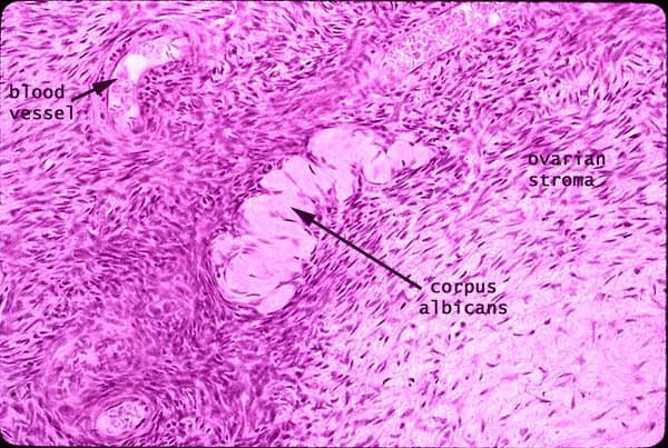
A glandular tissue called the corpus luteum is made by the granulosa cells.
The corpus luteum releases the following hormones: oestrogen, progesterone, and inhibin.
High levels of progesterone are produced compared to pre-ovulation (before ovulation)
About the corpus luteum:
- Size: It is commonly 2cm
- Shape:
- It varies under the microscope:
- It is round to serpiginous, larger, bright yellow, lobules with cystic centre.
Structure
It has luteinized granulosa and theca cells.
The corpus Luteum has additional structures:
- A central cavity.
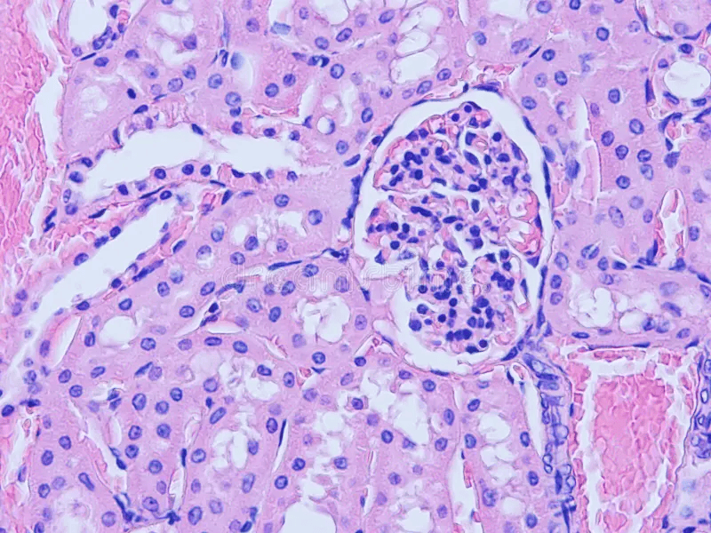
- Hyaline droplets: Hyaline droplets are a type of particle or inclusion body in the cytoplasm containing eosinophils. They are commonly found in the lining of the kidney tubules. They illustrate proteins accumulated alpha 2u-globulin or haemoglobin.
- Calcification (addition of calcium ions).
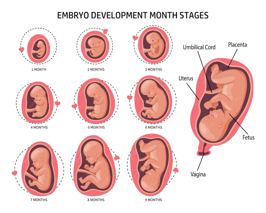
If Fertilisation Takes Place
If Fertilisation Does Not Take Place
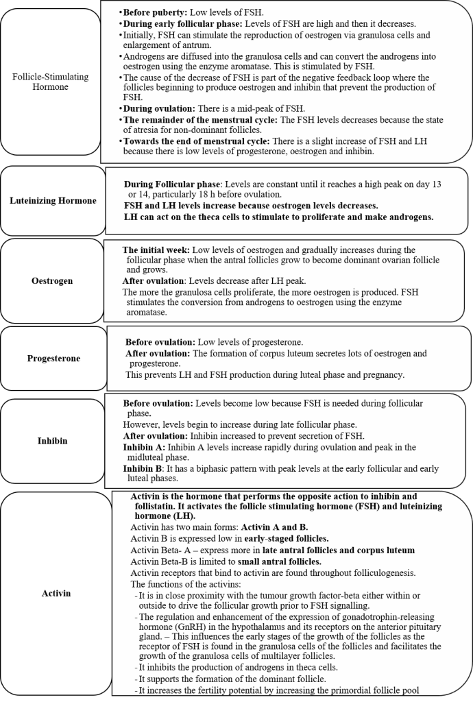
Overview Of The Hormones Involved In The Menstrual Cycle.
Uterine Changes During The Menstrual Cycle.
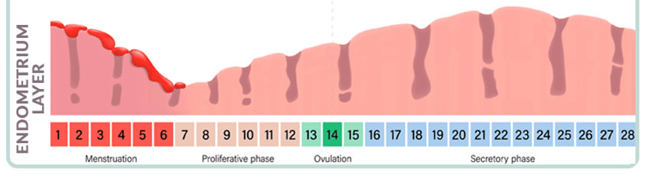
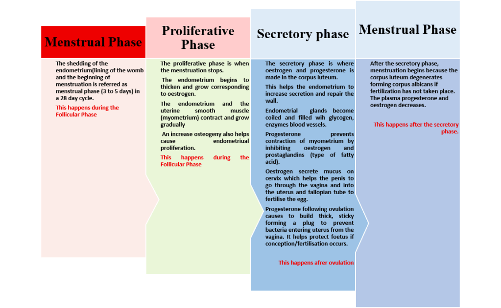
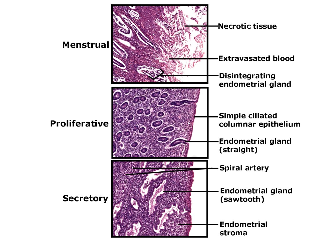
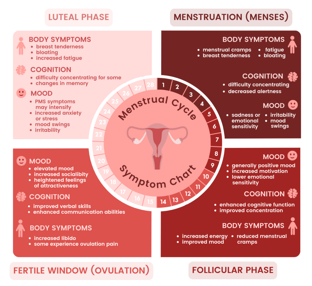
An Overview Linking Oogenesis, Folliculogenesis, Uterine Changes, And The Menstrual Cycle
References
Atwi, D., Ghofrani, M., and Hassell, L. (2023) General Anatomy & histology. Available at: https://www.pathologyoutlines.com/topic/ovarynontumornormalhistology.html (Accessed: 6th September 2026)
Chumduri, C. and Turco, M.Y. (2021). Organoids of the female reproductive tract. Journal of Molecular Medicine, 99(4), pp.531–553. doi:10.1007/s00109-020-02028-0.
Naidu, K., and Fredlund, K. (2023) Gestational Age Assessment. Available at: https://www.ncbi.nlm.nih.gov/books/NBK526000/ (Accessed: 9th September 2026)
Namwanje, M. and Brown, C.W. (2016). Activins and inhibins: Roles in development, physiology, and disease. Cold Spring Harbor Perspectives in Biology, 8(7), p.a021881. doi:10.1101/cshperspect.a021881.
Norman, A. (2026) An Overview of Metrorrhagia. Available at: https://www.verywellhealth.com/an-overview-of-metrorrhagia-4584380 (Accessed: 17th August 2026)
Rosner, J., Samardzic, T. and Sarao, M. (2024) Physiology, Female reproduction. Available at: https://www.ncbi.nlm.nih.gov/books/NBK537132/ (Accessed: 14th August 2026)
Seladi-Schulman, J. (2026) Everything to Know About Female Reproductive Organs. Available at: https://www.healthline.com/health/womens-health/female-reproductive-organs (Accessed: 7th August 2026)
The Calgary guide to understanding disease (2026). Polycystic ovary syndrome. Available at: https://calgaryguide.ucalgary.ca/?s=diabetes%20mellitus (Accessed: 10th August 2026)
Widmaier, E., Raff, H., Strang, K. (2006) Van der’s Human Physiology. New York: McGraw-Hill


Leave a comment