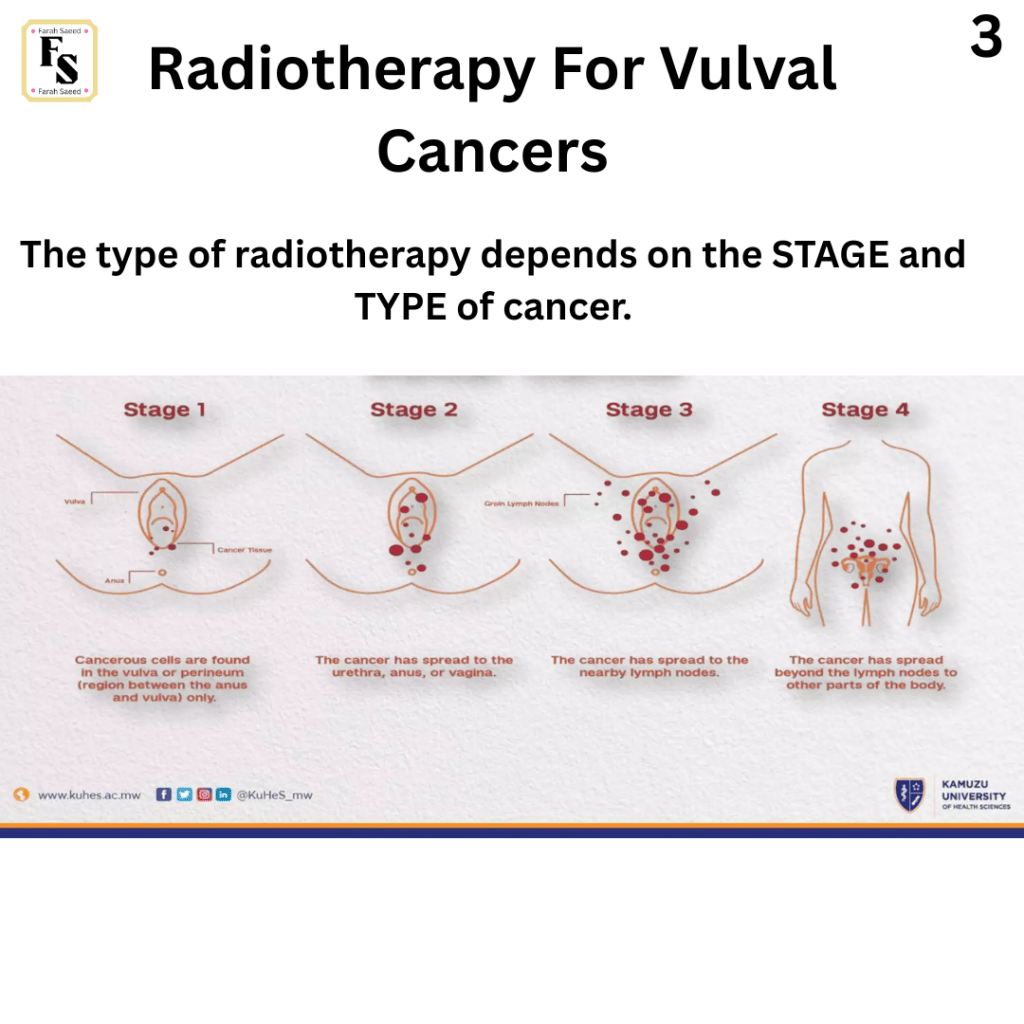
Below summarizes the main points for all treatments in accordance to British Gynaecological Cancer Society (BGCS) vulval cancer guidelines: An update on recommendations for practice 2023. Please read the guidelines attached for further details.
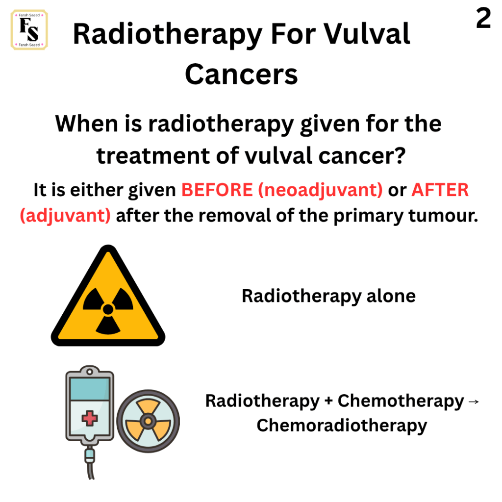
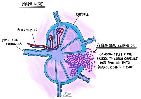
This is commonly performed after surgery to increase survival and lower recurrence if there are cancers that are > 5 mm or it has increased its size to affecting the extracapsular lymph nodes. For instance:
Ipsilateral groin nodes
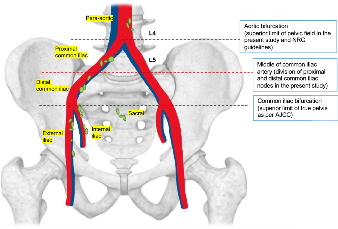
Distal part of the iliac nodes.
Bifurcation of the common iliac artery (bifurcation is the point at which division into two branches occurs)
It is also useful in cases where there is negative pelvic nodes.
It is also recommended when further resection margins is not possible because of higher grade and stage after finding positive surgical lymph nodes or at risk of damaging structures such as the anus, urethra and clitoris or unable to remove groin nodes due to risk of low morbidity.
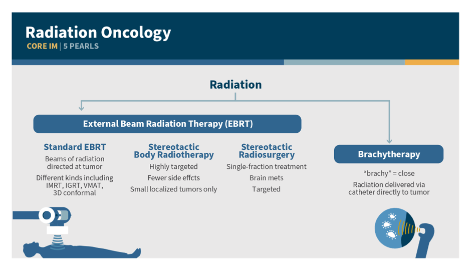
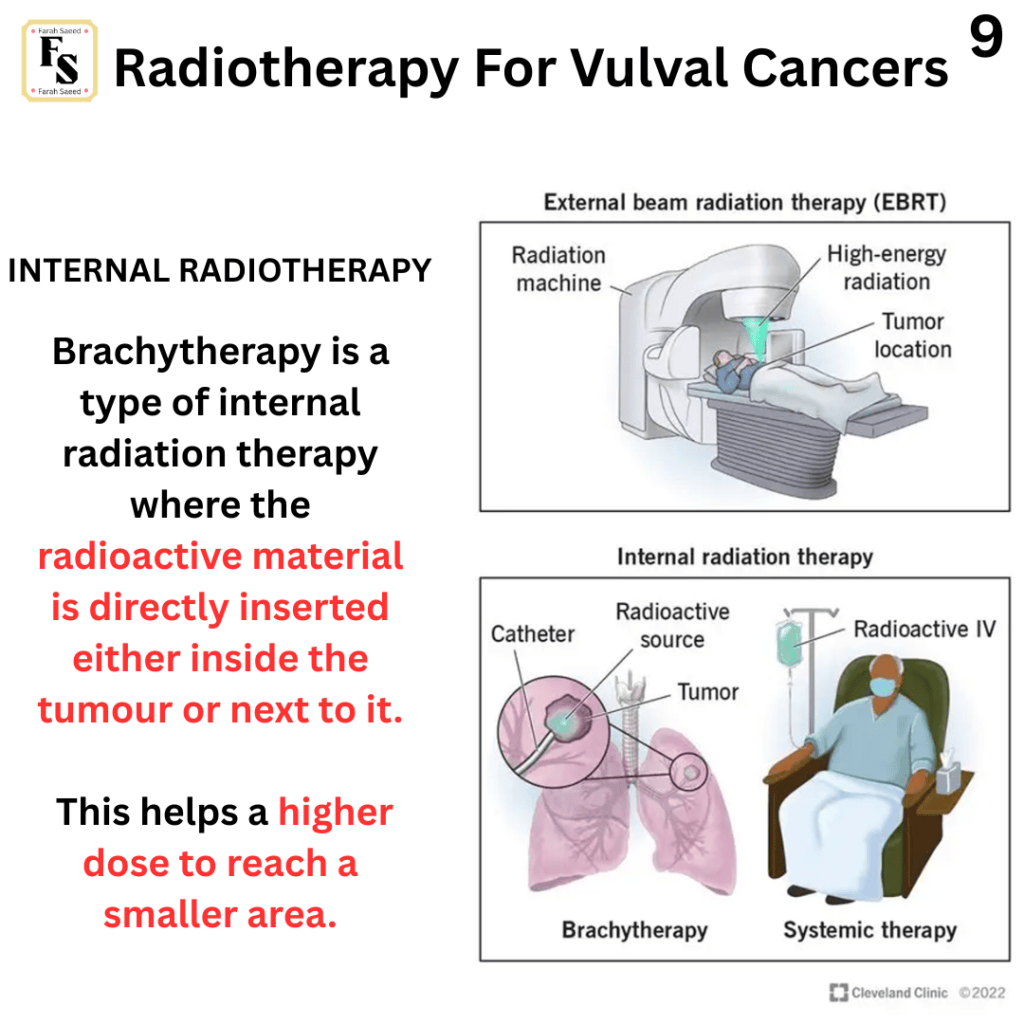
A common dose that is advised is 60 to 64 Gray this is equivalent dose in 2 Gray fractions (EQD2) with external beam radiotherapy. MRI-image guided brachytherapy may be considered as a boost for selected patients.
Assessment of response should be performed at 12 weeks following completion of treatment with clinical assessment and imaging.
Biopsy should be performed if residual disease is suspected.
A closer look at the blood supply in the pelvis. Here we can see two main groups of blood vessels: arteries (red) and vein (blue)’. The iliac blood vessels supply the lower limbs and pelvic region. The abdominal aorta divides into right and left common iliac arteries via its the terminal branches. Each branch is subdivided into the external iliac artery and the smaller internal iliac artery. On the other hand, the iliac veins drain most of the blood and is subdivided into right and left common iliac veins that unite to form the inferior vena cava. The unification of internal and external iliac veins are shown.
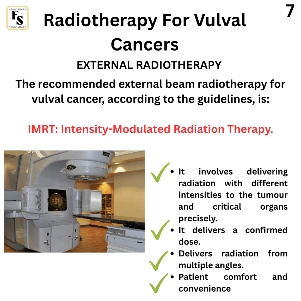
Importance Of External Beam Radiotherapy: IRMT
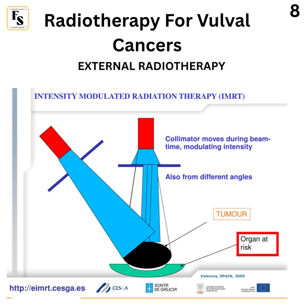
Intensity Modulated Radiation Therapy (IMRT) involves small radiation beams with different intensities delivered to the tumour precisely and to critical organs. It creates a confirmed dose.
The choice depends on the patient’s condition and the availability of the technique.
IMRT is MORE PRECISE than 3DCRT because it covers the planning target volume (PTV) using a BETTER DOSE. This minimizes damage to normal tissue,side effects, and secondary malignancies.
There is a boost with involved nodes that should improve outcomes.
IMRT should commonly occur within 8 weeks of surgery and completed within 105 days.
Understanding Radiotherapy On A Cellular Level
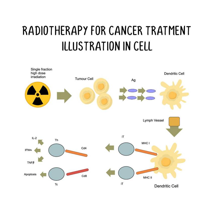
This image provides an example of how radiotherapy works and interacts with both the lymphatic system and the immune system. The lymphatic system consists of lymphatic vessels that carry lymph, which contains ions, water, and protein, bathes the tissues, and originates from blood. Lymph passes through lymph nodes, where it is filtered, and lymphocytes are produced to remove foreign particles. It drains into the small blood vessels called capillaries and passes into lymphatic vessels, which have valves to prevent backflow and lead to two large channels, the thoracic duct and right lymphatic duct, that return the lymph back to the blood circulation via the innominate veins.
A single dose of high-fraction radiation is aimed at the tumour cells. The antigens (Ag) are proteins found on the surface. The dendritic cells are an example of antigen-presenting cells that reside in the tissue. It helps to elicit the anti-tumour response of T-cells. The dendritic cells acquire the antigens by endocytosis and process the antigens via cleavage into smaller proteins called peptides by enzymes called proteases. The processed antigens are then translocated to the cell surface to be presented in association with other proteins from the major histocompatibility complex (MHC). This presentation takes place after they migrate to the lymphoid organs (spleen, thymus, bone marrow) to present them to the T cells.
There are two main types of MHC: MHC I and MHC II. There are also two types of T cells that are responsible for cell-mediated immunity: T-helper cells and cytotoxic T cells. MHC I targets CD8. CD8 is a surface antigen found on cytotoxic T cells. They destroy cancer cells and virus-infected cells. They recognise the peptide antigens attached to proteins and induce cell death. MHC II targets CD4. CD4 is a surface antigen found on T-helper cells and helps resist viruses. This is achieved by releasing protein molecules called cytokines.
The cytokines are released by cells when activated by antigens and help to boost the immune response when interacting with cell surface receptors. The interleukins are produced by leukocytes (IL-2), tumour necrosis factor-beta, and interferons (IFN-alpha). IL-2 stimulates the T-cells to become natural killer cells. The interferons inhibit viral growth and contain several forms: alpha from white blood cells, beta from fibroblasts, and gamma from lymphocytes. Interferon alpha is mainly used in the treatment of cancer.
CLINICAL TRIAL!
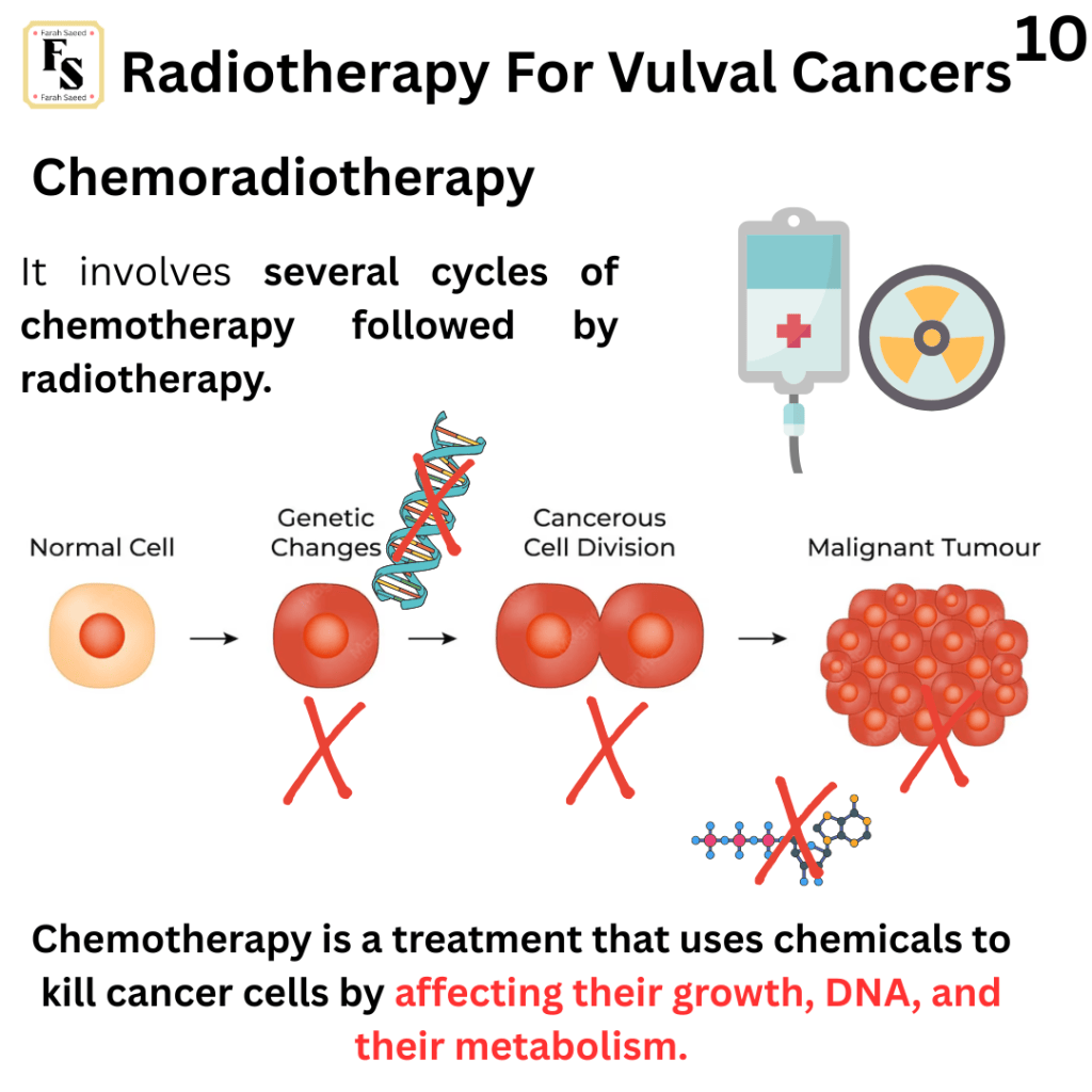
A case series using IMRT to escalate vulval dose to a median of 66 Gy and involved nodes to 60.6 Gy and concurrent weekly cisplatin chemotherapy had a complete clinical response rate of 88 %, compared to 63 % with a median vulval dose of 59.4 Gy. However, this was pre-operatively.
However, every treatment has a downside.
Radiotherapy may slow the ability to heal as it lowers cell division – reconstructive surgery techniques can be used to reduce tension on previously irradiated skin, or to introduce non- irradiated tissue into the wound bed.
Several side effects may appear after treatment and need to be seen in clinic:
Urinary symptoms: stricture (narrowing or constriction of a tubular structure), inflammation, contraction, detrusor muscle (muscle of the bladder) overactivity and urinary incontinence.
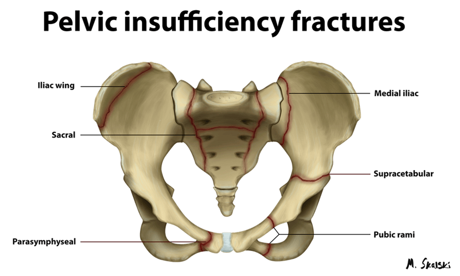
Endocrine: pelvic insufficiency fractures – weakens bones due to external beam. Other symptoms include pain, pain on weight bearing, immobility. MRI can be used to diagnose Pelvic insufficiency fractures and distinguish between bony metastases. Conservative management involves rest, pain management, and physiotherapy-led exercise for stable fractures.
The Downside Of Radiotherapy For Vulva
What Is Pelvic Insufficiency Fractures?
Pelvic insufficiency fractures can be pre-screened using the following:
Bone density measurement
Fracture risk assessment
Age above 65 years
Low Body Mass Index 20 kg/m
History of fragility
Smoking
Oral corticosteroids (anti-inflammatory drugs) are derived from steroid hormones that are subdivided into glucocorticoids and mineralocorticoids. The former is associated with metabolism of carbohydrate, fat, and protein and with normal response to stress. Mineralocorticoids help to regulate the salt, water, and ion balance.
These are commonly associated with patients with post-menopause-induced gynaecological tumours.
How To Prevent Pelvic Inflammatory Fractures?
Bisphosphonate therapies: They help overcome or prevent resorption of the bone (breakdown of the bone). This is achieved by blocking osteoclasts that are part of bone homeostasis to break down bone, where new bone cells can then be formed.
Denosumab: A type of monoclonal antibody that inhibits osteoclast formation, function, and survival, thereby decreasing bone resorption (breakdown of bone). A monoclonal antibody is a proteins that are produced in the laboratory. They increase sensitisation of the immune system by helping them recognise any microbes or cancer cells by recognising specific targets.
Calcium supplements: It help strengthen bones and teeth as the matrix of the bones is principally made of calcium phosphate. It is also useful for nerve function, muscle contraction and blood clotting.
Vitamin D supplements: This is a fat-soluble vitamin that helps to absorb calcium and phosphorus from the small intestine and deposits them in the bone.
However, both types of supplements show little evidence of effect.
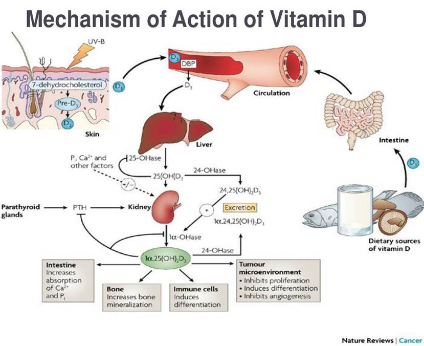
Vitamin D is produced within the body when the skin is exposed to the ultraviolet (UVB) radiation and it can also be established via dietary sources. Its main function is to regulation levels of calcium and phosphorus to build strong bones and teeth. There are different forms of Vitamin D: Vitamin D3 is called cholecalciferol derived from sunlight and works on cholesterol derivate called 7-dehydrocholesterol on the skin. The alternative form is Vitamin D2 called ergocalciferol derived from plants. The metabolism of vitamin D requires hormones and enzymes. Hormones are chemical messengers that can either be protein based or lipid/fat-based and are released into the blood to act on the target cells to maintain body function. Enzymes are proteins that speed up the chemical reaction, but it does not itself undergo any change during the reaction. Vitamin D is metabolised by the addition of hydroxyl groups (OH), it initially begins in the liver by the enzyme 25-hydroxylase (25-OHase) and also several kidney cells produce 1-hydroxylase (1alpha-OHase). The active product formed is 1,25-dihydroxyvitamin D [1-alpha25(OH)2D3], otherwise known as calcitriol, a hormone. Another way in how 1,25-dihydroxyvitamin D is produced is via the stimulation of the parathyroid hormone (PTH) in the parathyroid glands that stimulates the activity of 1-hydroxylase. The production of 1,25-dihydroxyvitamin D can also be produced via a two-step enzymatic action: firstly, the action of the enzyme 24-OHase where it converts 25(OH)D3 to 24,25(OH)2D3). Then the enzyme is 1-alpha OHase is then used to form 1,25-dihydroxyvitamin D. There are several functions of 1,25-dihydroxyvitamin D: It helps to increase the absorption of calcium and phosphorus in the intestines into the blood. It also helps to increase mineralization of the bone, induce differentiation of immune cells and help prevent cancer activity.
What Other Effects Does Radiotherapy cause?
Pelvic radiotherapy can not only affect the bones but also the heart.
Menopause: Pelvic radiotherapy may cause Iatrogenic menopause. Iatrogenic are conditions caused from treatment. In this case, pelvic radiotherapy causes menopause. Menopause is defined as the time where a woman’s life when ovaries stop to produce an egg cell every four weeks. And menstruation stops and women no longer bear. This is associated with vasomotor symptoms, mood, sleep and urinary changes.
Vasomotor symptoms: Vasomotor symptoms are sensory symptoms felt by women during their menopause. Examples are hot flushes where they feel heat. Sweating and wwaking up at night and reddening of the skin on the head , neck and chest. Their blood vessel also dilate (peripheral vasodilation). High body temperature and tachycardia (fast heart rate) but normal blood pressure.
The experts have also reported less but does exist – radiation-induced lumbosacral plexopathy. It increases with improved survival rates – This can be diagnosed with MRI after physical examination of lower limb pain, numbness, weakness, paresis or paralysis.
Paresis: nerve-induced muscular weakness/lethargy but to a lesser degree than paralysis. Paralysis is muscular weakness but can affect the spasticity (weakness of the limbs) and flaccidity (lacking in firmness) of the disease and how it affects the brain. Spinal cord, nerves and muscles.
Palliative Radiotherapy
This is commonly performed in higher stages and grade to provide relief for symptoms.
Examples of symptoms: pain, bleeding, ulcers, local invasion to the bladder or the rectum.
How Is It Commonly Given?
It is commonly given in short sections.
20 Gy in five fraction or 30 Gy in 10 fractions delivered over one or two weeks.
Hypofractionated regimens to a smaller volume including 30–36 Gy in six fractions over three to six weeks.
Frail patients with ongoing bleeding: A single fraction of 8 Gy or 10 Gy may be considered and to be repeated if required.
Hoppe-Seyler, K., Bossler, F., Braun, J.A., Herrmann, A.L. and Hoppe-Seyler, F. (2018). The HPV E6/E7 Oncogenes: Key Factors for Viral Carcinogenesis and Therapeutic Targets. Trends in Microbiology, [online] 26(2), pp.158–168. doi:10.1016/j.tim.2017.07.007.
Kochhar, R., Plumb, A.A., Carrington, B.M. and Saunders, M. (2012). Imaging of Anal Carcinoma. American Journal of Roentgenology, 199(3), pp.W335-W344. doi:10.2214/ajr.11.8027.
Lobna Ouldamer, Z. Chraibi, Flavie Arbion, Barillot, I. and Body, G. (2013). Bartholin’s gland carcinoma: Epidemiology and therapeutic management. 22(2), pp.117–122. doi:10.1016/j.suronc.2013.02.004.
Morrison, J., Baldwin, P., Hanna, L., Andreou, A., Buckley, L., Durrant, L., Edey, K., Faruqi, A., Fotopoulou, C., Ganesan, R., Hillaby, K. and Taylor, A. (2024). British Gynaecological Cancer Society (BGCS) vulval cancer guidelines: An update on recommendations for practice 2023. PubMed, 292, pp.210–238. Doi: 10.1016/j.ejogrb.2023.11.013.




Leave a comment