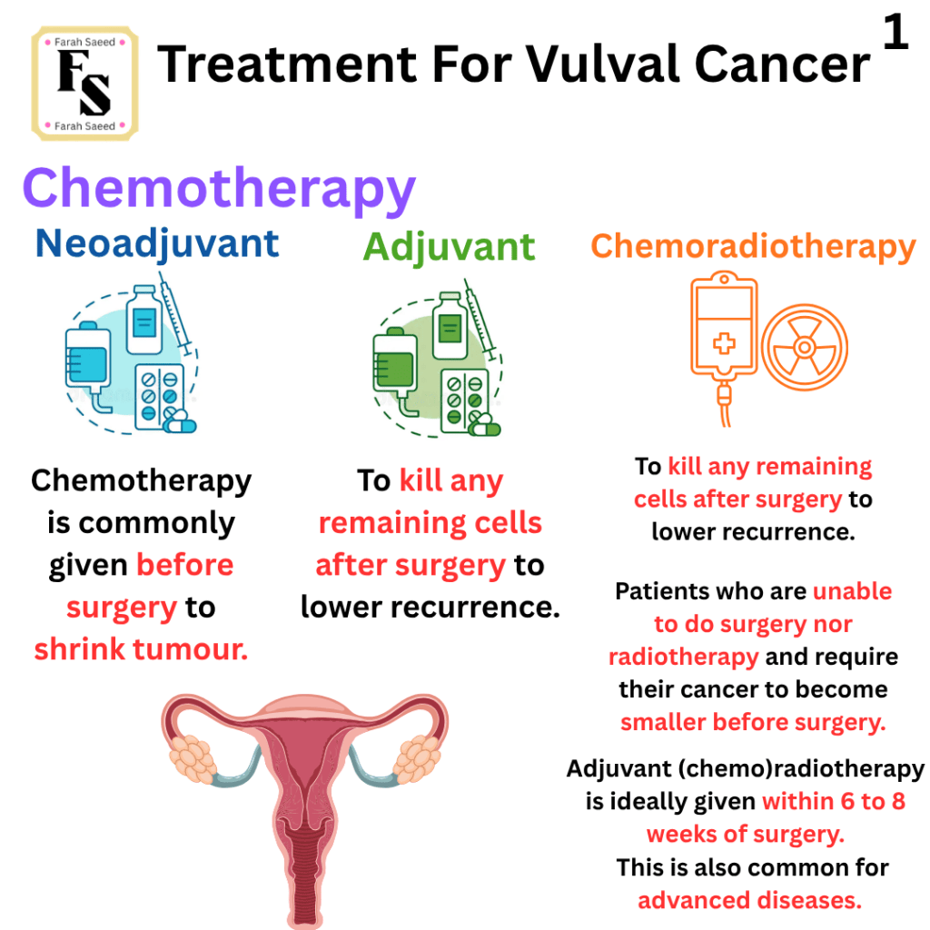
Below summarizes the main points for all treatments in accordance to British Gynaecological Cancer Society (BGCS) vulval cancer guidelines: An update on recommendations for practice 2023. Please read the guidelines attached for further details.
CHEMOTHERAPY = CHEMICALS USED TO KILL CANCER CELLS
A closer look…
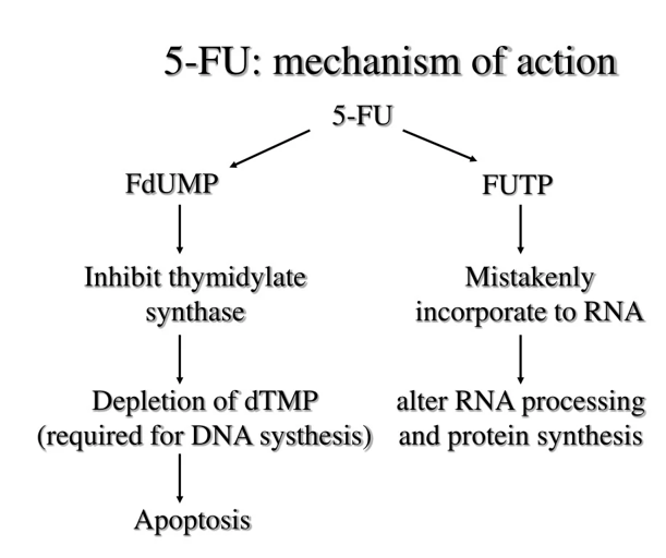
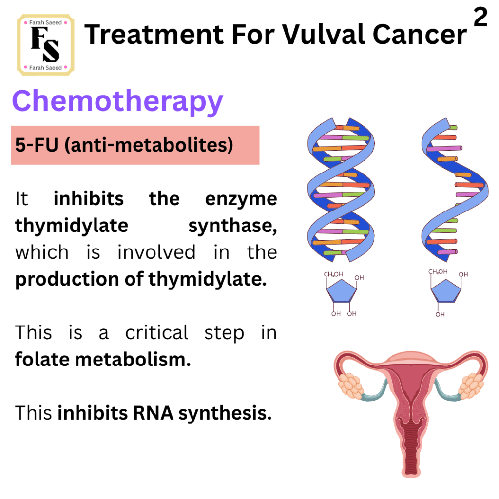
5-Fluorouracil
5-Fluorouracil (antimetabolites) acts on FdUMP (5-fluoro-2-deoxyuridine-5′-monophosphate) where, in the presence of CH2-FH4, it forms a stable complex, inhibiting thymidylate synthase.
Normally, thymidylate synthase is a rate-limiting step for thymidylate by converting dUMP (2′-deoxyuridine-5′-triphosphate) into dTMP.
dTMP converts to dTTP (2’oxythymidine 5’triphosphate) by removing a methyl group (CH2) from CH2-FH4. dTTP is needed for DNA.
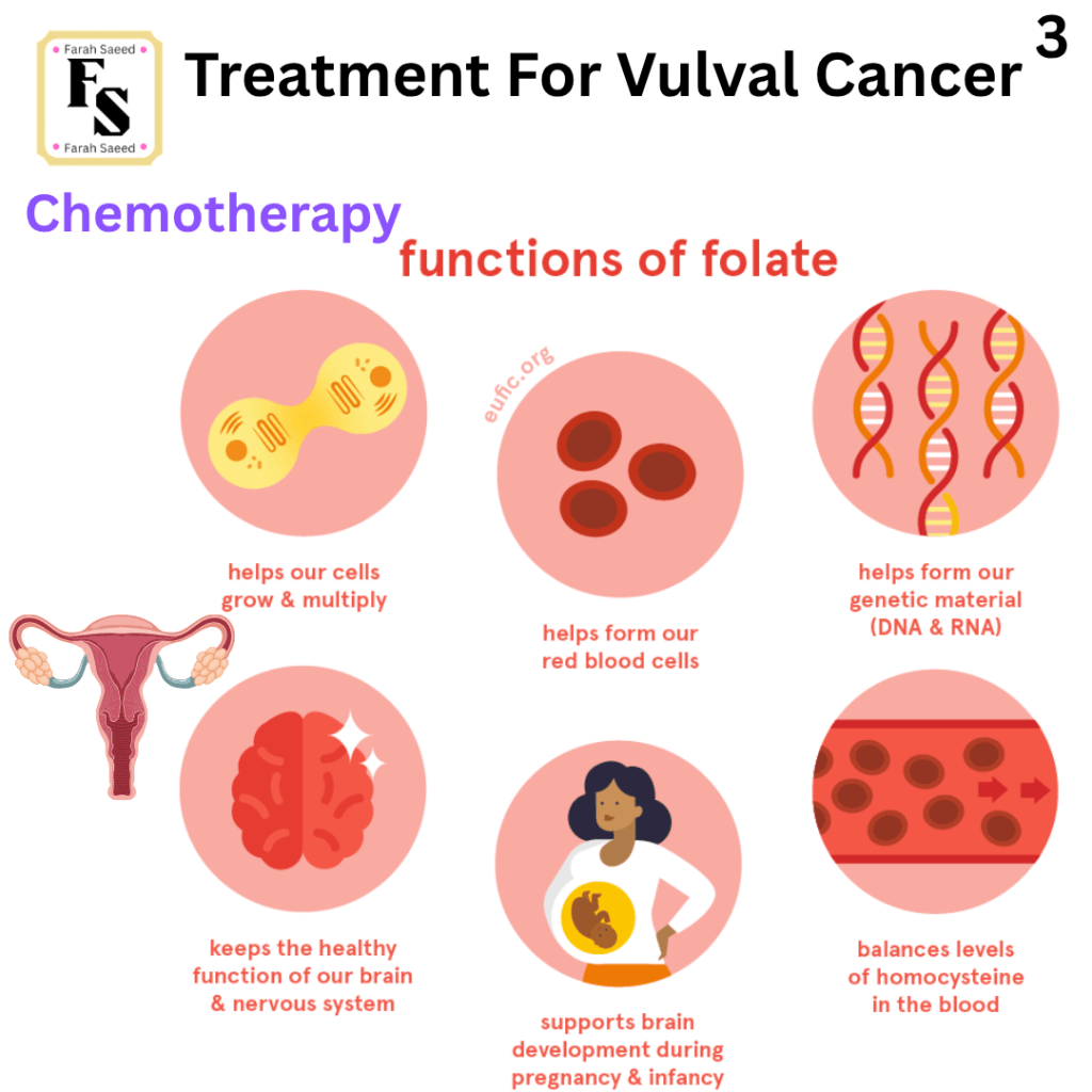
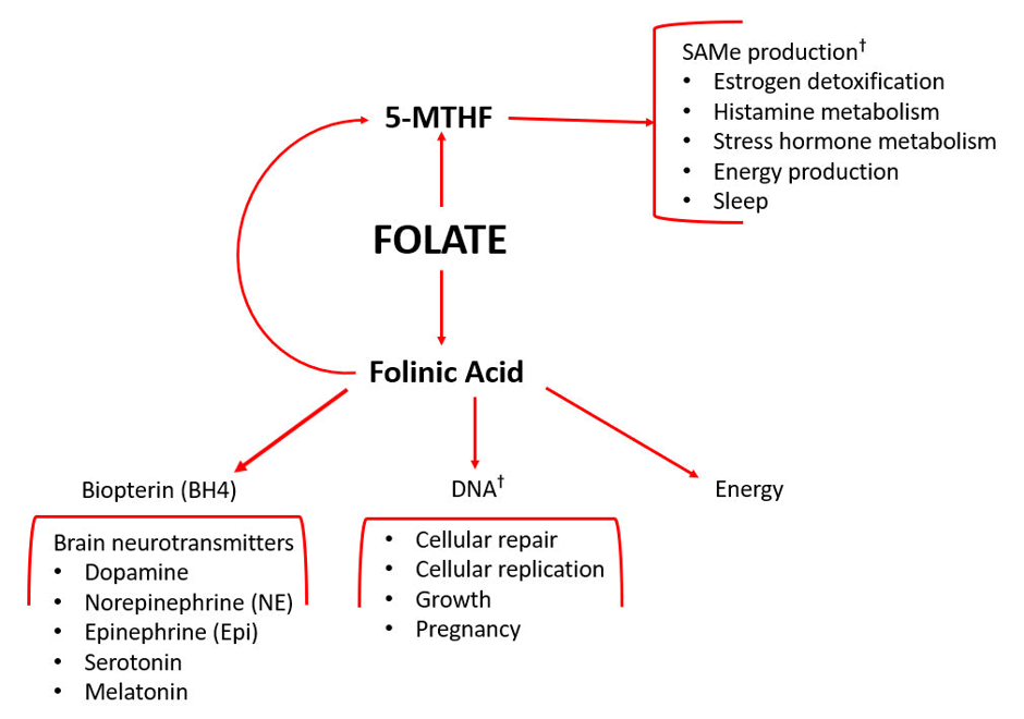
The enzyme dihydrofolate reductase (DHFR) maintains folate.
Cancer Research UK (2026) stated that between 2 and 8 out of 100 people (2 to 8%) have low levels of an enzyme called dihydropyrimidine dehydrogenase (DPD) in their bodies. Minimal levels of DPD lead to severe side effects and longer recovery from chemotherapy.
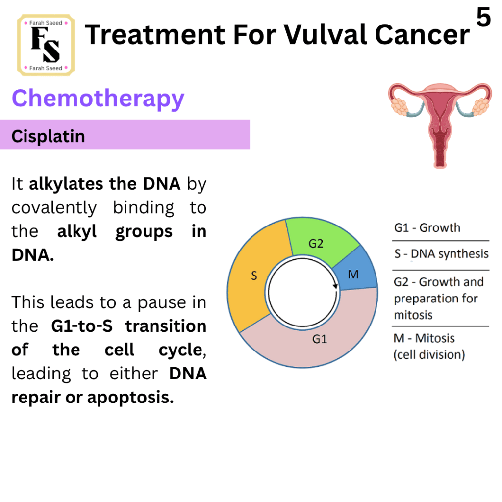
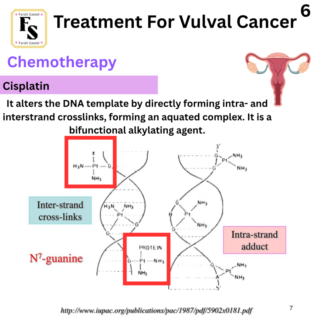
Cisplatin
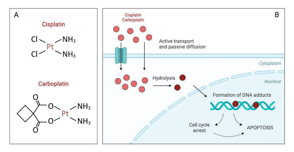
Cisplatin and carboplatin (DNA Alkylating agents) form crosslinks with N7 guanine residues (a type of base found in DNA and RNA). They intercalate DNA and produce toxic reactive oxygen species. It cleaves the transcription factor (CREB1) – this affects the transcription of target genes involved in cellular processes. This causes interstrand and intrastrand breaks in the DNA molecule. It affects how cells divide, copy (replicate), grow, and die (apoptosis).
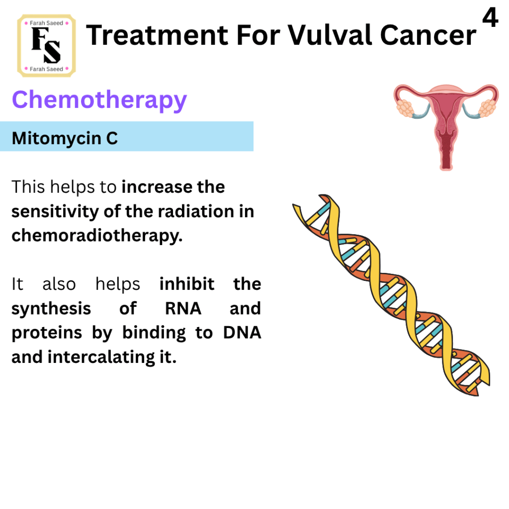
Mitomycin C
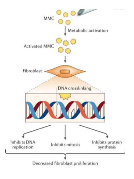
Mitomycin C (anthracycline antibiotic) – It binds with DNA and intercalates between the DNA nitrogenous bases. This causes the uncoiling of the structure of the DNA. The DNA consists of two strands of genetic material (polynucleotides) coiled in a spiral way. RNA is one strand of genetic material. Therefore, it affects the synthesis of DNA and RNA.
Paclitaxel
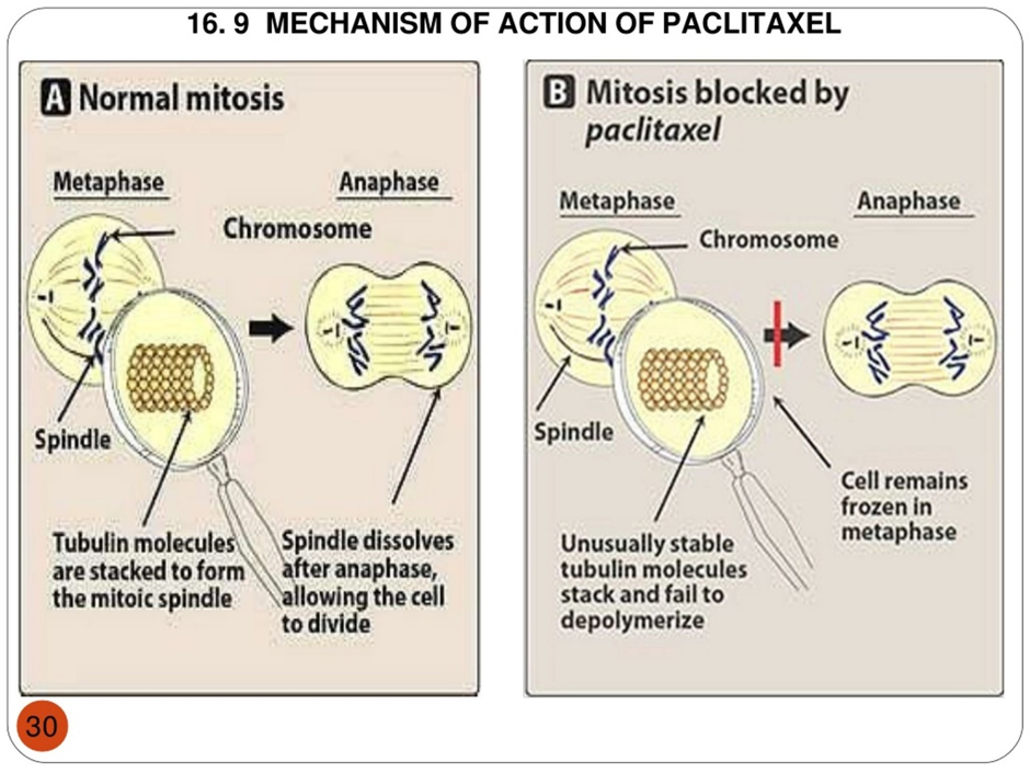
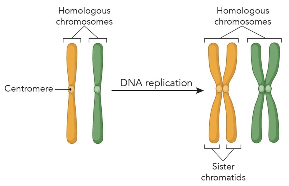
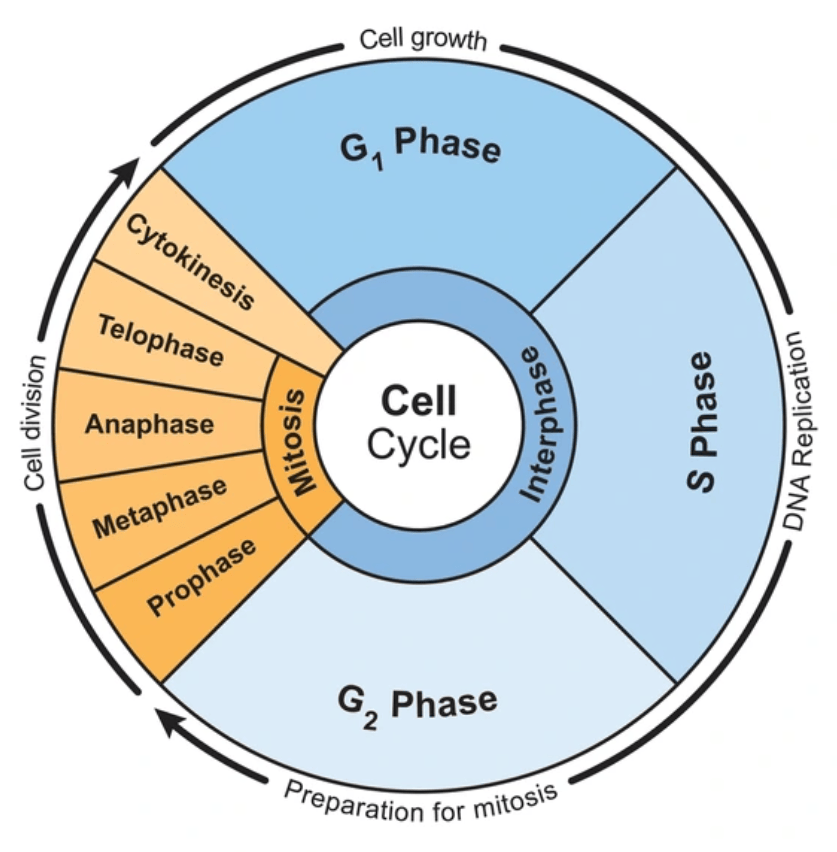
Paclitaxel (taxane) targets the tubulin protein involved in cell division and helps control growth. In normal mitosis, it is subdivided into prophase, metaphase, anaphase, and telophase. Collectively, this is known as the M phase in the cell cycle. Interphase is when the cell grows, and threadlike structures called chromosomes that carry DNA are replicated.
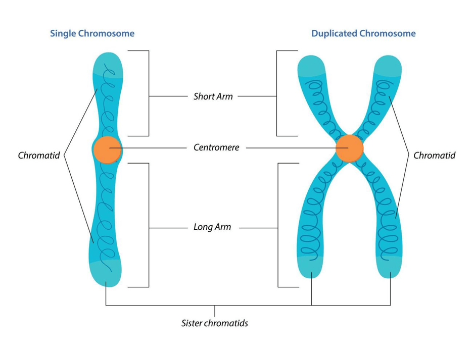
Prophase is when the sister chromatids condense, and spindles start to form. Spindles are made of protein fibers called microtubules. The microtubule itself is made from tubulin proteins. There are several types of microtubules. Some help with positioning the spindle within the cell (astral microtubules). Other microtubules are referred as polar microtubules that help separate the two poles. Kinetochore microtubules are found in the centre of each chromosome, called the centromere. Sister chromatids attach to the spindle via kinetochore microtubules.
Metaphase is when sister chromatids align in the metaphase plate.
Anaphase is when the sister chromatids move towards opposite poles with the aid of polar microtubules.
Telophase is when the nuclear membranes form after the chromosomes decondense and separate the two cells by a process called cytokinesis.
However, Paclitaxel blocks mitosis by keeping the cells in the metaphase stage and not polymerising the tubulin protein found in the spindle.
References
American Cancer Society (2026) Anal Cancer. Available at: https://www.cancer.org/cancer/types/anal-cancer.html (Accessed: 2nd October 2026)
British Association of Dermatologists (2026) Imiquimod cream. Available at: https://www.bad.org.uk/pils/imiquimod-cream (Accessed: 30th September 2026)
Cancer Research UK (2028) Chemotherapy for vulval cancer. Available at: https://www.cancerresearchuk.org/about-cancer/vulval-cancer/treatment/chemotherapy (Accessed: 30th September 2026)
Cleveland Clinic (2026) Volumetric Modulated Arc Therapy (VMAT). Available at: https://my.clevelandclinic.org/health/treatments/17626-volumetric-modulated-arc-therapy-vmat (Accessed: 30th September 2026)
Hoppe-Seyler, K., Bossler, F., Braun, J.A., Herrmann, A.L. and Hoppe-Seyler, F. (2018). The HPV E6/E7 Oncogenes: Key Factors for Viral Carcinogenesis and Therapeutic Targets. Trends in Microbiology, [online] 26(2), pp.158–168. doi:10.1016/j.tim.2017.07.007.
Kochhar, R., Plumb, A.A., Carrington, B.M. and Saunders, M. (2012). Imaging of Anal Carcinoma. American Journal of Roentgenology, 199(3), pp.W335-W344. doi:10.2214/ajr.11.8027.
Lobna Ouldamer, Z. Chraibi, Flavie Arbion, Barillot, I. and Body, G. (2013). Bartholin’s gland carcinoma: Epidemiology and therapeutic management. 22(2), pp.117–122. doi:10.1016/j.suronc.2013.02.004.
Morrison, J., Baldwin, P., Hanna, L., Andreou, A., Buckley, L., Durrant, L., Edey, K., Faruqi, A., Fotopoulou, C., Ganesan, R., Hillaby, K. and Taylor, A. (2024). British Gynaecological Cancer Society (BGCS) vulval cancer guidelines: An update on recommendations for practice 2023. PubMed, 292, pp.210–238. Doi: 10.1016/j.ejogrb.2023.11.013.
National Cancer Institute (2026) Vulvar Cancer Treatment (PDQ®)–Health Professional Version. Available at: https://www.cancer.gov/types/vulvar/hp/vulvar-treatment-pdq (Accessed: 17th August 2026)
Royal College of Obstetricians and Gynaecologists (2014) Guidelines for the Diagnosis and Management of Vulval Carcinoma. Available at: https://www.rcog.org.uk/media/dqwnw2a1/vulvalcancerguideline.pdf (Accessed: 1st October 2026)
The Eve Appeal (2026) Vulval cancer Available at; https://eveappeal.org.uk/information-and-advice/gynaecological-cancers/vulval-cancer/ (Accessed: 7th August 2026)


Leave a comment