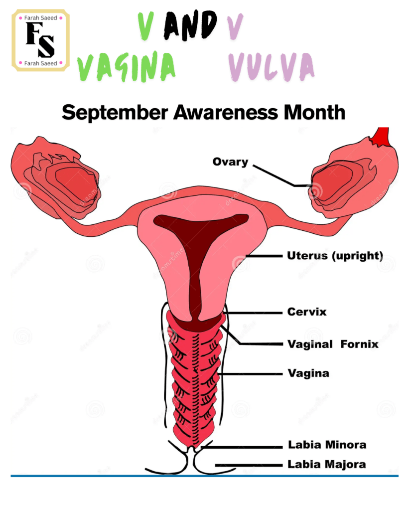
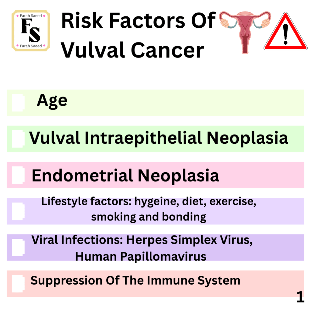
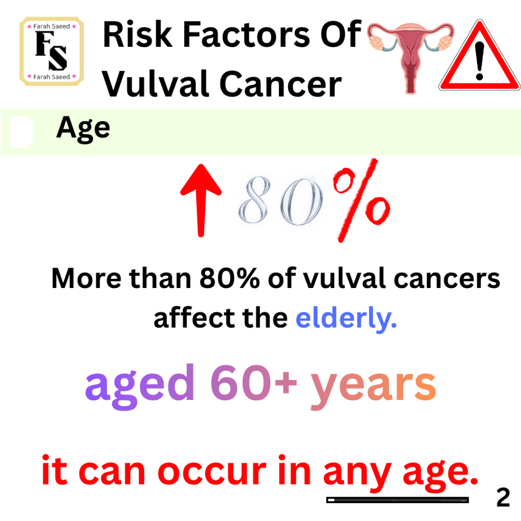
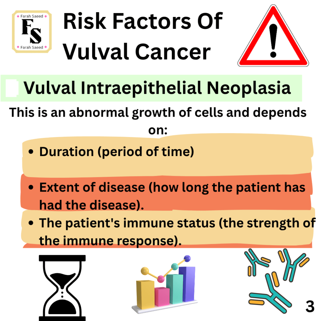
Risk Factors Of Vulval Cancer
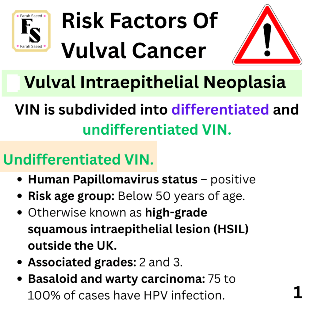
Undifferentiated VIN
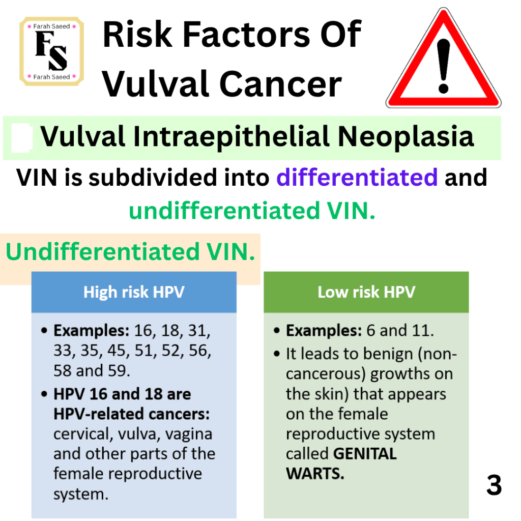
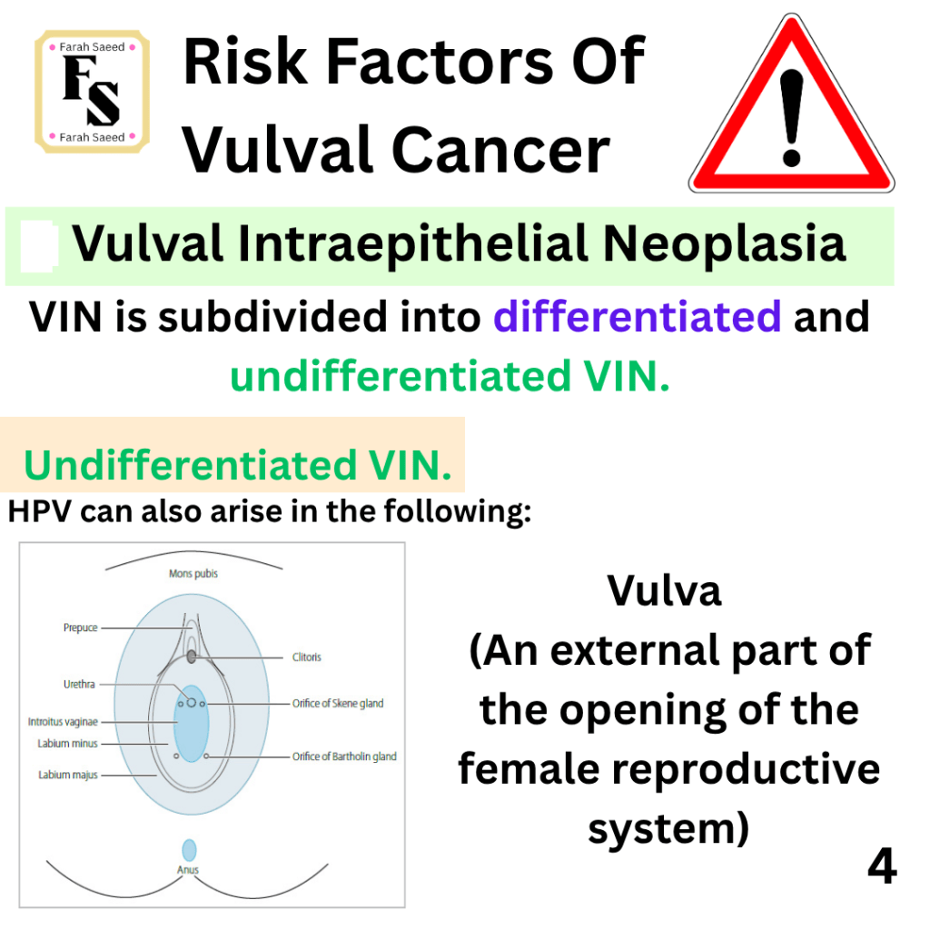
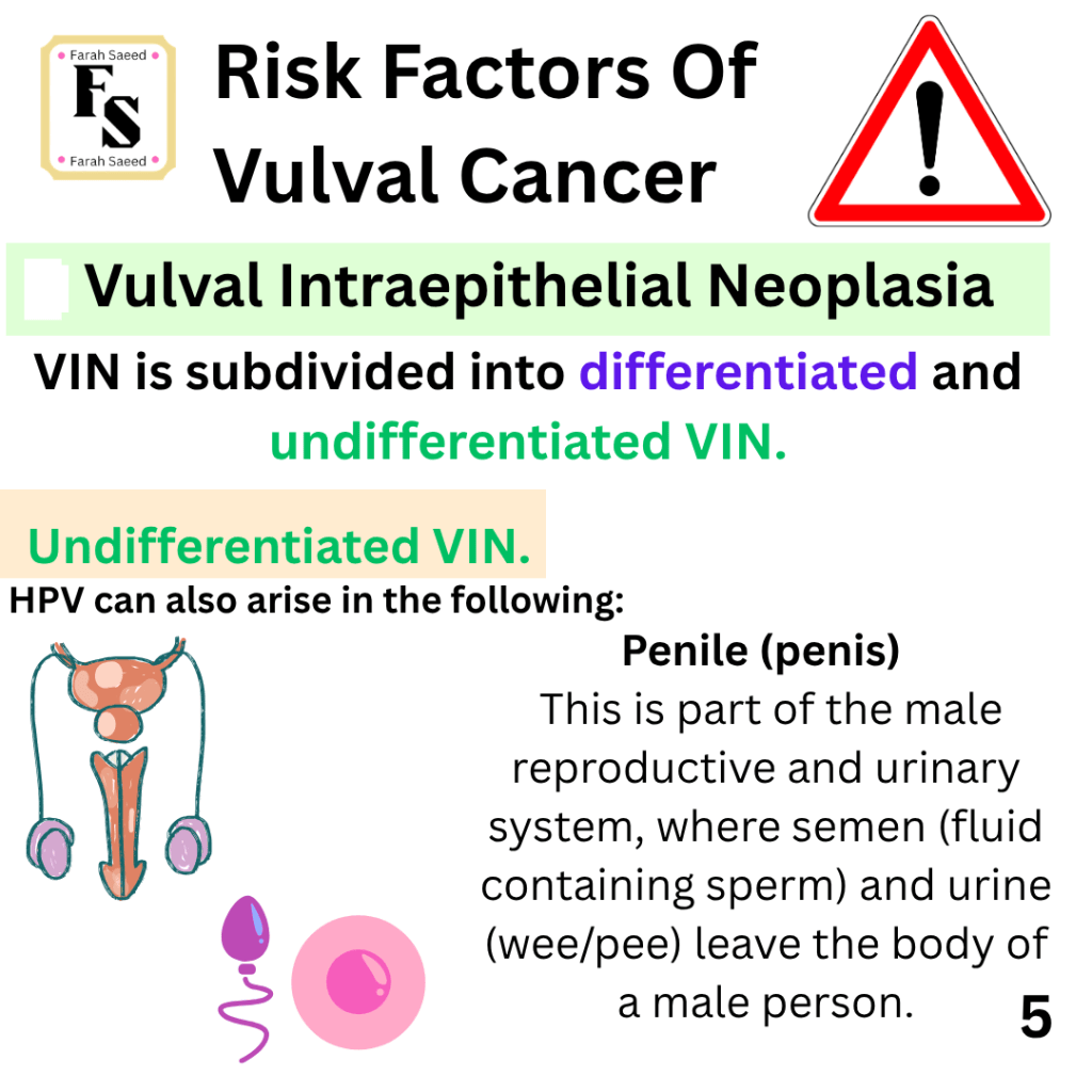
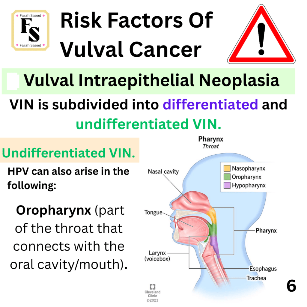
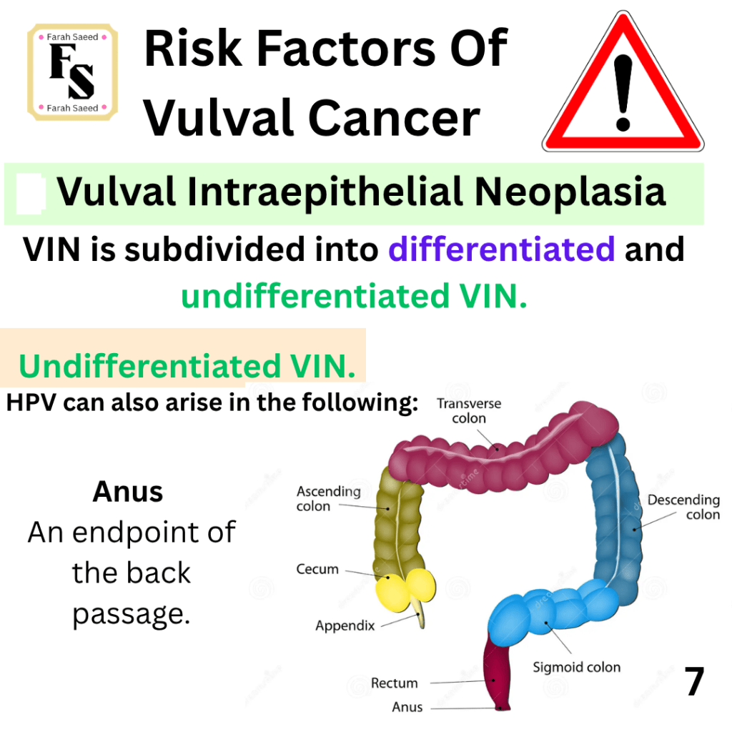
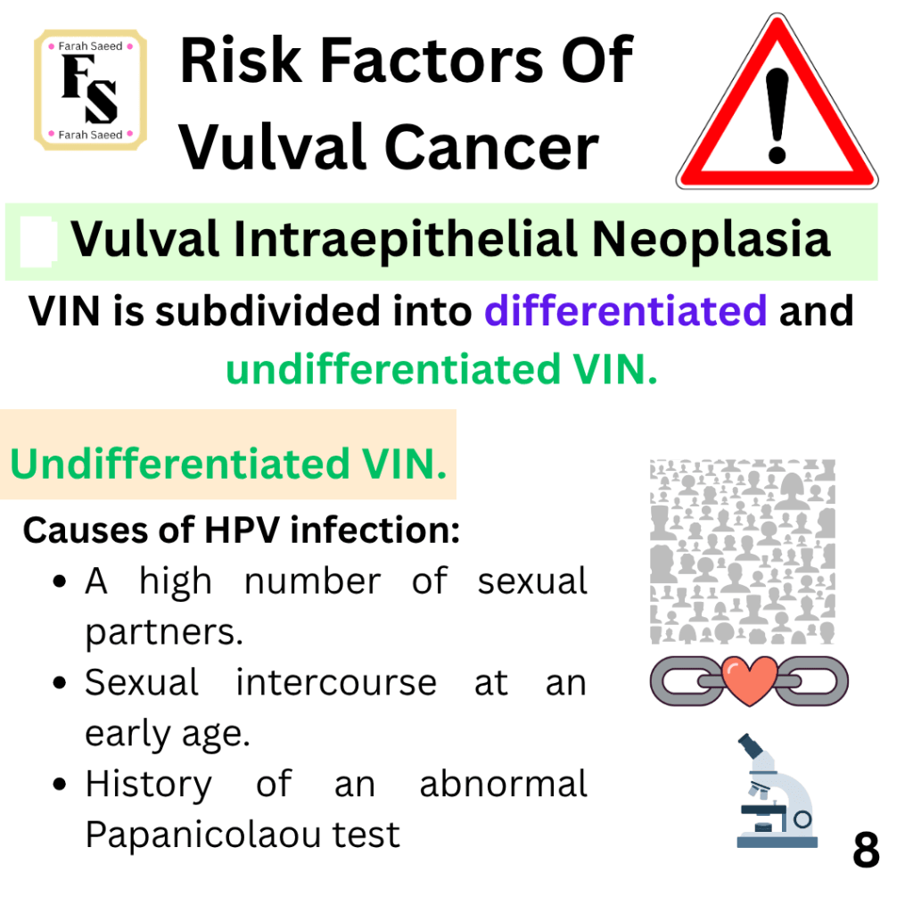
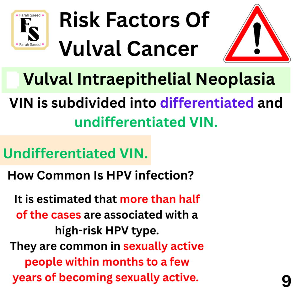
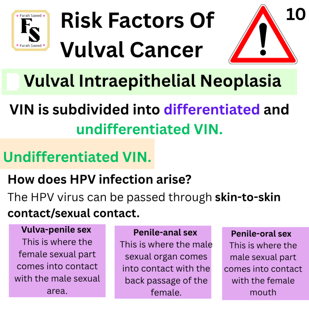
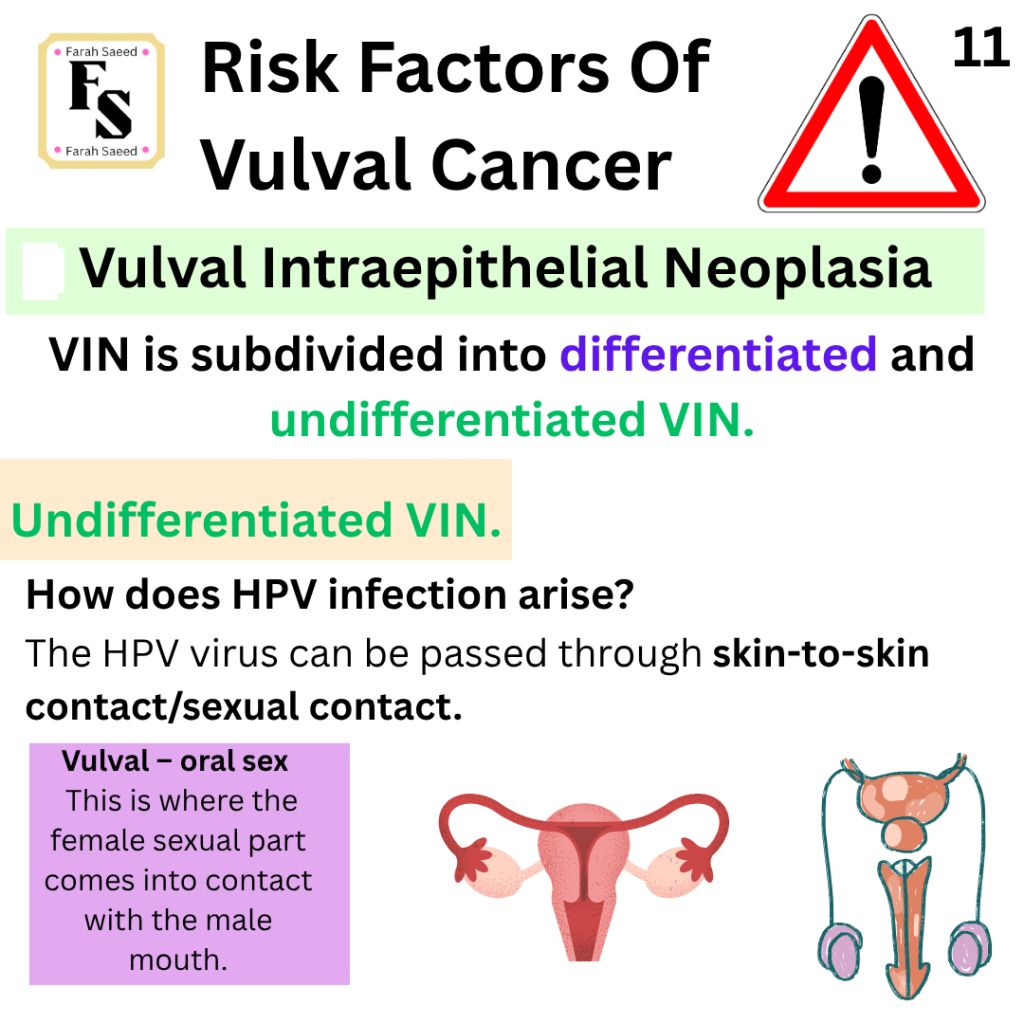
High-risk HPV to cancer – how does it occur?
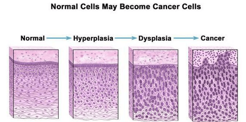
A change in the abnormal growth of the squamous cell lining is called dysplasia. Dysplasia may arise in the cervix, vulva and vulva. This process is called intraepithelial neoplasia. It can transition from hyperplasia to low-grade dysplasia (non-cancerous) to high-grade dysplasia (non-cancerous) and then become cancerous. If the viral HPV is not cleared.
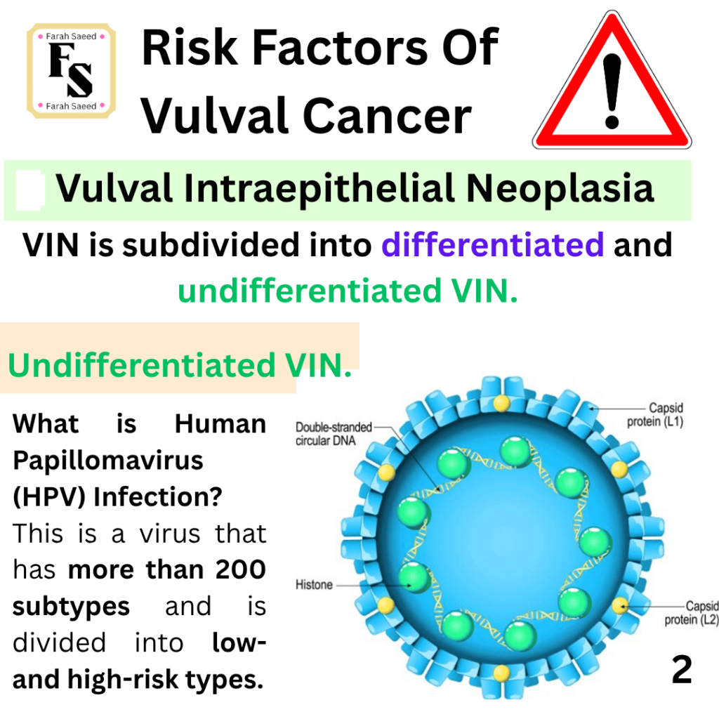
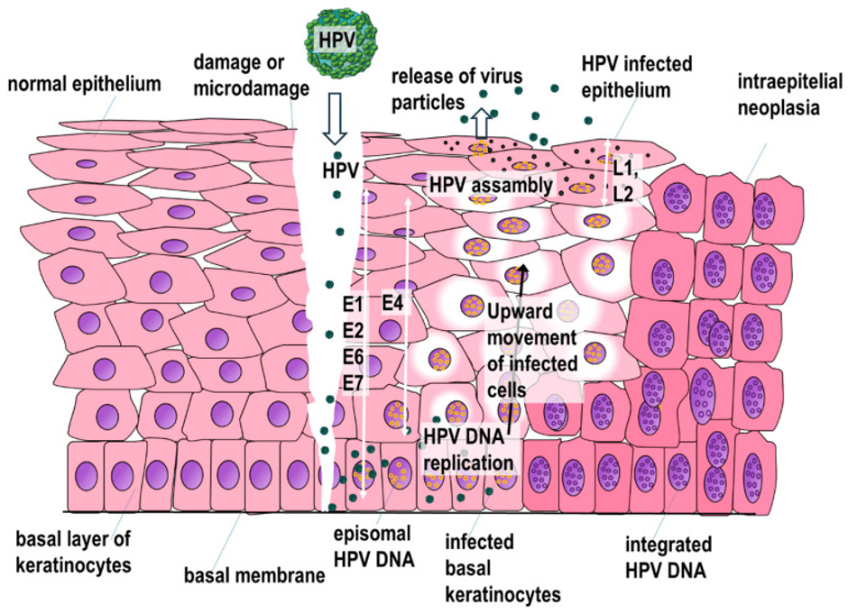
How does the HPV grow?
At first, the protein coat (capsid) of the virus binds to cell and becomes attached. Then it enters the cell, targets the DNA, replicates (copies DNA) and divides to make little viruses (virions) and releases the new viruses to affect other cells in the lining uncontrollably. This is recognised by the immune cells. However, some remain and continue to grow and become precancerous and if not treated, cancer. Researchers estimated that 5 to 10 years HPV-infected cervical cells can become cancerous and 20 years to become cancerous.
There are several viral oncoproteins (proteins that promote premalignant transformation and cancer) that are overexpressed. The E6 oncoprotein deactivates the tumour suppressor protein p53 which helps overcome cell cycle arrest and increase proliferation. The E7 oncoprotein increases overgrowth of host cells (hyperproliferation) by inducing an inactive tumour suppressor gene product called retinoblastoma. It also overexpresses the marker p16.
Supported Images of Undifferentiated VIN
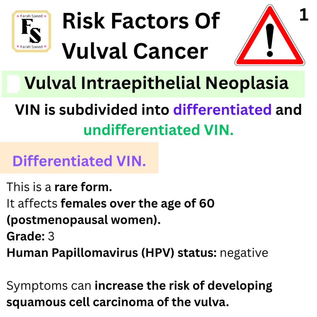
Differentiated VIN
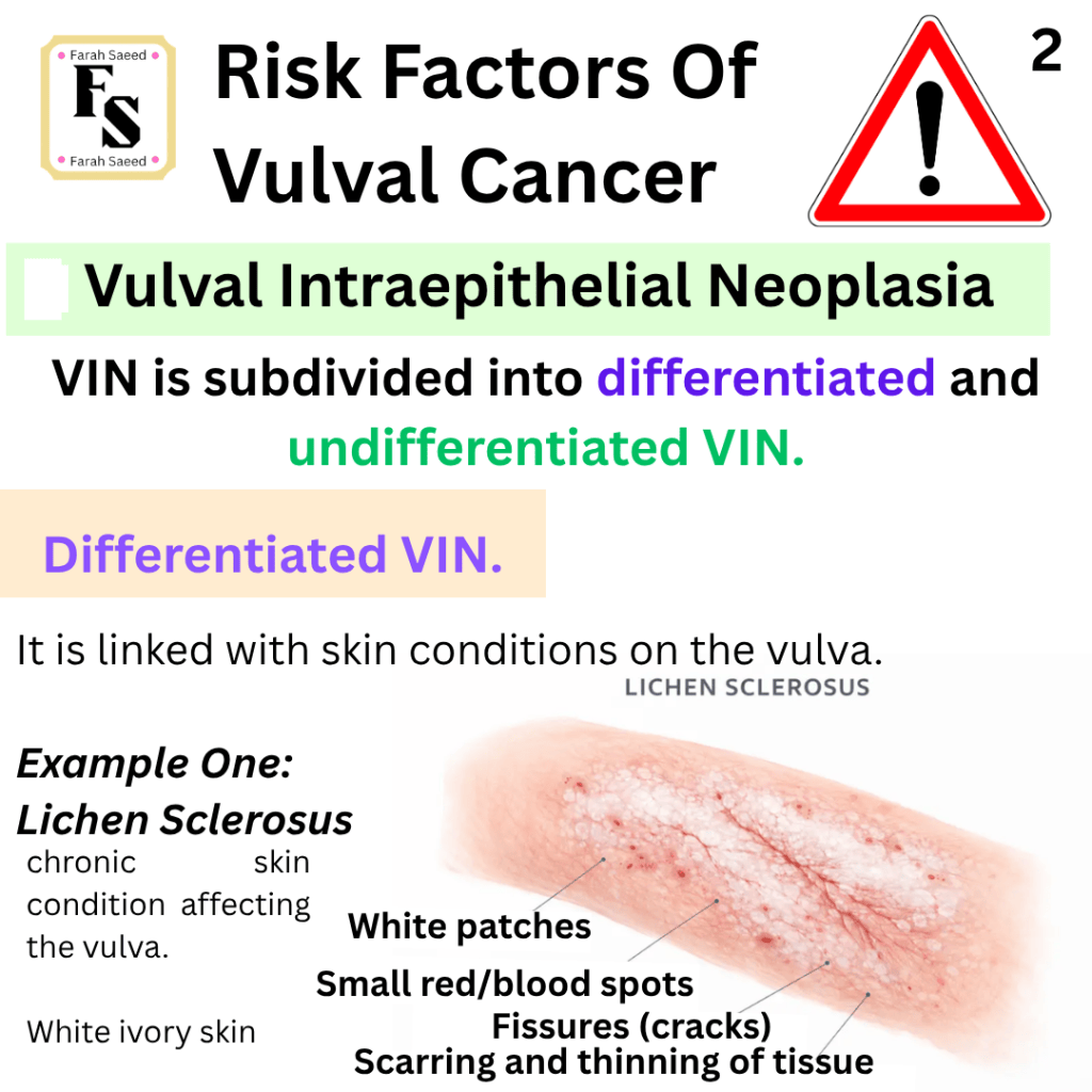
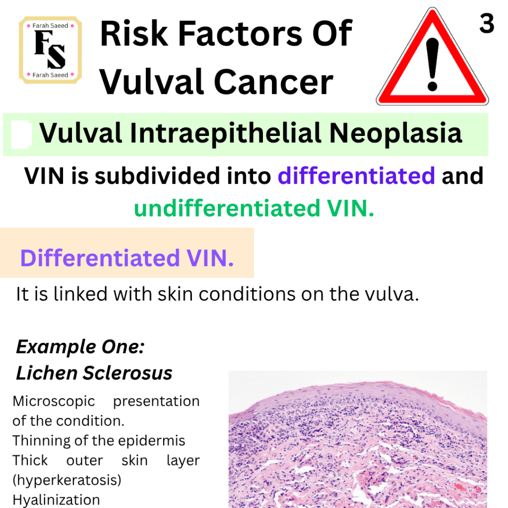
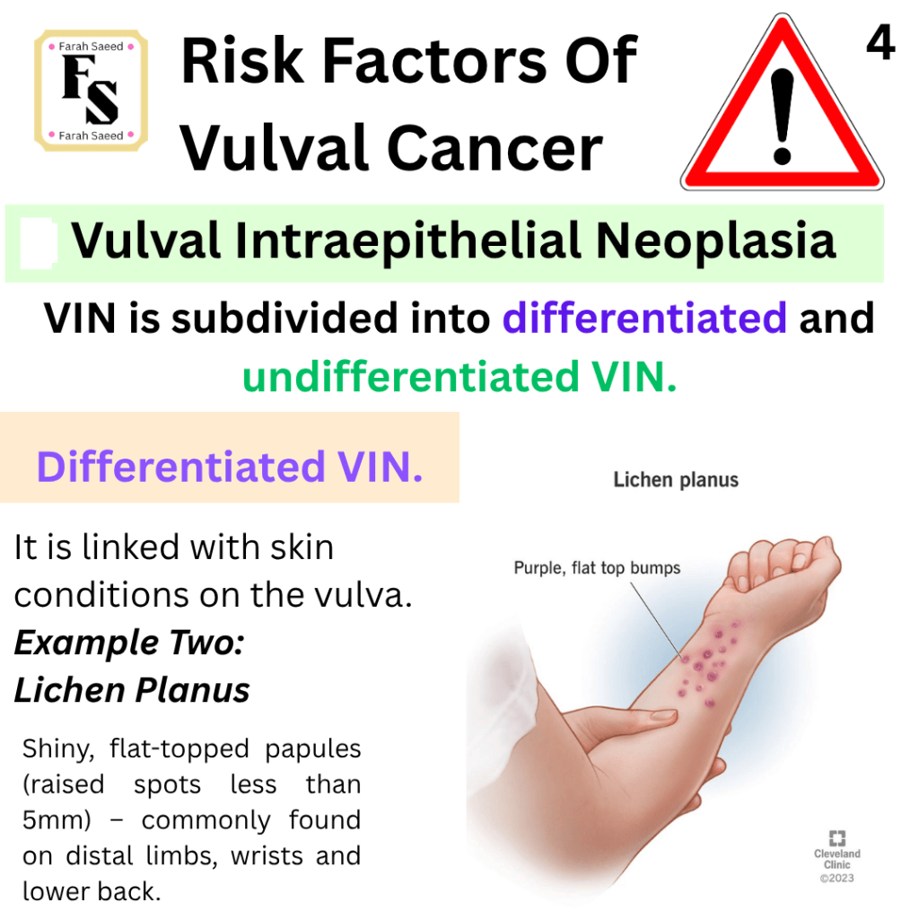
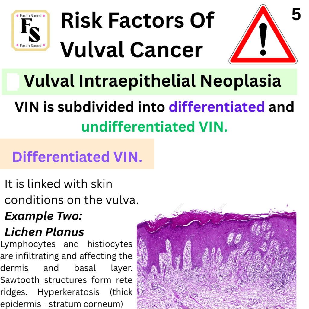
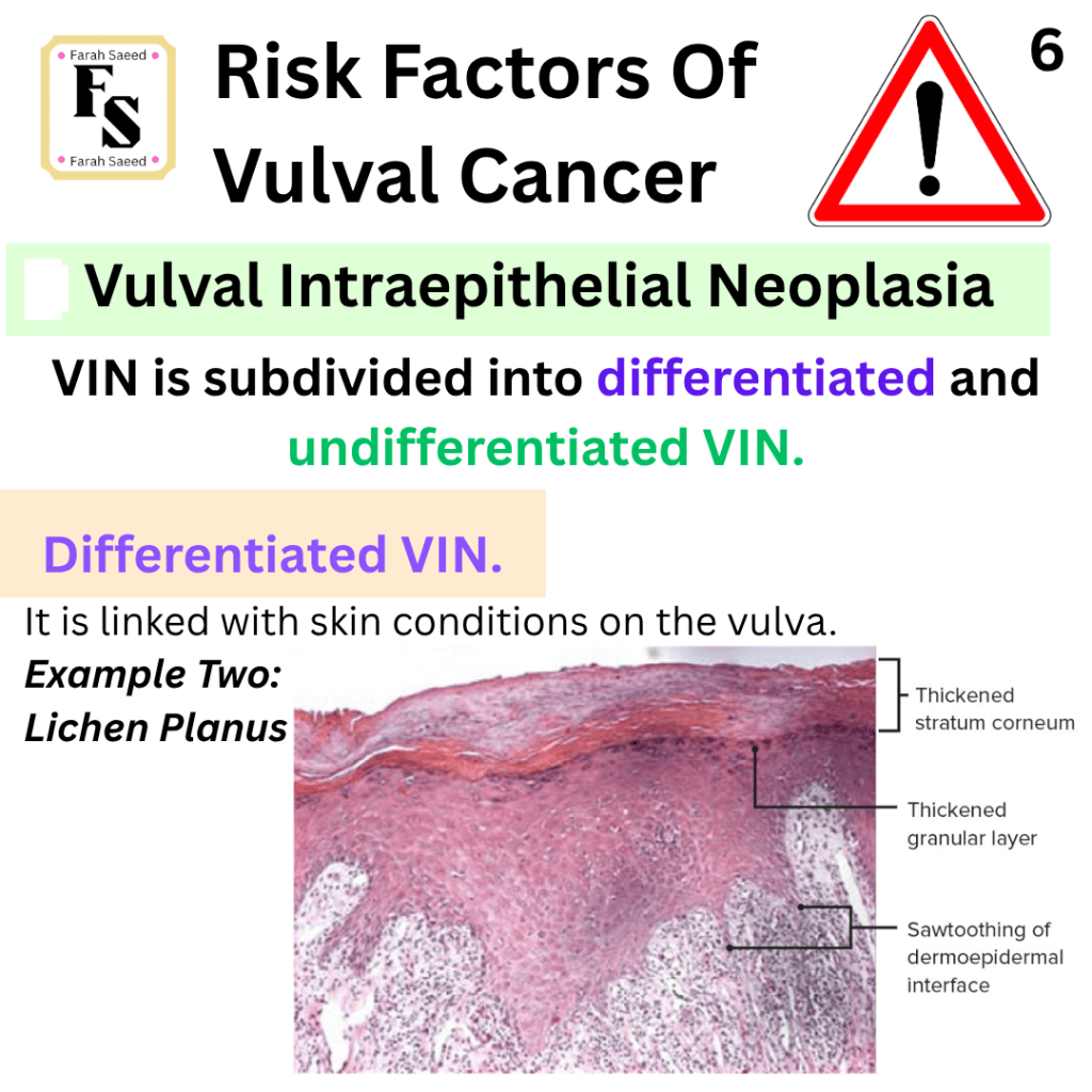
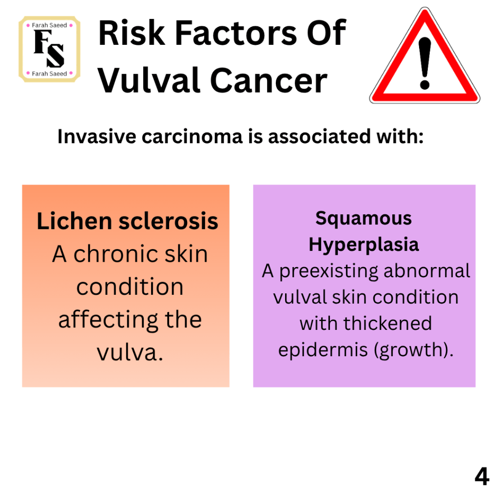
Further Information On Lichen Sclerosus
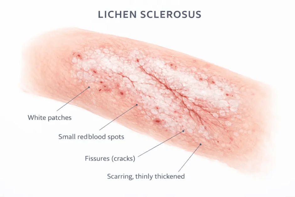
- It has smooth white, ivory plaques or macules (flat areas with altered skin) that increase in size and coalesce to form a surface that is similar to porcelain or parchment.
- It can cause atrophy of the labia minora, where it decreases, the vaginal opening/orifice constricts, and there is possible itching in the area.
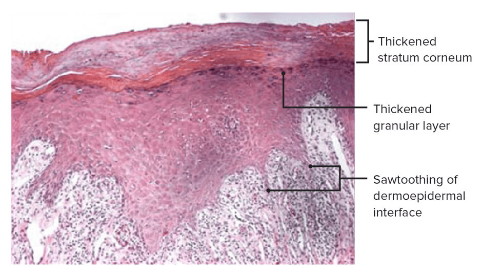
- Thinning of the epidermis.
- Atypia of basal layer of the squamous epithelium (Degeneration of the basal epithelial cells)
- Maturation of superficial layers (outermost layer).
- Hyperkeratosis (excessive production of keratin) – central keratin pearls.
- Squamous hyperplasia – preexisting abnormal vulval skin conditions that have thickened epidermis.
- Lymphocytes (T cells) entering under the skin (dermis).
- Increased frequency of autoimmune disorders in affected women. Studies have shown that the possible cause of Lichen sclerosus is the immune system attacking itself, and this causes a breakdown in the elastin protein whose normal role is to stretch the skin and an increase in fibrin that causes less elasticity.
Mutations (change in the genes)
- TP53: diffuse, strong staining in the basal and parabasal zone. A null p53 staining when there is lack of staining. TP53 is a tumour suppressor gene that maintains integrity of the DNA and regulates cellular events like cell proliferation, cell cycle and apoptosis.
- PTEN (phosphatase and tensin homolog in chromosome 10): a tumour suppressor gene that regulates activity of oncogenic kinases.
Hypermethylation of MGMT, RAASF2A or TSP1.
- MGMT (O-6-methylguanine-DNA methyltransferase is a gene that is involved in the repair of DNA
- RAASF2A: a tumour suppressor gene against the Ras oncogene – It induces cell cycle arrest by blocking cell survival pathways and prevents them from forming tumours.
- TSP1 (thrombospondin-1): glycoprotein that regulates formation of blood vessels (angiogenesis), wound healing, platelet aggregates, tissue repair and cell-to-cell communication.
p53 staining variation in squamous cell carcinoma and in situ lesions
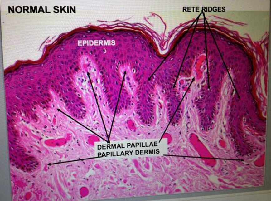
The conventional VSCC are characterised by having malignant squamous cells that vary in the keratin content and have definite stromal invasion. Verrucous carcinoma is a well-differentiated squamous tumour with minimal cells that are abnormal (cytologic atypia), bullous epithelial pegs, and a broad pushing front into the stroma. VSCC with sarcomatoid features has long, thin, spindle-like structures that are poorly differentiated. dVIN has abnormal basal nuclear cells (basal nuclear atypia) that have stronger staining (hyperchromasia), karyomegaly (abnormal enlargement of the nucleus), large nucleoli, abnormal cell division (atypical mitoses, dyskeratosis, and elongated and anastomosing (linking) rete ridges. Dyskeratosis is the impaired overgrowth of the skin (keratinization of cells below stratum granulosum)
HSIL/uVIN have basaloid-shaped cells, hyperchromasia (staining), crowding, and anisonucleosis. Excessive variation in the size of the nucleus.
deVIL has prominent acanthosis or verrucous changes, lack of basal atypia, and abnormal changes in keratinocytes (hypogranulosis, hyperkeratosis, parakeratosis, dyskeratosis). Hypogranulosis is a decreased presence of granules. Hyperkeratosis is increased overgrowth of the skin. Parakeratosis is the retention of nuclei in the stratum corneum (top layer). Dyskeratosis is the impaired maturation of the skin (keratinization of cells below stratum granulosum).
VAAD has acanthosis with verrucous changes, loss of granular layer, superficial epithelial cell pallor, and multilayered parakeratosis.
Therefore, the alternative lesions linked to differentiated VIN (dVIN) are: differentiated exophytic vulvar intraepithelial lesion (DE-VIL) and Vulvar acanthosis with altered differentiation (VAAD). However, they lack the following features: Significant basal atypia and TP53 mutations. DE-VIL and VAAD are more associated with verrucous carcinoma.
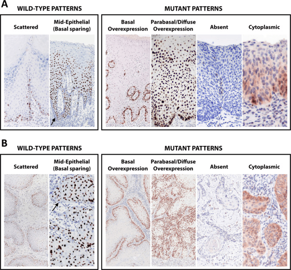
The tissues were stained with p53 immunohistochemistry and were grouped based on the staining intensity and percentage of cells staining counted over 100 cells. There were five groups of staining: absent, uniformly weak, uniformly moderate, uniformly strong, or heterogeneous staining. For the parabasal staining, we divided staining into no parabasal extension, 1/3 thickness, 2/3 thickness, or 3/3 (full) thickness, and intensity (uniformly none, weak, moderate, or strong, or heterogeneous staining of varying intensities).
For vulvar squamous cell carcinoma (VSCC), the basal layer was considered for peripheral cells in the invasive squamous nests, whereas the parabasal layer was considered for the central part of the squamous nests. The nuclear and cytoplasmic evaluation was also performed. Scores were made based on the area with the strongest staining. The results were found in 95% of VSCC (58/61) and 93% (39/42) of in situ lesions and were consistent. Cases that exhibited strong scattered staining and those with a weak basal overexpression pattern could be easily confused.
For TP53 wild-type patterns, there are two major patterns: scattered and mid-epithelial (basal sparing). The scattered pattern has varied nuclear staining because of the different intensities in the basal and parabasal layers. Mid-epithelial (basal sparing) has strong staining in the epithelial cells present in the middle. There is sparing of basal cells and lower basal cells.
For the TP53 mutant patterns, there are four types: basal overexpression, parabasal/diffuse overexpression, absent, and cytoplasmic expression. The Basal overexpression had uniform, strong nuclear staining in a minimum of 80% of basal cells without parabasal staining. The sample with parabasal/diffuse overexpression also showed uniform, strong nuclear staining in at least 80% of basal cells, with strong parabasal staining and extension. Staining extended into the parabasal layers in dVIN, but not in the other entities. The absent (null) had completely absent staining, with a positive internal control, particularly in inflammatory and stromal cells. The cytoplasmic staining, whether with or without nuclear staining, had a positive internal control in inflammatory and stromal cells (Tessier-Cloutier et al., 2020)
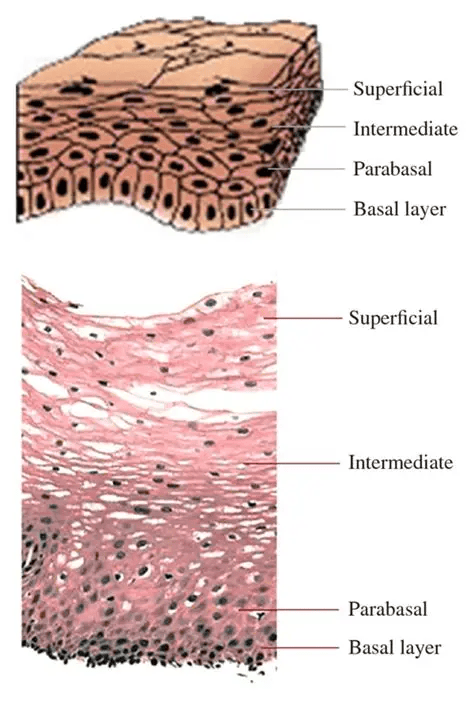
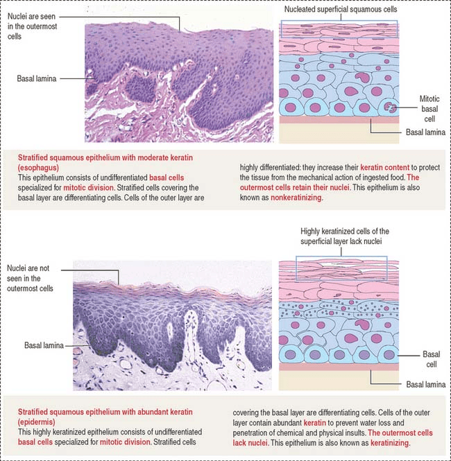
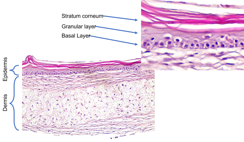
Supporting Images To Help With Evaluation of Histological Images:
Common Misdiagnoses with Differentiated VIN
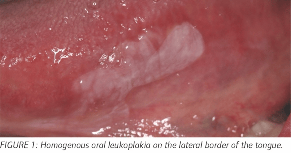
Leukoplakia is a white patch on the mucous membrane lining of the mouth.
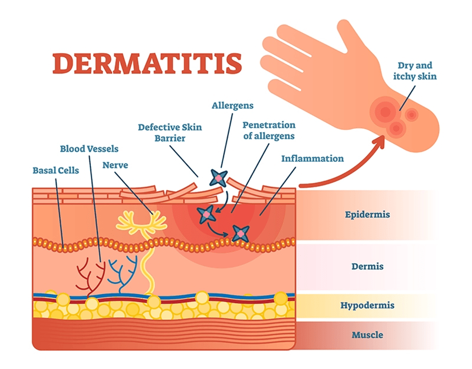
Dermatitis is the chronic inflammation of the skin
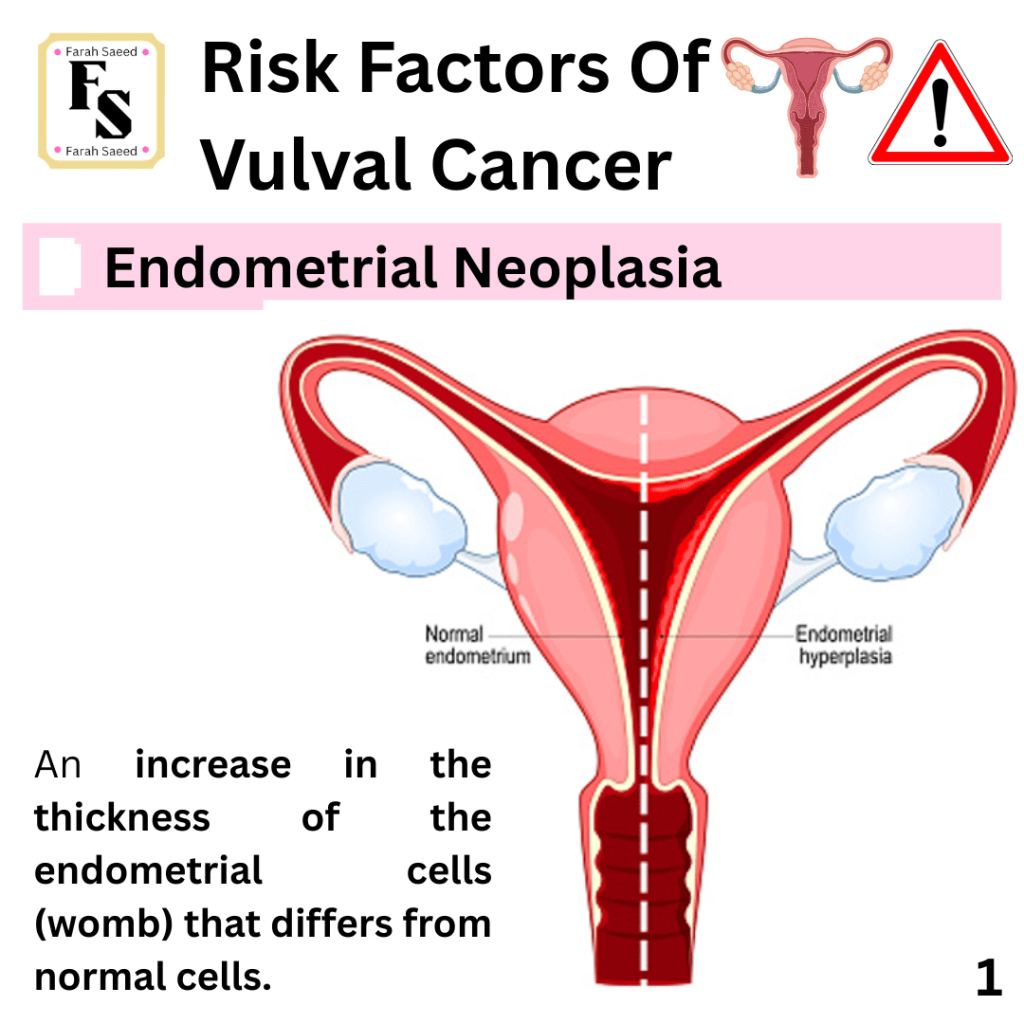
Endometrial Hyperplasia
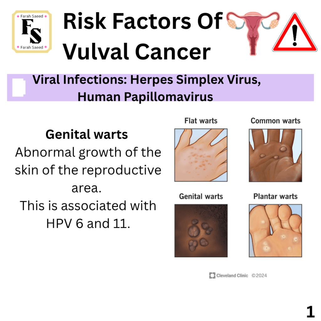
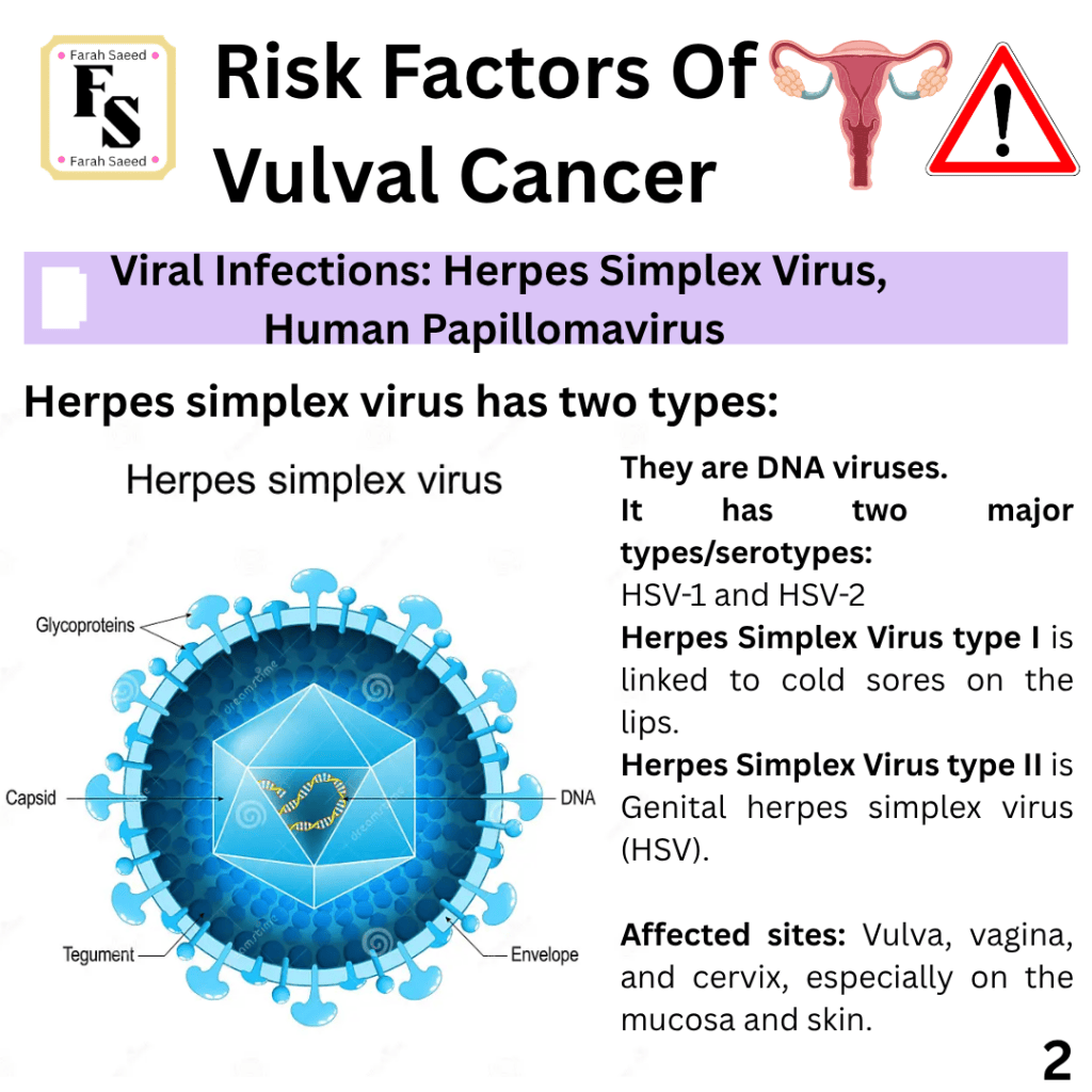
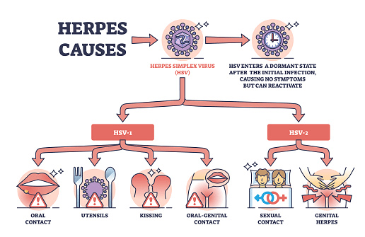
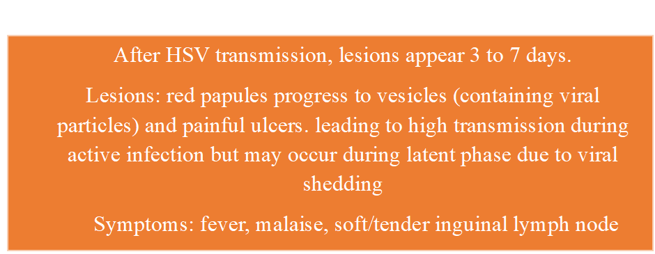
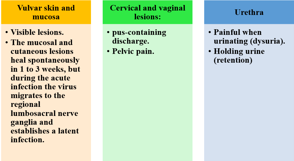
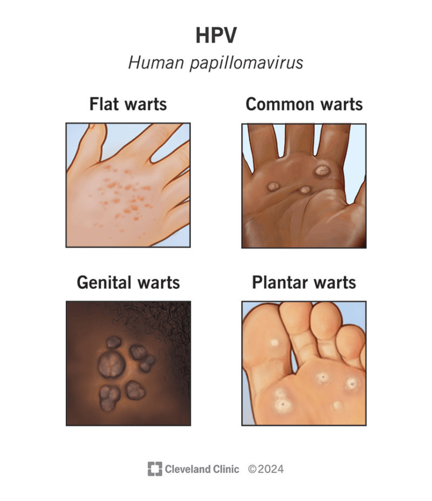
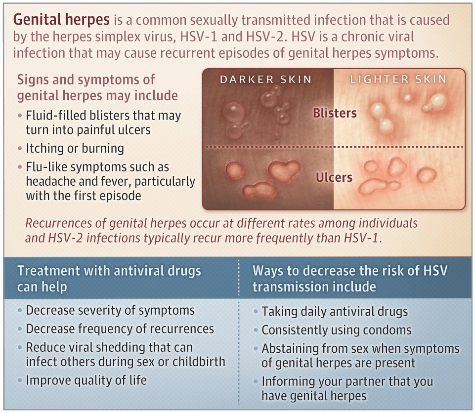
Other Infections: HPV and HSV
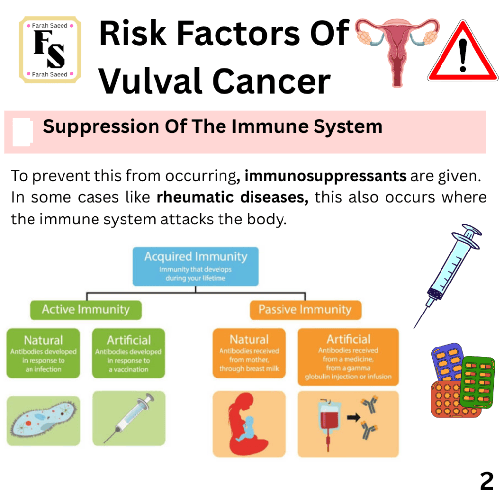
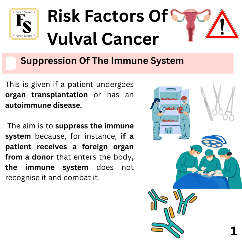
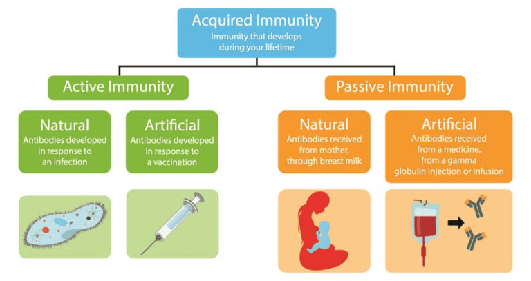
Suppression of the Immune System
References
Cancer Research UK (n.d) Vulval cancer statistics. Available at: https://www.cancerresearchuk.org/health-professional/cancer-statistics/statistics-by-cancer-type/vulval-cancer#heading-Zero (Accessed: 20th September 2026)
Cancer Research UK Cambridge Centre (n.d.) Clinical research on gynaecological cancer. Available at: https://crukcambridgecentre.org.uk/patient-care/clinical-research/gynaecological (Accessed: 10th August 2026)
National Cancer Institute (2024) Vulvar Cancer Causes and risk factors. Available at; https://www.cancer.gov/types/vulvar/causes-risk-factors (Accessed: 7th August 2026)
National Cancer Institute (2025) HPV and Cancer. Available at: https://www.cancer.gov/about-cancer/causes-prevention/risk/infectious-agents/hpv-and-cancer (Accessed: 6th August 2026)
National Cancer Institute (2026) Vulvar Cancer Treatment (PDQ®)–Health Professional Version. Available at: https://www.cancer.gov/types/vulvar/hp/vulvar-treatment-pdq (Accessed: 17th August 2026)
National Institute for Health and Care Excellence (2026) Gynaecological cancers – recognition and referral:Presentation. Available at: https://cks.nice.org.uk/topics/gynaecological-cancers-recognition-referral/background-information/presentation/ Accessed 7th August 2026.
Robbins and cotran pathologic basis of disease (2023) The Female Genital Tract. Availale at: https://clinicalpub.com/the-female-genital-tract/ (Accessed 12th September 2026)
Tessier-Cloutier, B., Kortekaas, K.E., Thompson, E., Pors, J., Chen, J., Ho, J., Prentice, L.M., McConechy, M.K., Chow, C., Proctor, L., McAlpine, J.N., Huntsman, D.G., Gilks, C.B., Bosse, T. and Hoang, L.N. (2020). Major p53 immunohistochemical patterns in in situ and invasive squamous cell carcinomas of the vulva and correlation with TP53 mutation status. Modern Pathology, 33(8), pp.1595–1605. doi:10.1038/s41379-020-0524-1.
Updated September 2026; Next Review September 2028

