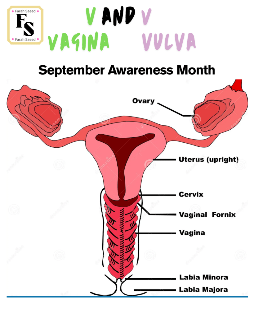
Symptoms And Diagnosis Of Vulval Cancer
What Symptoms May Arise With HPV-associated Cancers i.e. Vulva?
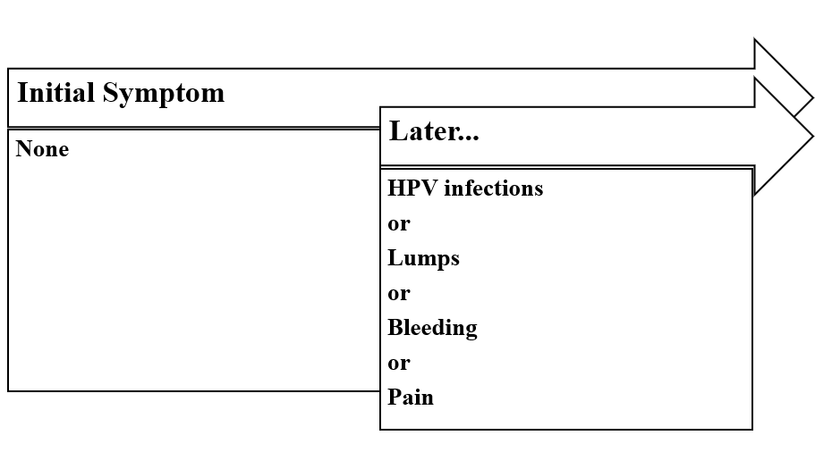
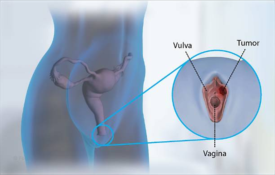
According to the National Institute for Health and Care Excellence (NICE) guidelines, the main symptoms are:
Unexplained vulval bleeding, lump or ulcer, all require referral on a suspected cancer pathway. There is also vulval itching or redness.
What Other Symptoms May Arise?
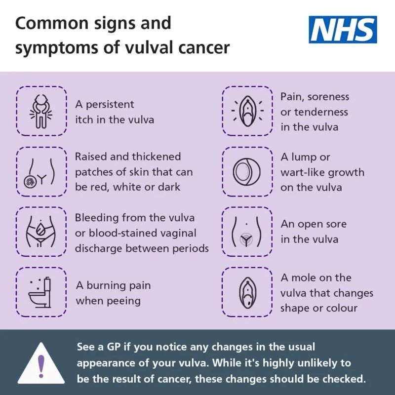
The most common symptoms are the following indicated by Centre of DiseControl and Prevention and National Cancer Institute:
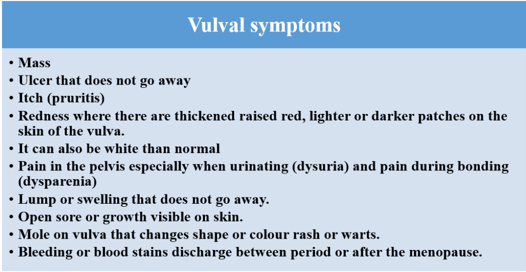
Specific Symptoms For Some Types Of Vulval Cancers
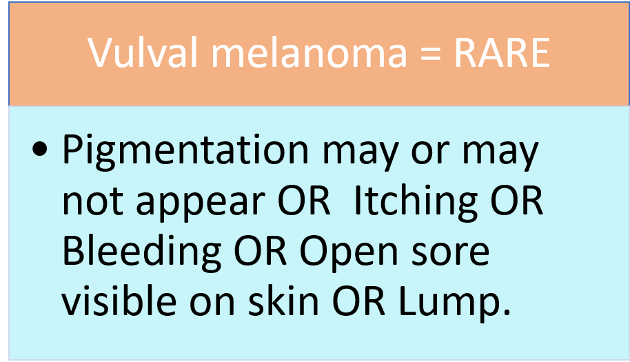
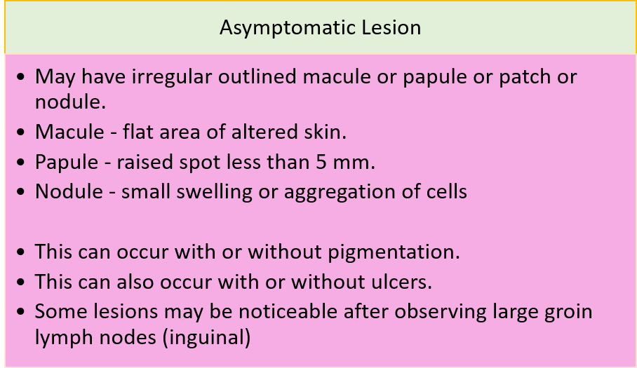
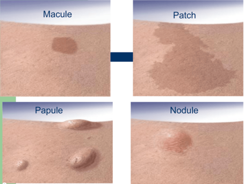
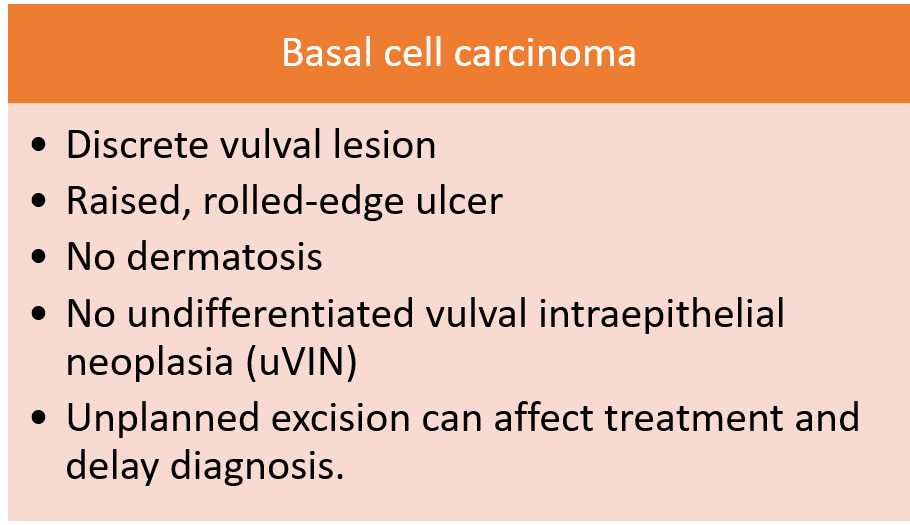
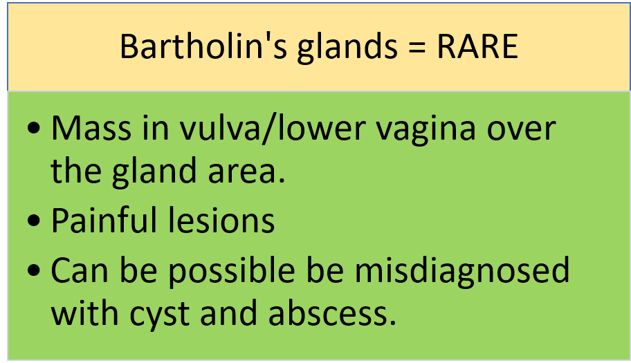
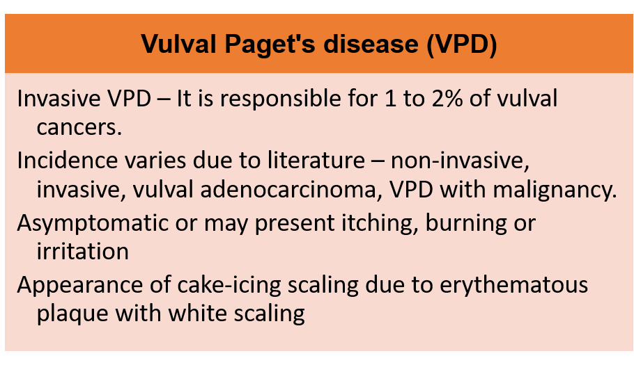
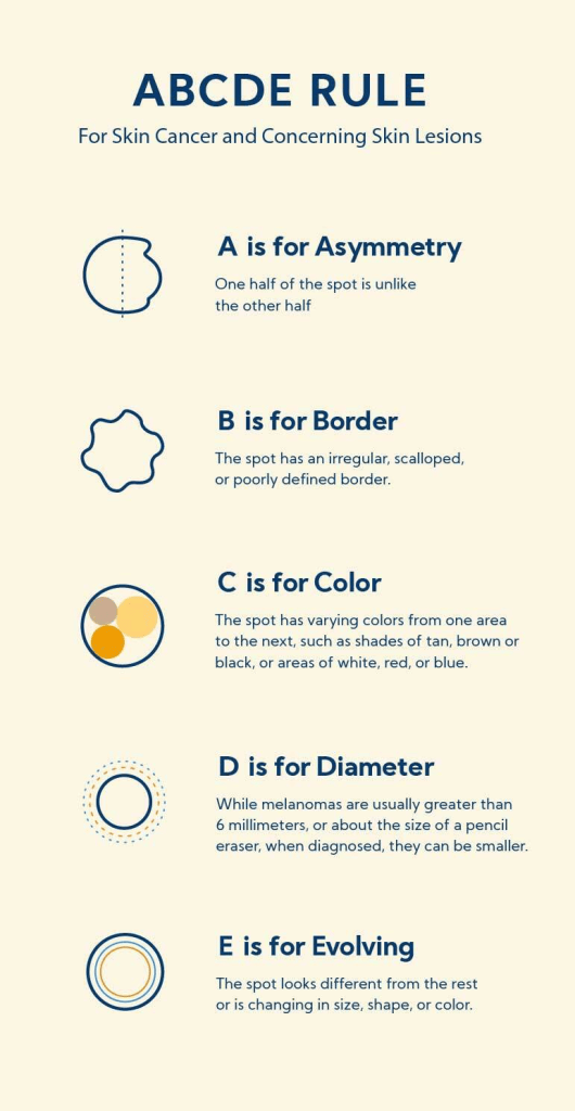
Further Information About Signs And Symptoms Of Types of Skin Lesions And Melanoma
Simple steps for diagnosis
Speak with GP for examination and referral.

Additional notes:
A female over the age of 40 years of age, may have a biopsy of the cyst if there are solid components due to swelling. This helps to exclude vulval carcinoma.
If a patient has fungating lesion with or without palpable groin nodes, cancer referral is needed without requiring biopsy results.
Endocervical and high vaginal swabs to be taken if there are indications of sexually transmitted infection.
Colposcopy (Example of Pelvic Examination)
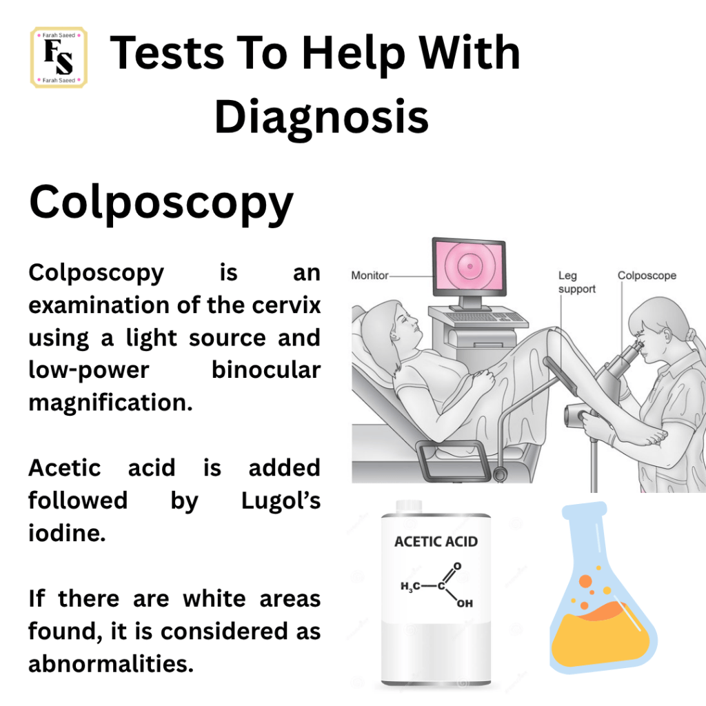
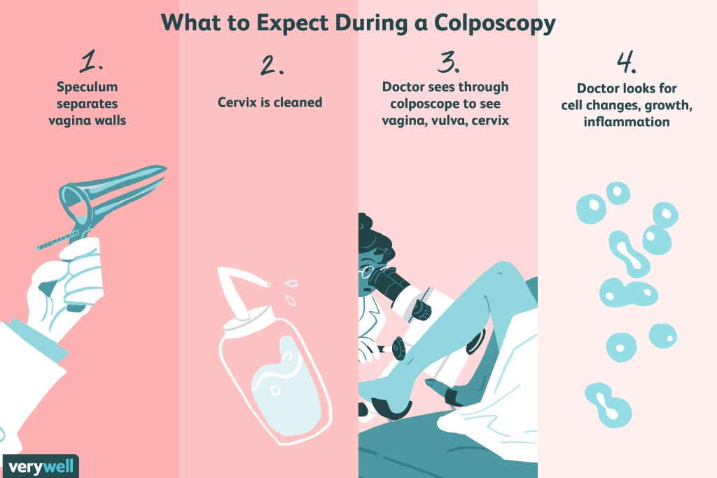
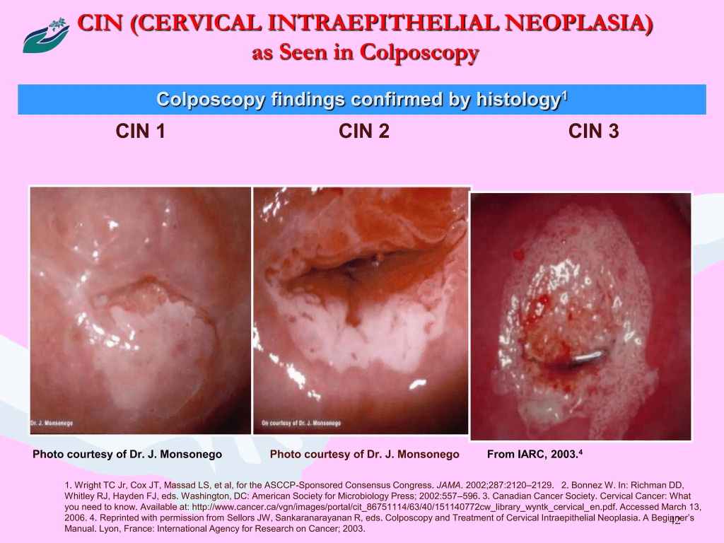
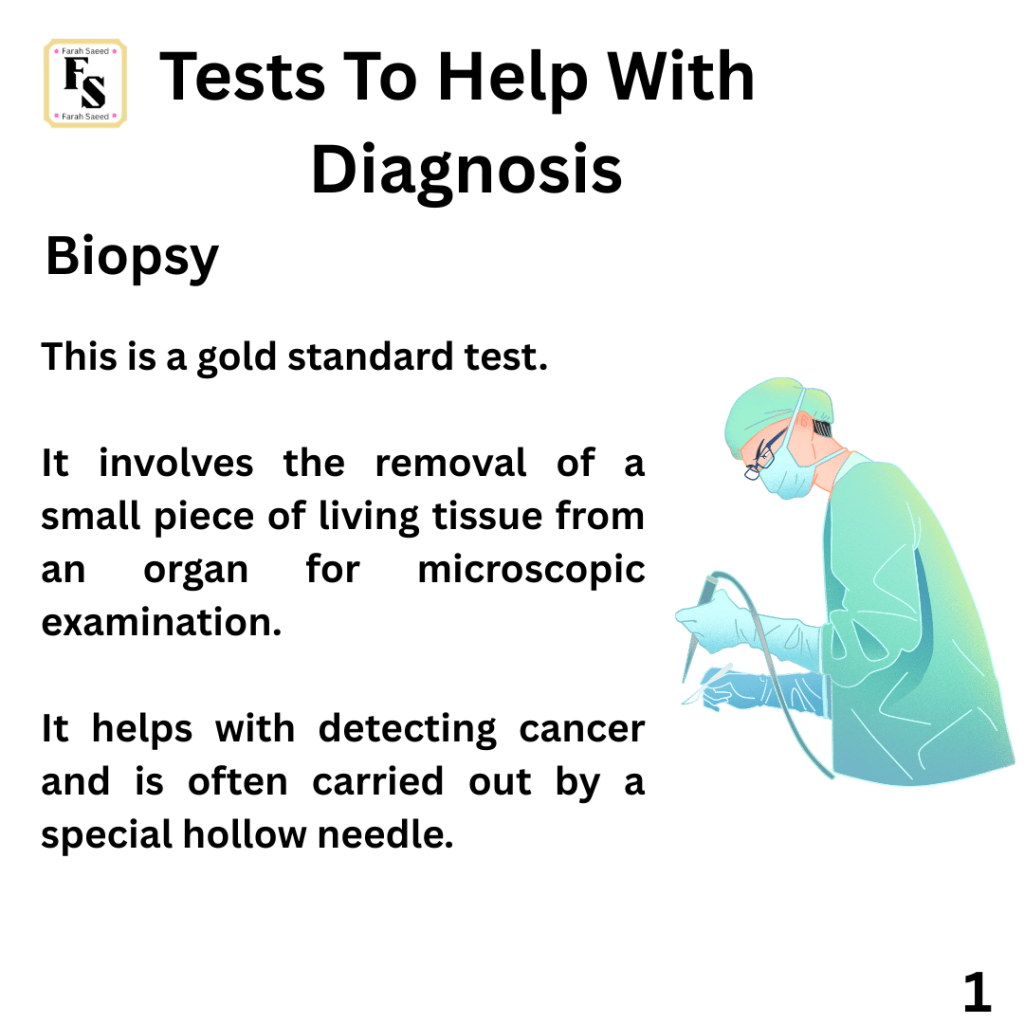
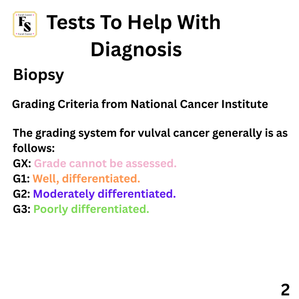
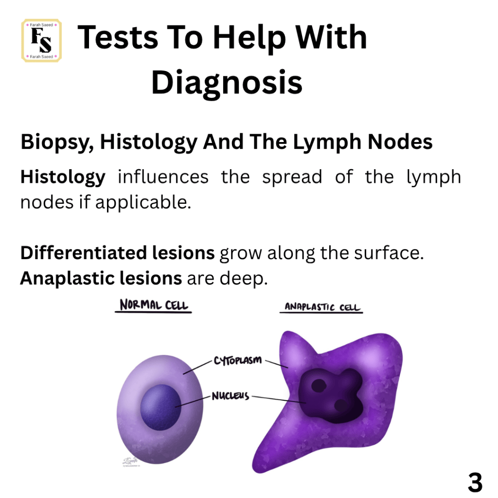
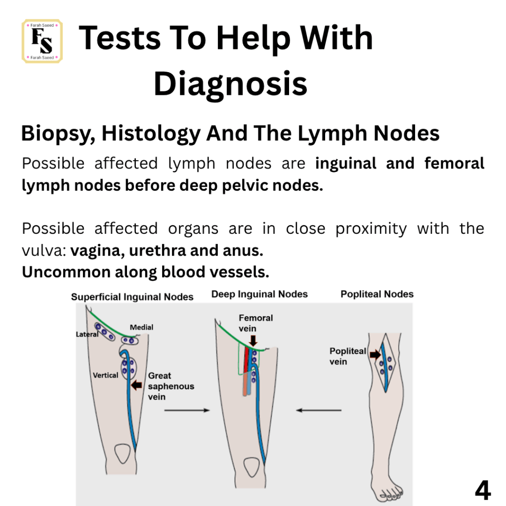
Biopsy
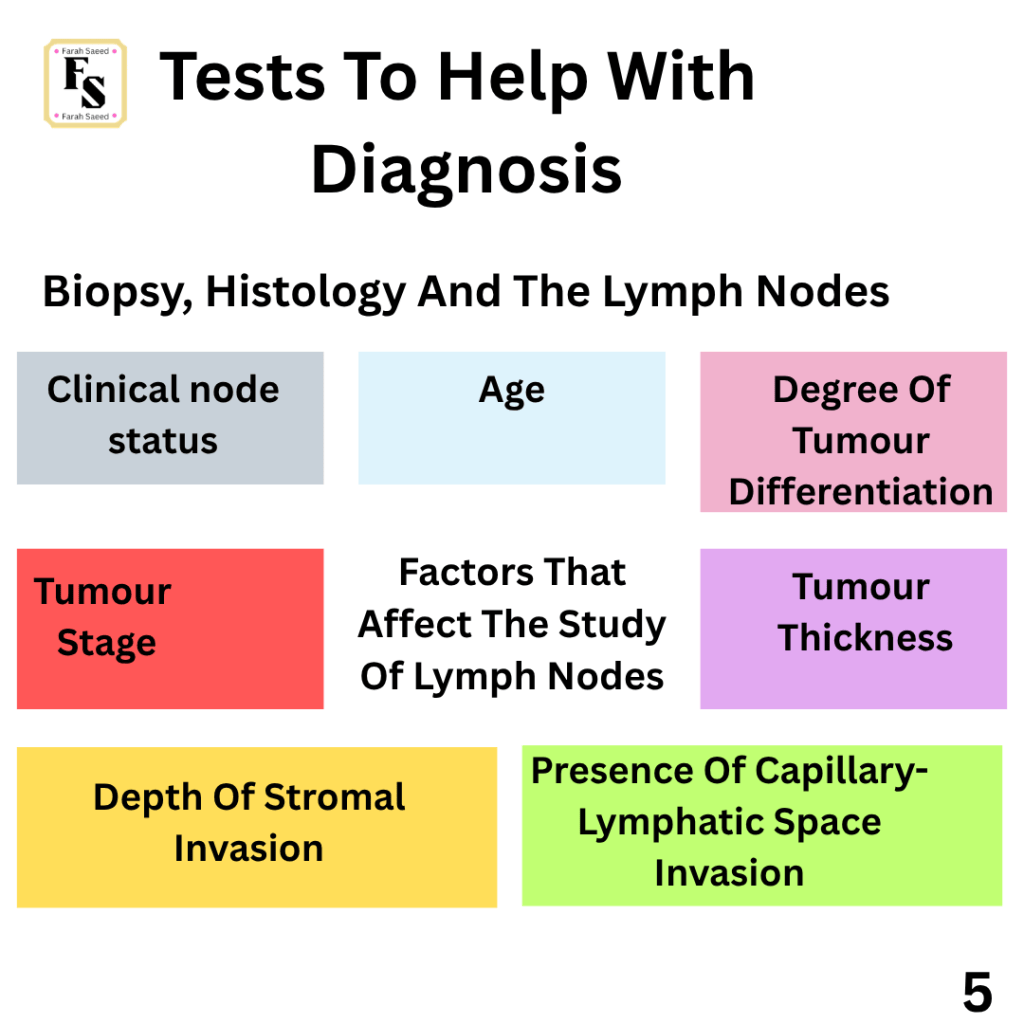
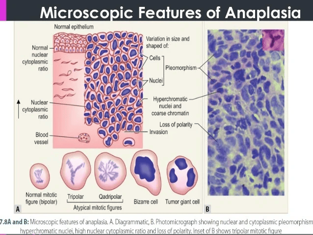
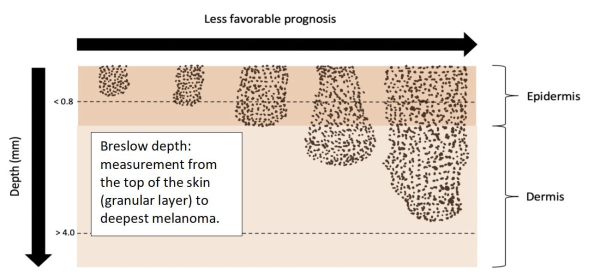
What is the difference between Clark level and Breslow’s thickness?
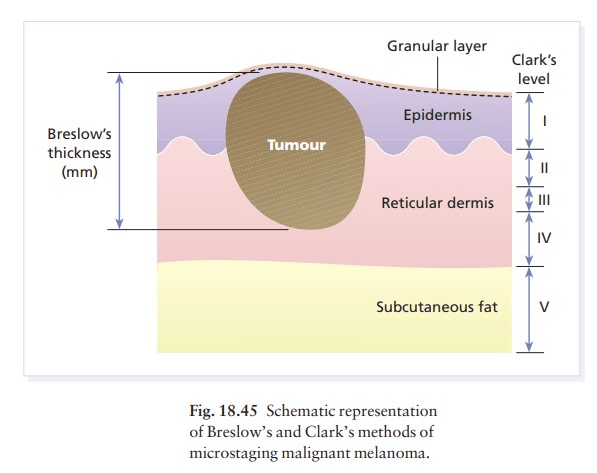
They measure the depth of invasion:
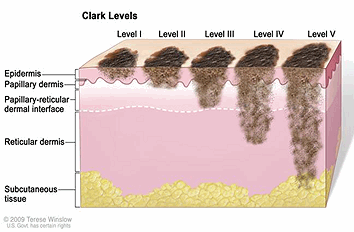
Clark – The anatomical extent of the depth of the invasion (Level 1 is epidermis and Level 5 is subcutaneous adipose tissue).
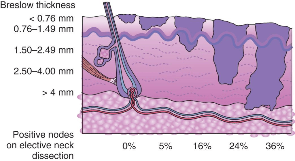
Breslow’s thickness: Vertical depth of invasion
Breslow’s thickness is suitable for cutaneous melanoma that arises from the vulva but not mucosal type.
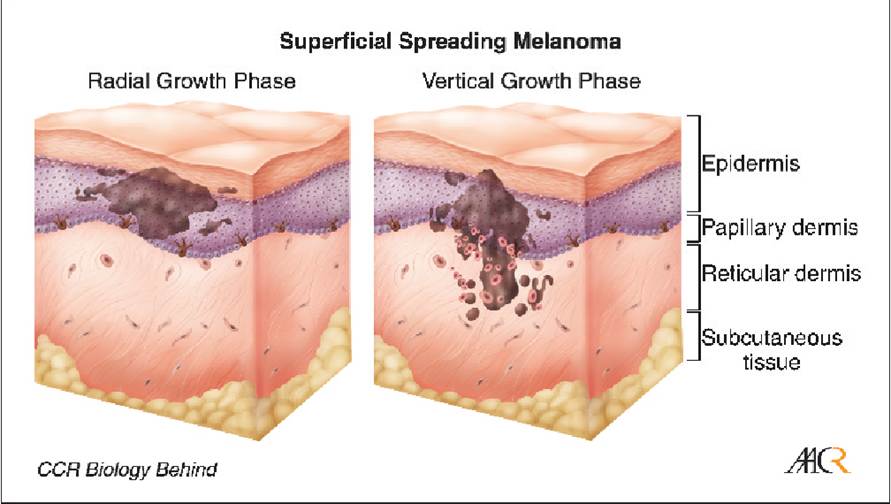
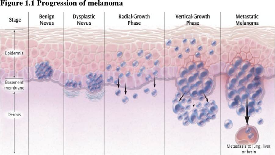
Understanding the difference between radial and vertical growth phase.
The radial growth phase applies to all early stages from the in-situ stage. It may also when growth of melanoma progresses to the dermis or submucosa tissue where single cells or nests do not grow (no mitotic activity) but remain embedded within the dermis.
Vertical growth helps to evaluate when the dermal nests enlarge or when mitotic activity can be measured with the melanocytes (melanin-producing cells) in the dermis. It is also a measurement of metastasis but is associated with the rate of mitosis rather than the size of the tumoral nests.
How is cell growth (mitotic activity) measured?
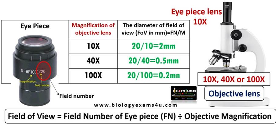
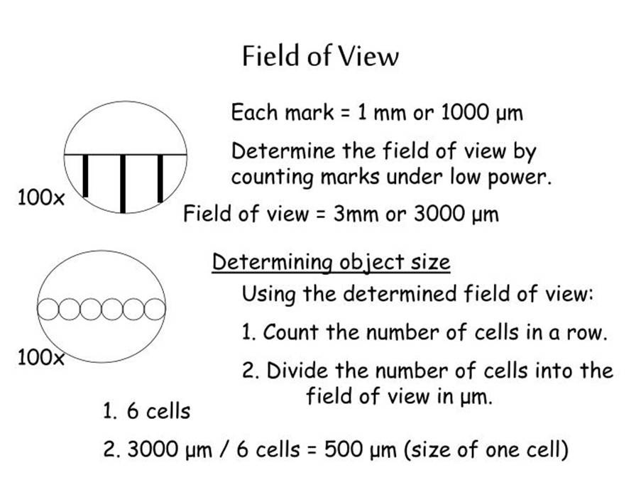
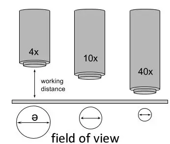
Mitotic activity is reported as mitotic count per square millimetre. The average reported was 6.6 and depends on the field of the microscope (visible area seen in the microscope). The most common reference is: 4 and ½ consecutive high-power fields at a magnification of 40×.
Supporting Images for mitotic cell count

Further information on the Clark level to determine melanoma invasion
A recall of level of melanoma invasion – Clark level critieria

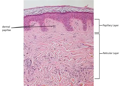
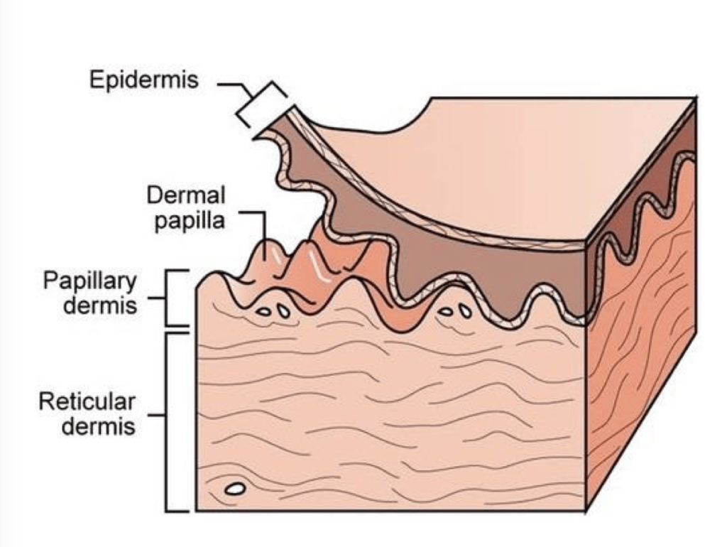
Supporting Images to help understand the difference between Clark level and Breslow’s thickness
Points To Consider
Clark levels II, III, IV – they vary in observation.
Clark level is not a recommended staging technique as mitosis (number of cell divisions for growth and repair) is not included in the analysis.
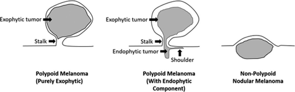
Polypoid growth pattern in melanoma: Breslow thickness may indicate it is invasive but still has Clark level III. – no prognosis (course of disease) as the level of thickness represents clinical behaviour.
Perineural invasion (affecting neurones/nerves) is commonly seen in mucosal forms of melanoma and experts indicate it needs to be reported as part of Breslow thickness.
A Further Insight Into Histological Features Of Vulval Cancers and Immunohistochemistry markers
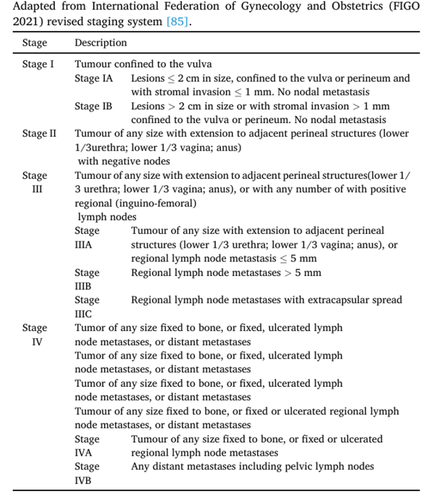
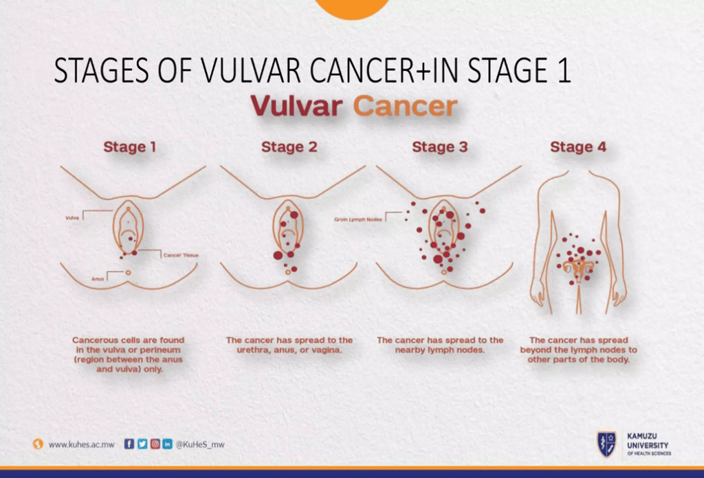
Staging of Vulval Cancers
Staging is achieved either through histology and/or radiology.
British Gynaecological Cancer Society (BGCS) vulval cancer guidelines: An
update on recommendations for practice 2023
Investigation Of Vulval Lesions
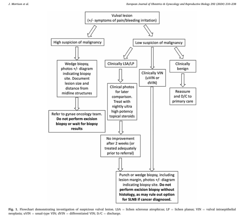
Further Insight Into Vulval Lesion Flow Chart
What is punch biopsy?
It is a type of incisional technique that involves the removal of a small piece of tissue from an organ for microscopic examination. In this case, superficial sampling.
It can help with diagnosis of the issue.
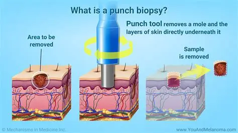
What is the relation between punch biopsy and vulval lesion?
It may not be suitable to take sample from if it is large or deep and may cause delay for diagnostic biopsy.
Features where punch or wedge biopsy is suitable?
The edge of a lesion where there is a change or transition from normal to abnormal tissues.
Avoid central ulceration as it does not help with detection.
Adequate depth: superficial invasion> 1 mm.
Avoid excision biopsy because it limits options for surgical techniques that involve wide local excision and sentinel node biopsy.
If the Lesion is small, vulva can heal well.
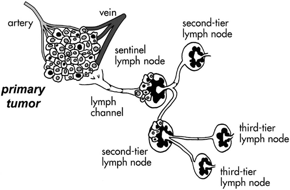
What is sentinel lymph node biopsy?
The sentinel lymph node is the first lymph node from the primary site that is used to present evidence of metastasis of a malignant tumour. The absence of cancer cells in this location will suggest that the more distal lymph nodes are cancer-free.
This can influence the treatment choices and is used for palliation and improve the quality of life.
Other incisional biopsies help to diagnose deeper lesions.
Radiological Examination of Vulval Cancer
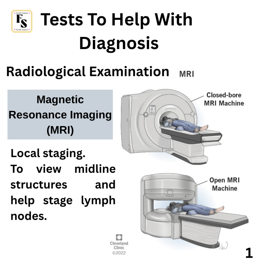
Further Information on Radiological Techniques
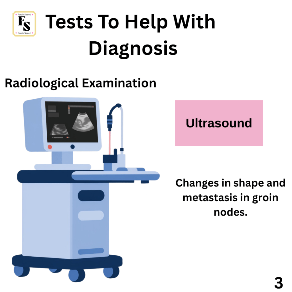
Ultrasound
Ultrasound is a good assessment of groin nodes in patients with vulval cancer.
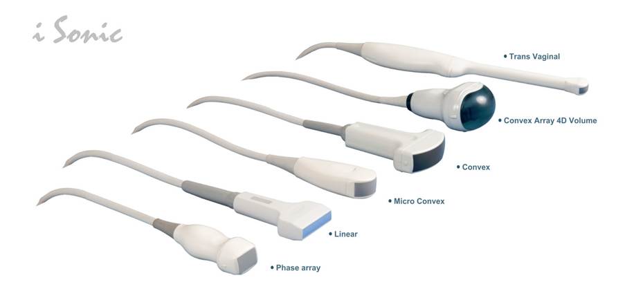
Ultrasound has different types of probes.
Linear Probe
A linear probe otherwise known as a linear array transducer has a flat rectangular surface that has piezoelectric elements in a straight line. The piezoelectric elements are what emit the ultrasound waves especially between 5 to 15 MHz perpendicular to the tissue. It is used for superficial masses especially in the peripheral lymph nodes as there is a limit of penetration between 2 to 6 cm beneath the skin. The sound waves or echoes that are reflected back using the piezoelectric elements create images when transmitted to the computer creating images with axial and lateral resolution. Thus pelvic MRI and ultrasound are the first line method to assess local regions. This is also in accordancwithto the European Society of Gynaecological Oncology guidelines.
Convex Probe
A convex array probe is predominantly used to view abdominal surfaces, organs and lymph nodes (visceral and retroperitoneal) when performing a transabdominal ultrasound. This helps to assess disease resectability/removal.
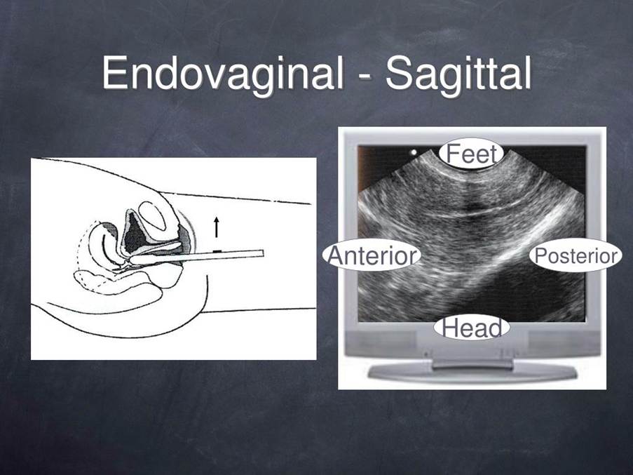
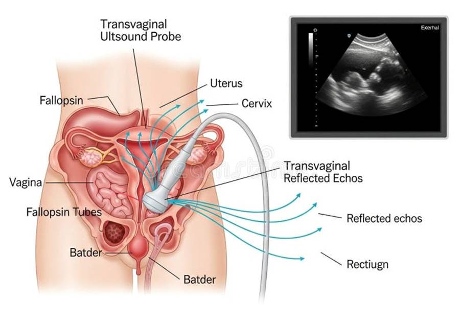
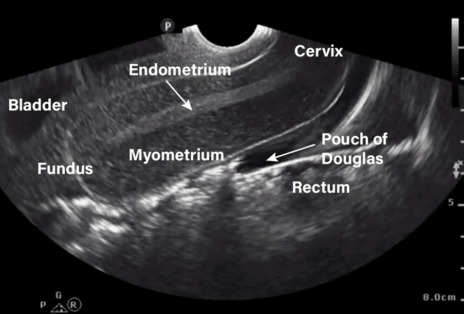
Endovaginal Probe
An endovaginal probe can help to view the pelvis. During the staging of the nodes (inguinofemoral), ultrasound is recommended as it has a sensitivity of 76 to 90% and specificity 60 to 96%.
The link between lymph nodes, biopsy and ultrasound
Suspected cases of groin nodes based on clinical examination/and or imagine may be further investigated with USS-guided fine needle aspiration or core biopsy where node positivity would alter management care.
What does this mean?
Ultrasound can help detect if there are any changes in shape (morphology) and metastasis in the lymph nodes when the healthcare professional performs fine-needle aspiration or core needle biopsy. Core needle biopsy is preferred in order to obtain sufficient sample to perform assessment via histology. However, if the lymph nodes are small, then fine-needle aspiration is recommended (Fischerova et al 2024).
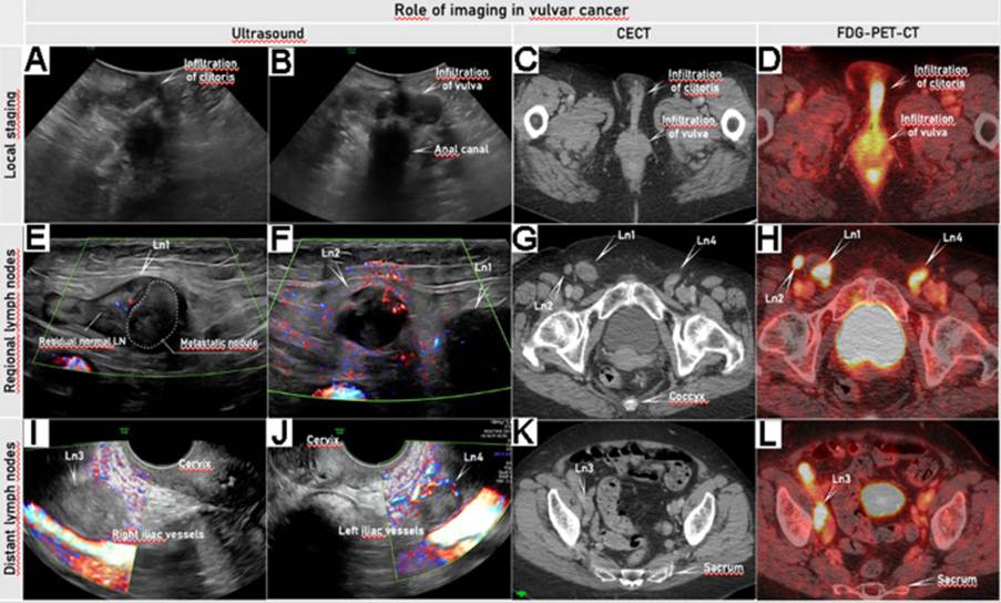
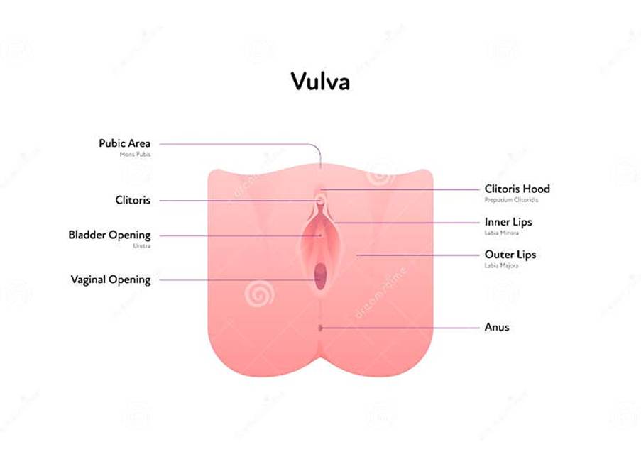
Fischerova et al. (2024) originally presented this image to help the general public understand the different features of the female reproductive system. Here, the scholars have presented an elderly patient with squamous cell vulvar cancer, International Federation of Gynecology and Obstetrics (FIGO) stage IVB. The images were obtained by ultrasound, contrast-enhanced CT (CECT), and 18F-fluorodeoxyglucose positron emission tomography combined with CECT (FDG-PET-CT). A convex array probe was used to perform a transperineal ultrasound in the transverse position to see how the tumour has affected the female reproductive system in Images A and B. Image A presents the tumour infiltrating the clitoris, as seen on ultrasound. Image B presents the tumour infiltrating the labia majora bilaterally (both sides).
Image C presents the infiltration of the clitoris and labia majora on CECT, where there is hyperdense tissue.
Image D presents the hyperdense tissue of the same vulvar pathology with high FDG-PET-CT. The illustration of the redness is a reflection of the fluorodeoxyglucose, which is a variant of normal glucose (sugar) that is not metabolised but is aggregated or joined together in areas where there is high metabolism and is often accompanied by fluorine-18. Metabolism is how the body is able to break down organic substances like carbohydrates to release energy. Therefore, it is a measure of the total biochemical and physical changes that are performed within the body.
An alternative ultrasound probe is being applied for Images E and F. A transcutaneous ultrasound using a linear array probe is applied to evaluate the regional lymph nodes (Ln) in the groin (inguinofemoral) as part of the Vulvar International Tumour Analysis. Two abnormal lymph nodes were identified and are labelled as Ln1 and Ln2, present above the fascia lata and femoral vessel on the right-hand side. They vary in the degree of infiltration, where Ln1 is partially infiltrated, and Ln2 is completely infiltrated.
The pathological lymph nodes (Ln1 and Ln2) are presented on CECT in Image G. They are also intensely FDG-avid on Image H.
To assess the lymph nodes in the pelvis, an endoluminal probe was inserted transvaginally to observe any metastasis. This is because of the location and size of the vulvar lesion.
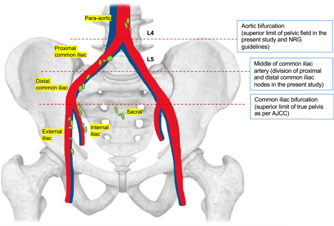
Metastatic lymph nodes can be found in Ln3 on the right iliac vessel in Image I. Image J presents a metastatic lymph node in Ln4 on the left iliac vessel.
For an in-depth evaluation of the iliac lymph nodes, Ln3 in the right iliac vessel is hyperintense on CECT in Image K.
The FDG has intensely labelled Ln3, illustrating high metabolic activity in the area via FDG-PET-CT in Image L.
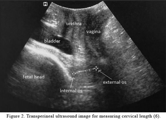
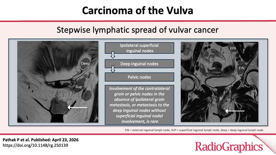
Images To Facilitate Understanding the Ultrasound Images

Cross-sectional imaging
Cross-sectional imaging is performed for further evaluation of pelvic nodes, groin nodes and median structures (urethra, vagina, anus and rectum).
In other words, if patients cannot undergo sentinel lymph node biopsy or if they have tumours in different areas (multifocal) or one area (unifocal) with a size that is larger than 4 cm (≥4 cm) or if there are suspected inguinal-femoral nodes detected before operation (pre-operative evaluation) then further imaging besides ultrasound or pelvic MRI. It helps to see the loco-regional location and exclude distant metastases. This can be achieved via one of the following:
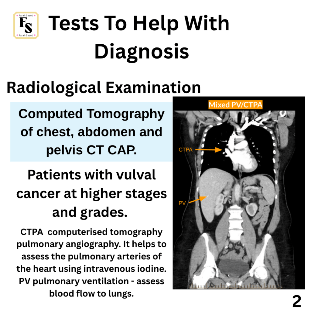
Computerised tomography of chest, abdomen and pelvis (CT CAP)
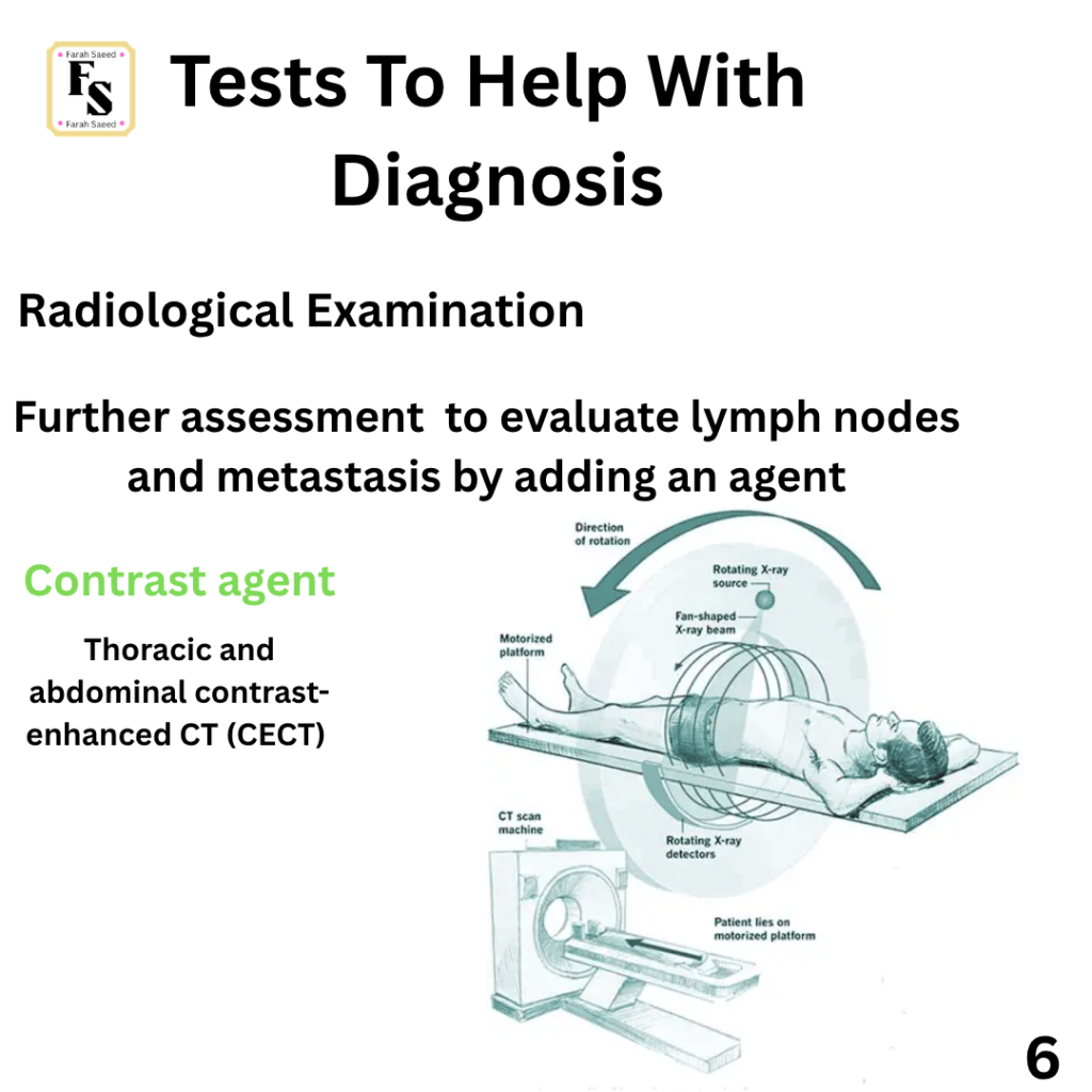
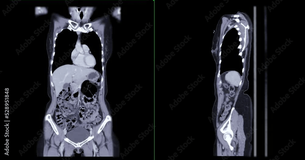
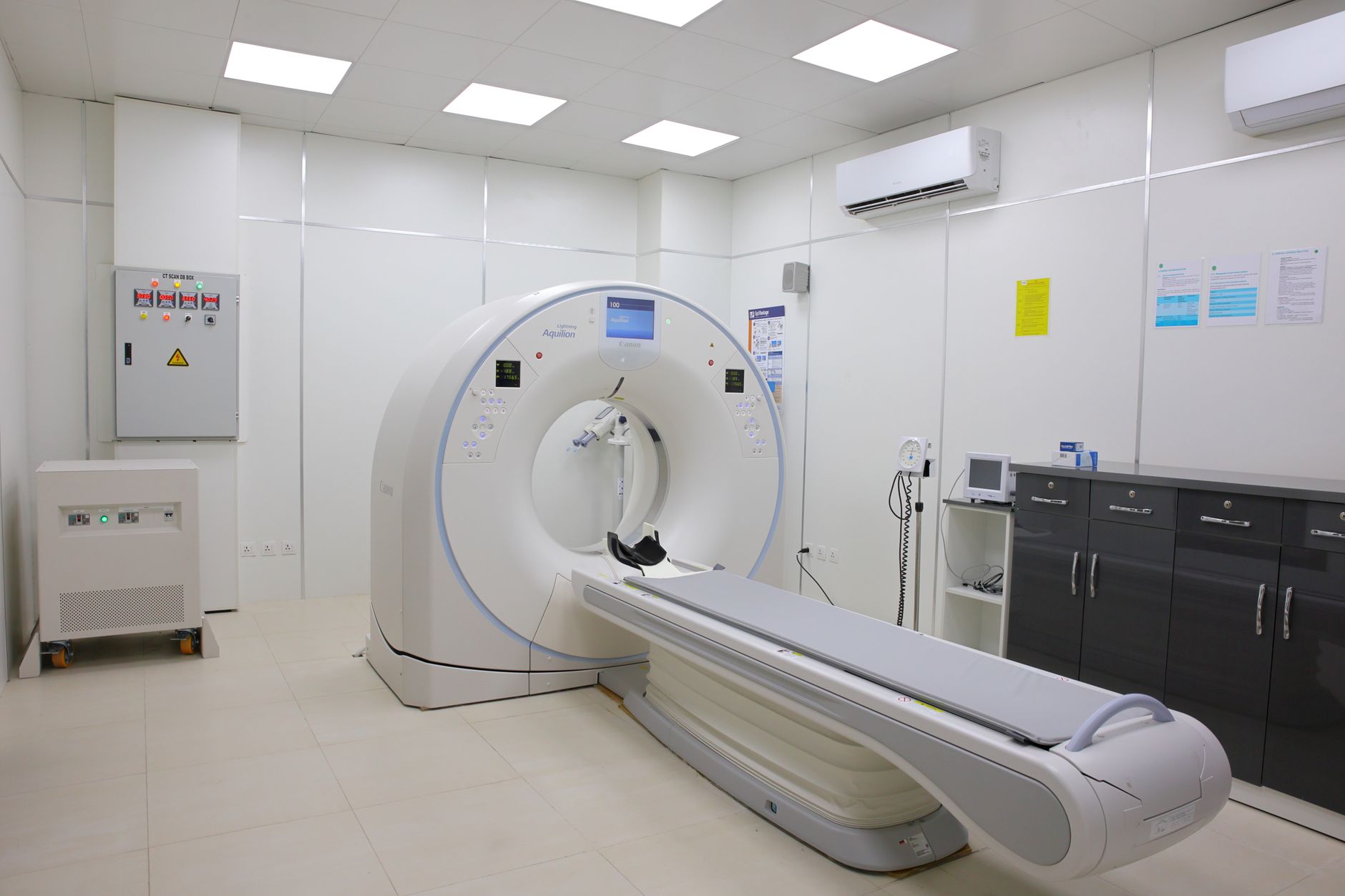
Computerised tomography of chest, abdomen and pelvis (CT CAP) is considered for patients with higher stage (III) and/grades to have a full-body scan. Computed tomography is a form of X-ray examination where the CT scanner, which contains the X-ray source and detector, rotates around the patient. The information is then transmitted to form cross-sectional images by a computer.
CT is also suggested for patients who are not recommended to have radical treatment e.g. surgery and to facilitate in discussion and planning treatmenttreatmenttreatment planning.
Magnetic Resonance Imaging (MRI)
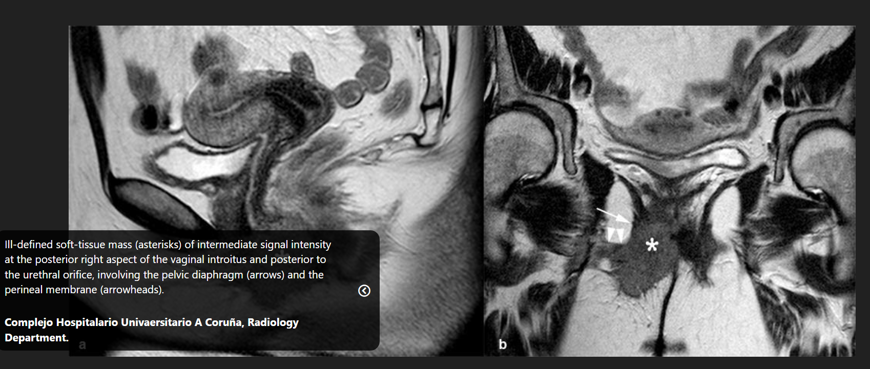
Magnetic Resonance Imaging (MRI) is suitable for patients with tumours with clear or equal involvement of midline structures (urethra, vagina, anus and rectum) and helps manage surgery. It can also help with local staging of vulval/vagina. Magnetic resonance imaging utilises a combination of strong magnetic field and radiofrequency radiation. The strength of the magnetic field is measured in teslas and most rely on a signal from hydrogen in water.
MRI is also applied for the staging of lymph nodes. It has variable sensitivity (40 to 89%) and this depends on what assessmentcriteria ares being applied. There are new MRI techniques for instance the whole-body diffusion-weighted imaging (DWI), DCE (dynamic contrast-enhanced), and high-resolution T2WI (T2-weighted imaging) series) that helps to assess the upper abdomen (belly) and distant sites for metastases. This helps to improve the staging of the local region. Experts have indicated that ultrasound or MRI should have a structured imaging report to present information to healthcare professionals.
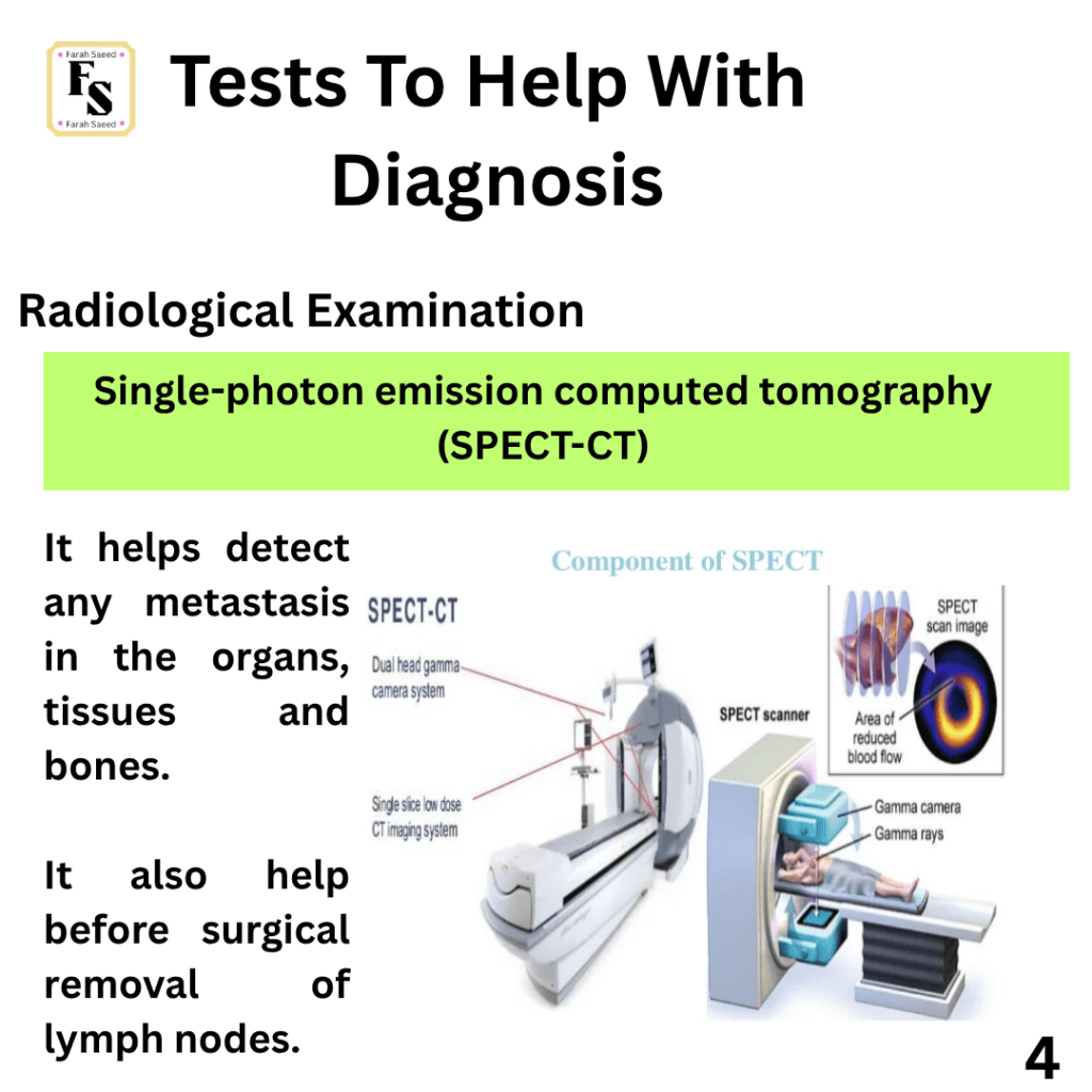
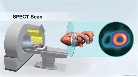
Single-photon emission computed tomography (SPECT-CT)
Single-photon emission computed tomography (SPECT-CT) is a type of imaging test where a chemical called radiotracer is injected in the vein and a special gamma camera takes 3D images of the organs, tissues and bones. In some cases the radiotracer is inhaled or swallowed. The main types of SPECT scans are brain, heart and bone. The SPECT machine takes the images and transmits them to the allocated computer.
One of the primary differences between all the scans is most focus on identifying organs, tissues and bones but other scans like Positron Emission Tomography (PET) help understand how it works. It also helps before surgical removal of lymph nodes if necessary (lymphadenectomy).
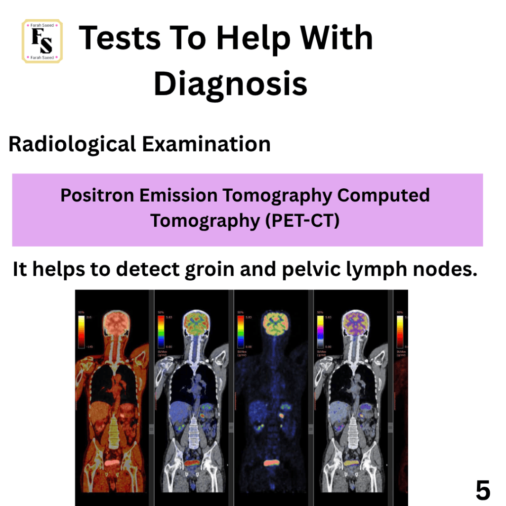
Positron emission tomography (PET-CT)
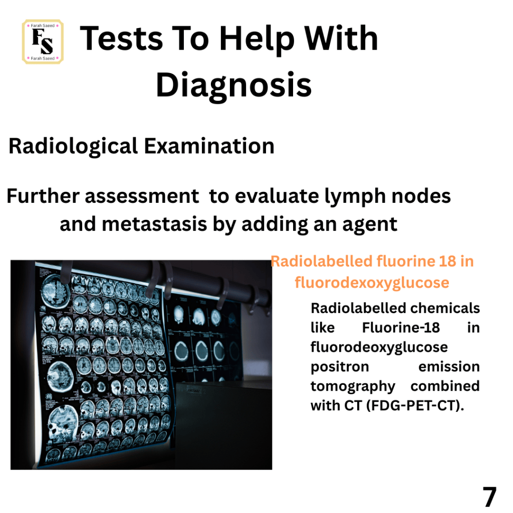
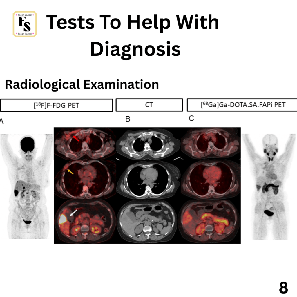
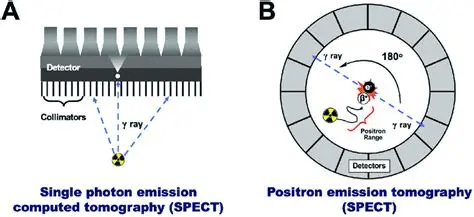
Positron emission tomography (PET-CT) has sensitivity ranging from 50 to 100% and specificity from 67 to 100% to evaluate the inguinal lymph nodes. This is why it has limited value in detecting lymph node metastasis that is less than 5 mm and in necrotic nodes. It is also not recommended for staging vulval cancers. PET helps to evaluate the metabolic activity and applies cross-sectional imaging. A positron is emitted by a radioisotope and this leads to annihilation of the positron when colliding or binding with an electron. A positron is an electrically charged particle. It has the same mass an electron but a positive charge. The two gamma rays travel opposite directions and the PET scanner helps detect the arrival of both the gamma rays. Chemicals are added and labelled to examine metabolism. Common examples are carbon -11, oxygen-15, nitrogen-12 and fluorine-18.
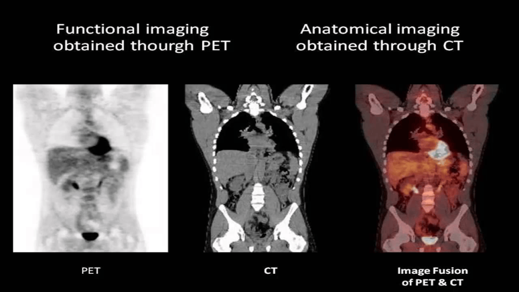
Thoracic and abdominal contrast-enhanced CT (CECT) or whole-body 18F-fluorodeoxyglucose positron emission tomography combined with CT (FDG-PET-CT)
Both are recommended to assess lymph nodes and metastasis. Other roles of CECT are to detect cause of symptoms, (coughing, pain in the chest and breathing difficulties like shortness of breath). It helps to guide fine-needle biopsy and congenital abnormalities.
Radiological Diagnosis Of Vulval Melanoma

One of the challenges faced when diagnosing melanoma is the risk of metastasis is high because the locally advanced lesion is difficult to visualise and it correlates with depth of invasion (Breslow thickness).
Younger age is linked with improved overall survival.
Mutational testing for c-KIT and BRAF mutations though they are rare.
Intracranial lesions are not uncommon.
Recommended imaging for Vulval Melanoma
Computerised tomography of chest, abdomen and pelvis (CT CAP)
Computed Tomography (CT)
Magnetic Resonance Imaging (MRI)
Radiology and Basal Cell Carcinoma (Vulva)
Distant disease is rare.
Itching and bleeding are the main symptoms.
No specific imaging needed unless there is suspicion of nodal disease.
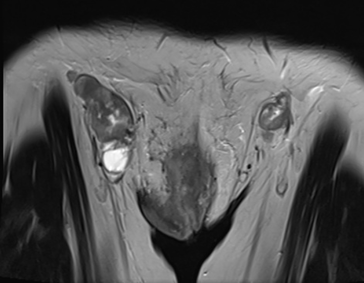
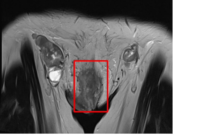
Magnetic Resonance Imaging (MRI) – it helps to evaluate growth of vulvar cancer by taking a coronal T2 image. MRI is considered the imaging modality of choice for evaluating local growth of vulvar cancer.
Vulval Paget’s Disease
Tests for urological, colorectal, uterine and breast cancers hence examination via cystoscopy, colonoscopy, hysteroscopy, CT and breast examination required.
Primary cutaneous VPD needs to have routine screening for secondary malignancies.
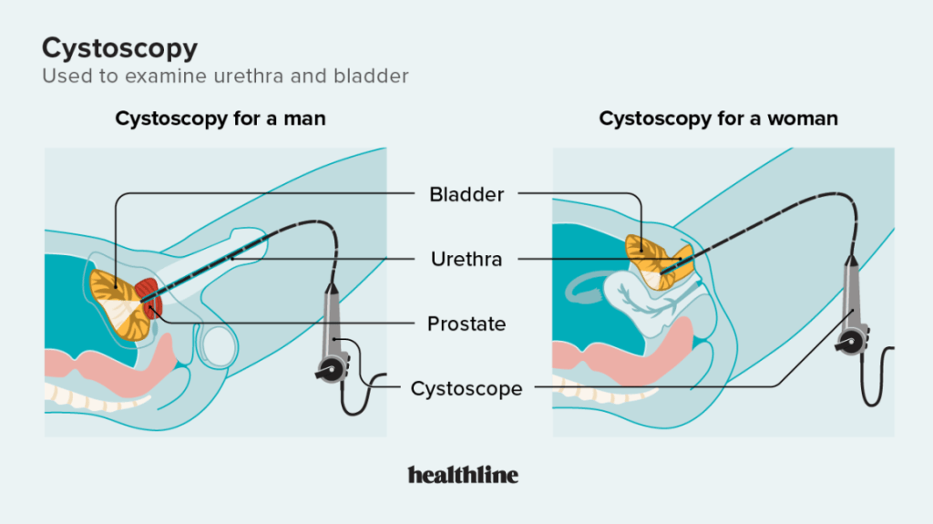
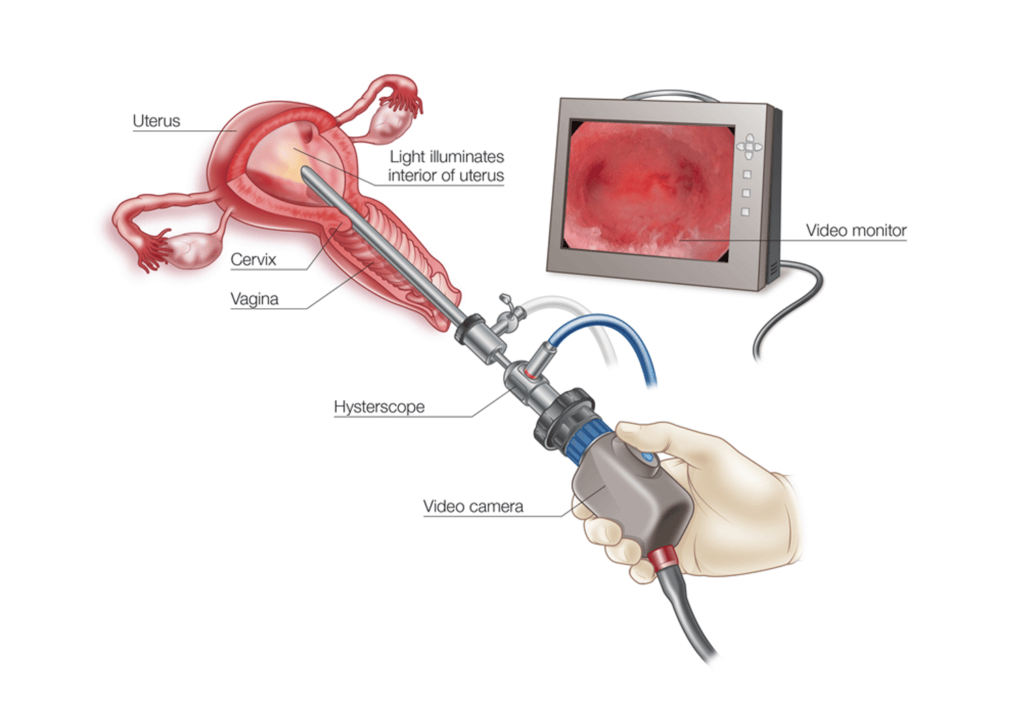
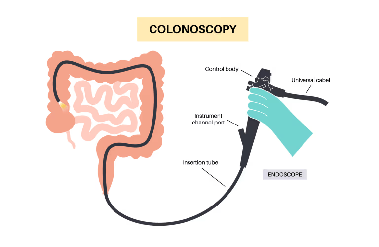
Primary cutaneous VPD need to have routine screen for secondary malignancies.
Cancer of the Bartholin’s glands

High rate of advanced disease due to their location: deep into the skin and less obvious.
Prior to operations, CT-CAP recommended because not suitable for sentinel lymph node as it increases risk of local-regional spread at diagnosis.
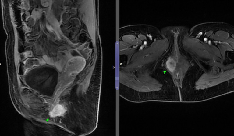
MRI pelvis to gain an idea or delineate of the extent it has been affected.

Recommended Article
Ultrasound Findings Of The Lymph Node Using Vulva International Tumour Analysis By Fisherova et al. 2021
References
Ablaza, A.C., Gupta, R. and Fielder, T. (2026) Stains (IHC and special) p16. Available at: https://www.pathologyoutlines.com/topic/stainsp16.html (Accessed: 23rd September 2026)
Addley, S., Sadeghi, N., Smyth, S.L., Johnson, C., Damato, S. and Majd, H.S. (2023). Bartholin’s gland carcinoma—the diagnostic and management challenges of a rare malignancy—a case report and review of current literature. Translational Cancer Research, [online] 12(1). doi:10.21037/tcr-22-612.
Almadani, N., Thompson, E.F., Tessier-Cloutier, B., Pors, J. and Hoang, L. (2020). An update of molecular pathology and shifting systems of classification in tumours of the female genital tract. Diagnostic Histopathology, 26(6), pp.278–288. doi: 10.1016/j.mpdhp.2020.03.007.
Al-Sajee, D. and Huizinga, J. D (2012). Interstitial Cells of Cajal : Pathology , Injury and Repair = خلايا كاجال الخلالية : علم الأمراض و الإصابات و الإصلاح. 12(4), pp.411–421. doi:10.12816/0003165.
Anton, C., Vicentini, A., Carvalho, F.M., Baracat, E.C. and Carvalho, J.P. (2011). Clinical treatment of vulvar Paget’s disease: a case report. Clinics, 66(6), pp.1109–1111. doi:10.1590/s1807-59322011000600033.
Basic Medical Key (n.d.) Malignant Melanoma of the Vulva. Available at: https://basicmedicalkey.com/malignant-melanoma-of-the-vulva/ (Accessed: 22nd September 2026)
Cancer Research UK (2025) Vulval Melanoma. Available at: https://www.cancerresearchuk.org/about-cancer/vulval-cancer/stages-types-grades/types/vulval-melanoma (Accessed: 20th August 2026)
Cancer Research UK Cambridge Centre (n.d.) Clinical research on gynaecological cancer. Available at: https://crukcambridgecentre.org.uk/patient-care/clinical-research/gynaecological (Accessed: 10th August 2026)
Cassidy, J., Bissett, D., Spene, R. and Payne, M. (2010) Oxford Handbook of Oncology. Oxford: Oxford University Press.
CDC (2024) Symptoms of Vaginal and Vulvar Cancers. Available at: https://www.cdc.gov/vaginal-vulvar-cancers/symptoms/index.html (Accessed: 10th August 2026)
Chen-Yost and Antic (2026) Stains (IHC and special) Epithelial membrane antigen (EMA). Available at: https://www.pathologyoutlines.com/topic/stainsema.html (Accessed: 22nd September 2026)
Cleveland Clinic (2023) SPECT Scan. Available at: https://my.clevelandclinic.org/health/diagnostics/spect-scan (Accessed: 23rd September 2026)
Clinical Tree (2024) Vulvovaginal melanoma. Available at: https://clinicalpub.com/vulvovaginal-melanoma/ (Accessed: 27th September 2026)
Diaz Angulo, C., Perez Ramos, T., Mendez Diaz, C., Rodriguez Garcia, E., Soler Fernandez, R. and Teijo Quintans, A. (2015) Bartholin’s gland carcinoma. Available at: https://www.eurorad.org/case/12314 (Accessed: 26th September 2026)
Fischerova, D., Smet, C., Umberto Scovazzi, Diana Natacha Sousa, Hundarova, K. and Ingfrid Salvesen Haldorsen (2024). Staging by imaging in gynecologic cancer and the role of ultrasound: an update of European joint consensus statements. International Journal of Gynecological Cancer, 34(3), pp.363–378. doi:10.1136/ijgc-2023-004609.
Frauenfeld, L. and Schurch, C. (2022) Stains and CD markers: CD117/KIT. Available at: https://www.pathologyoutlines.com/topic/cdmarkerscd117.html (Accessed: 22nd September 2026)
Honest Pathology (2026) Clark Level in Melanoma: Understanding Its Role in Staging, Prognosis, and Follow-Up. Available at: https://honestpathology.com/is-clark-level-still-relevant/ (Accessed: 26th September 2026)
Jones, O. (n.d.) Bartholin’s Cyst and Abscess. Available at: https://teachmeobgyn.com/gynaecology/vaginal-vulval/bartholins-cyst-abscess/ (Accessed: 17th August 2026)
Kamungo, A. (n.d.) Female Genital Tract Pathology. Available at: https://www.slideshare.net/slideshow/female-genital-tract-pathology-and-pptx/280526961 (Accessed: 23rd September 2026)
Kösemehmetoğlu, K. and Johnson, G. (2022) Stains and CD markers Actin, alpha smooth muscle type. Available at: https://www.pathologyoutlines.com/topic/stainsalphasmoothmuscleactin.html (Accessed: 22nd September 2026)
Mahjabin, F., Ordulu, Z., Martinez-Duarte, E. and Falzarano, S.M. (2023). Sebaceous differentiation can occur in vulvar adenoid cystic carcinoma. International Journal of Gynecological Cancer, 33(11), pp.1830–1832. doi:10.1136/ijgc-2023-004323.
Mohamed, K.S., Means, A., and Chen, H. (2026) Cervix: Other epithelial tumours. Available at: https://www.pathologyoutlines.com/topic/cervixadenoidcystic.html (Accessed: 27th September 2026)
Morrison, J., Baldwin, P., Hanna, L., Andreou, A., Buckley, L., Durrant, L., Edey, K., Faruqi, A., Fotopoulou, C., Ganesan, R., Hillaby, K. and Taylor, A. (2024). British Gynaecological Cancer Society (BGCS) vulval cancer guidelines: An update on recommendations for practice 2023. PubMed, 292, pp.210–238. Doi: 10.1016/j.ejogrb.2023.11.013.
National Cancer Institute (2024) What is Vulvar Cancer? Available at; https://www.cancer.gov/types/vulvar/what-is-vulvar-cancer (Accessed: 7th August 2026)
National Cancer Institute (2025) HPV and Cancer. Available at: https://www.cancer.gov/about-cancer/causes-prevention/risk/infectious-agents/hpv-and-cancer (Accessed: 6th August 2026)
National Cancer Institute (2026) Vulvar Cancer Treatment (PDQ®)–Health Professional Version. Available at: https://www.cancer.gov/types/vulvar/hp/vulvar-treatment-pdq (Accessed: 17th August 2026)
National Cancer Institute (n.d.) What Does the Ki-67 score mean? Available at: https://cancerexplained.org/report-terms/what-does-ki-67-mean/ (Accessed: 22nd September 2026)
National Institute for Health and Care Excellence (2026) Gynaecological cancers – recognition and referral: Presentation Available at: https://cks.nice.org.uk/topics/gynaecological-cancers-recognition-referral/background-information/presentation/ (Accessed: 7th August 2026)
National Institute for Health and Care Excellence (2026) What symptoms are suggestive of gynaecological cancers? Available at: https://cks.nice.org.uk/topics/gynaecological-cancers-recognition-referral/background-information/presentation/ (Accessed: 21st September 2026)
Ohashi, R. (2026) Stains (IHC and Special) Vimentin. Available at: https://www.pathologyoutlines.com/topic/stainsvimentin.html/ (Accessed: 22nd September 2026)
Pathak, P., Lundgren, M., Green, I., Abdalla, I., Thampy, R., Hunt, C.R. and Behnia, F. (2026). Carcinoma of the Vulva. Radiographics : a review publication of the Radiological Society of North America, Inc, [online] 46(5), p.e250139. doi:10.1148/rg.250139.
Rasmy, A., Amal, A., Mashiaki, M. and Alanizi, B. (2019) PD-1 Inhibitors: To Go or To Stop? Review Articles. Cancer Stud Ther J Volume 4(2): 1–6.
Samba, S., Guerrouaz, M.A., Bensghier, A., Berhili, S., Moukhlissi, M. and Mezouar, L. (2024). Adenoid Cystic Carcinoma of the Vulva: A Case Report. Cureus. doi:10.7759/cureus.56048.
The Eve Appeal (2026) Cervical Cancer. Available at: https://eveappeal.org.uk/information-and-advice/gynaecological-cancers/cervical-cancer/ (Accessed: 7th August 2026)
The Eve Appeal (2026) Vulval cancer Available at; https://eveappeal.org.uk/information-and-advice/gynaecological-cancers/vulval-cancer/ (Accessed: 7th August 2026)
Vargas-Hernández, V.M., Manuel, V., Abraham, M., Xochilt, P. and Ferrer, G.M. (2024). Basic cell carcinoma of the vulva. Case report and literature review. Obstetrics & Gynecology International Journal, [online] 15(3), pp.132–135. doi:10.15406/ogij.2024.15.00748.
Vera‐Kellet, C. and Hasbún, C. (2020). Extramammary Paget’s disease of the vulva: Persistent vulvar itching requires a skin biopsy! Clinical Case Reports, [online] 8(7), pp.1325–1326. doi:10.1002/ccr3.2862.
Xing, D. and Lu, J. (2018). Distinctive clinicopathological features and disease‑specific survival of adenoid cystic carcinoma and adenoid basal carcinoma in the lower female genital tract. Oncology Reports. doi:10.3892/or.2018.6927.
Xu, B. (2025) Salivary glands Primary salivary gland neoplasms: Malignant Adenoid cystic carcinoma. Available at: https://www.pathologyoutlines.com/topic/salivaryglandsadenoidcystic.html (Accessed: 23rd September 2026)
Additional Educational Resources
An overview of Vulval Cancer By South Tyneside and Sunderland NHS Foundation Trust.
What to expect: Vulval Cancer Referral By North East London Cancer Alliance.

