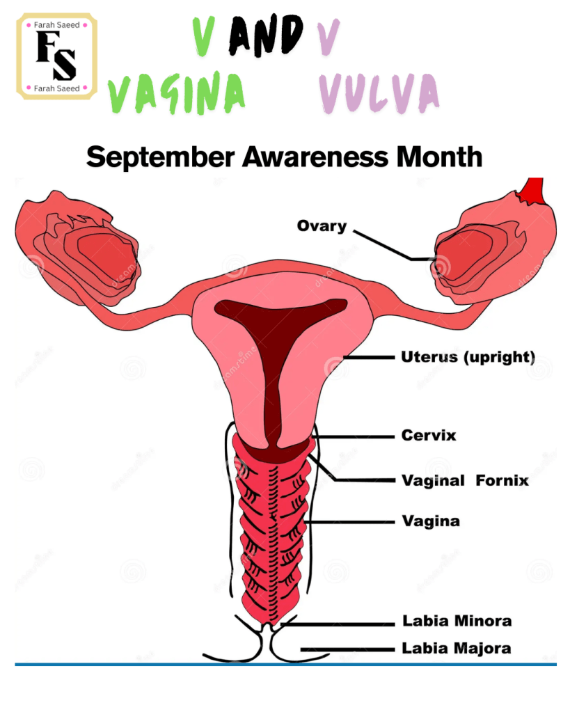
Treatment Of Vulval Cancer
Below summaries the main points for all treatments in accordance to British Gynaecological Cancer Society (BGCS) vulval cancer guidelines: An update on recommendations for practice 2023. Please read the guidelines attached for further details.
Just like with any cancer, the treatment depends on the location, size, stage, grade, age, general health and medical history of the patient.

Surgery
Overview And Preparation
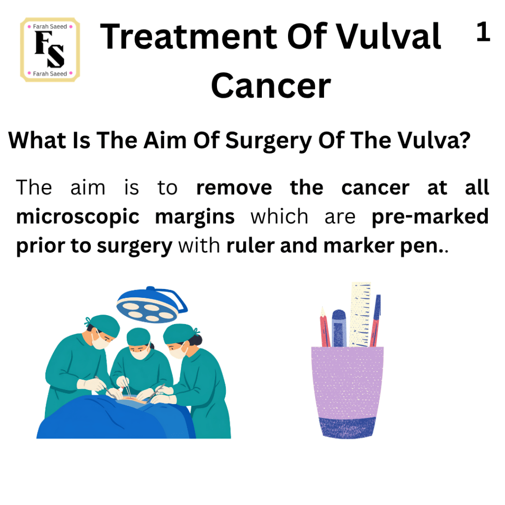
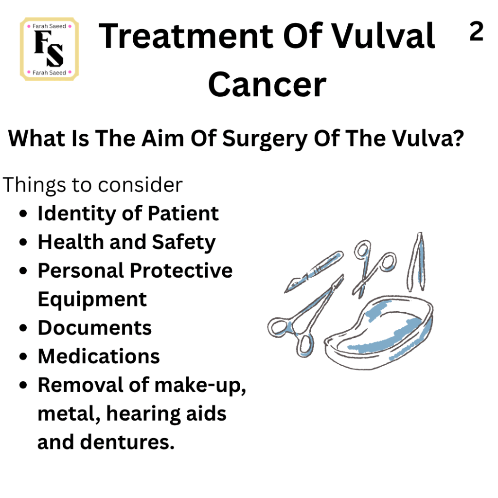
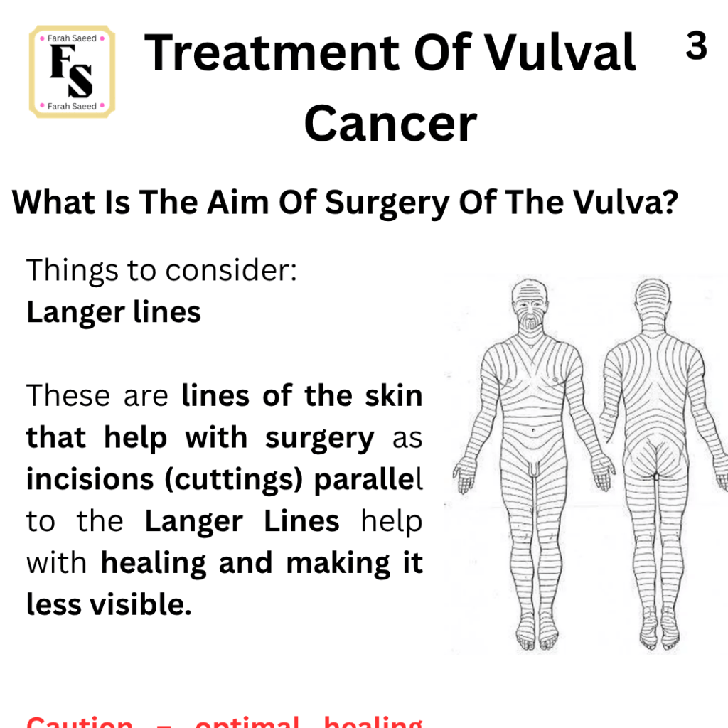
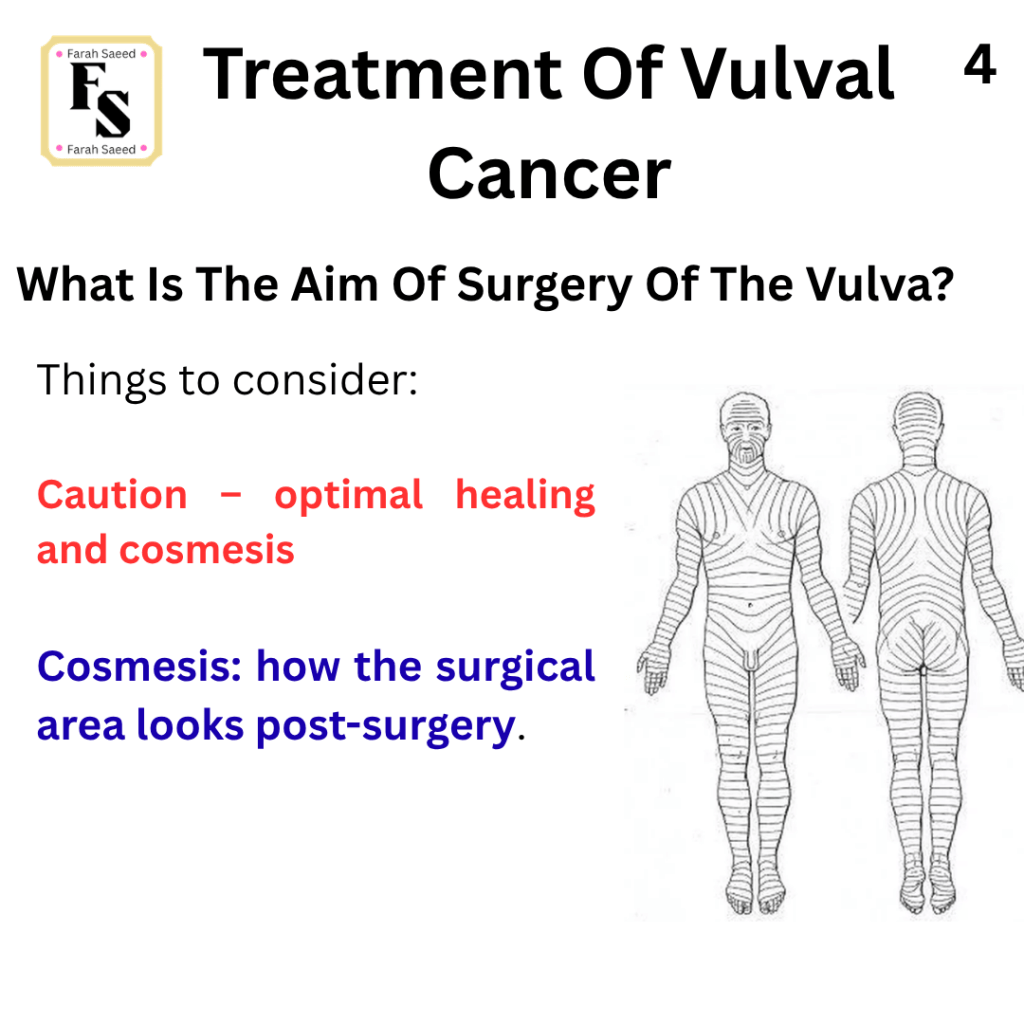
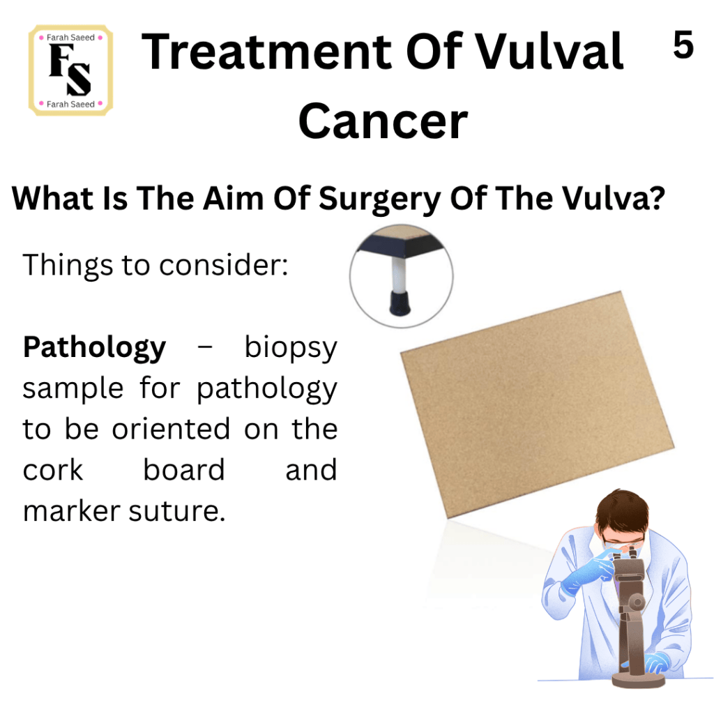
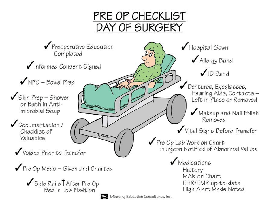
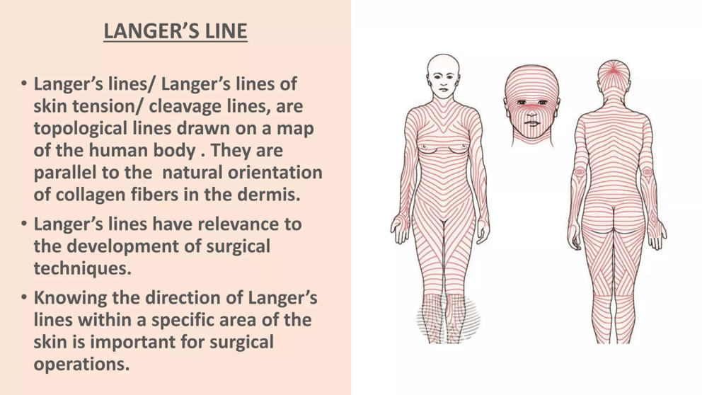
Additional Resources For Preparation For Surgery
Main Types Of Surgery Of The Vulva
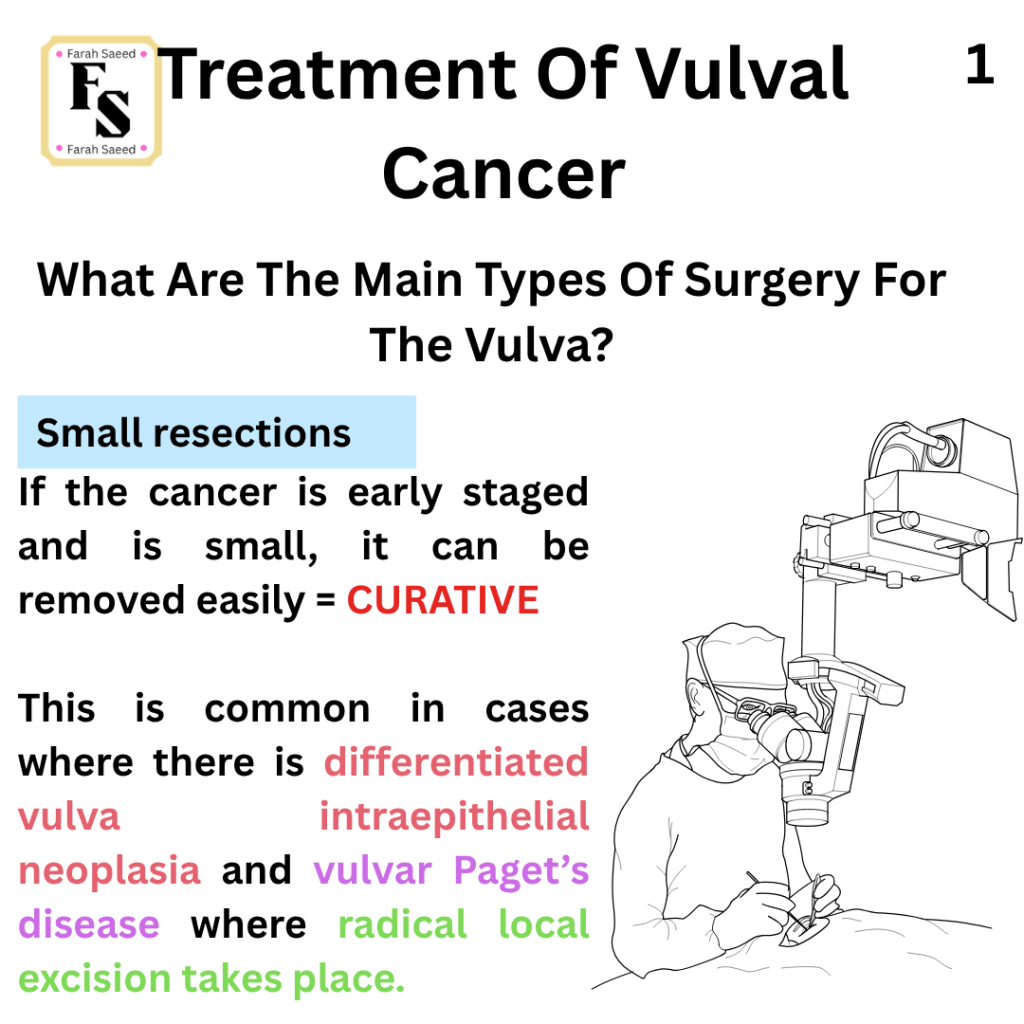
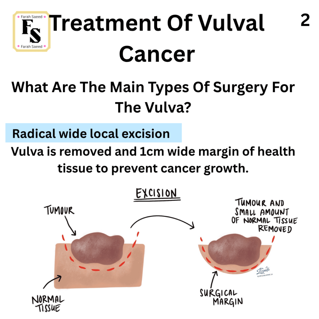
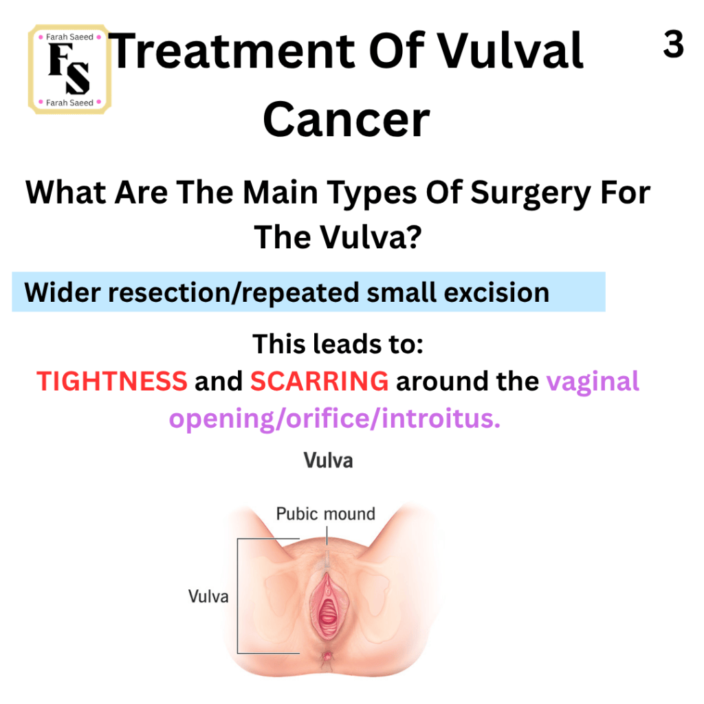
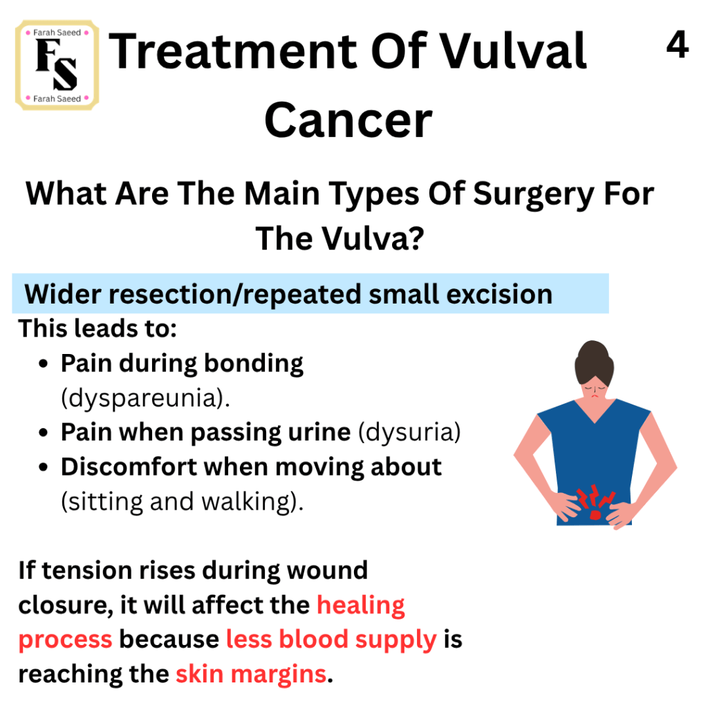
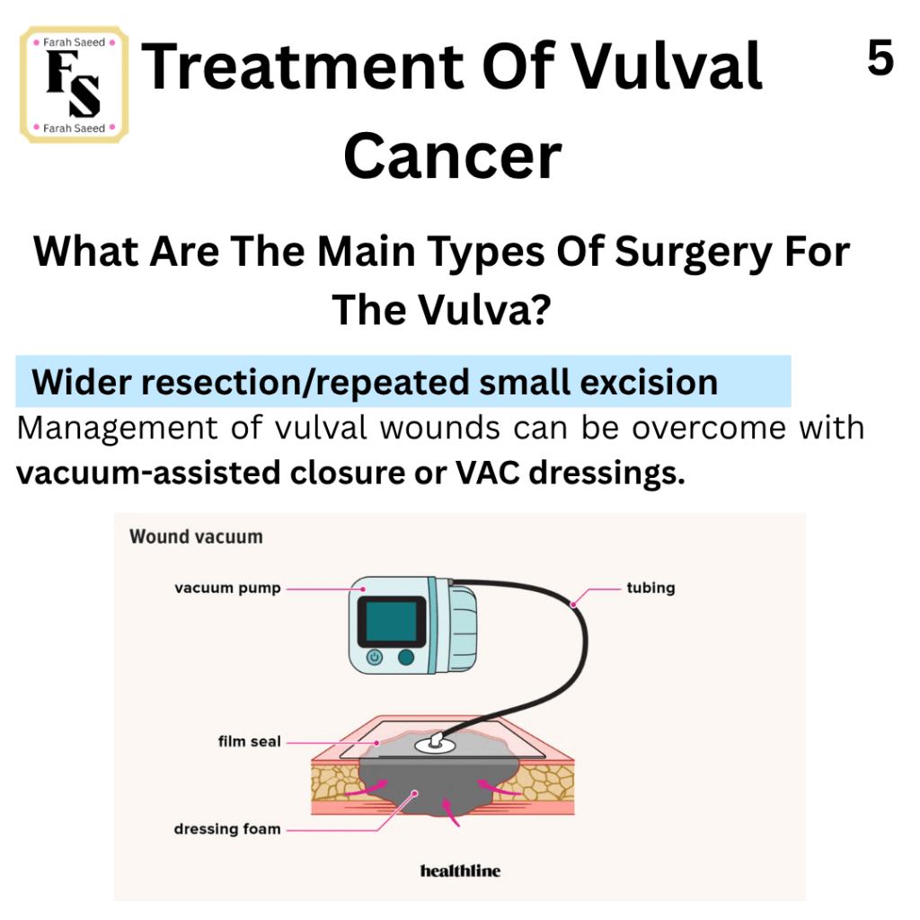
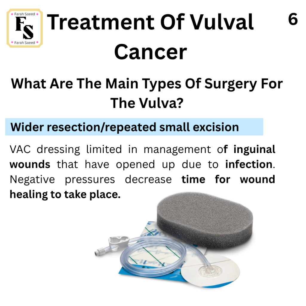
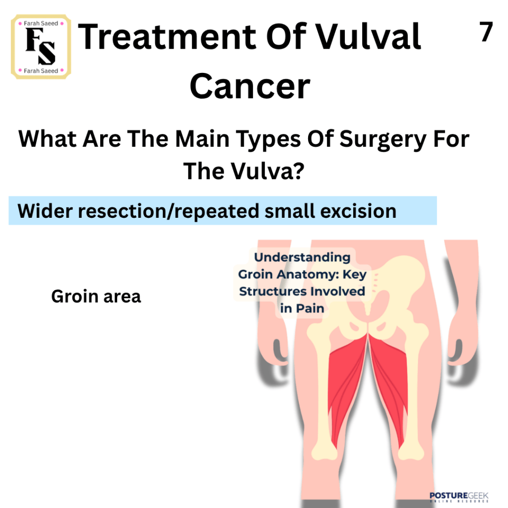
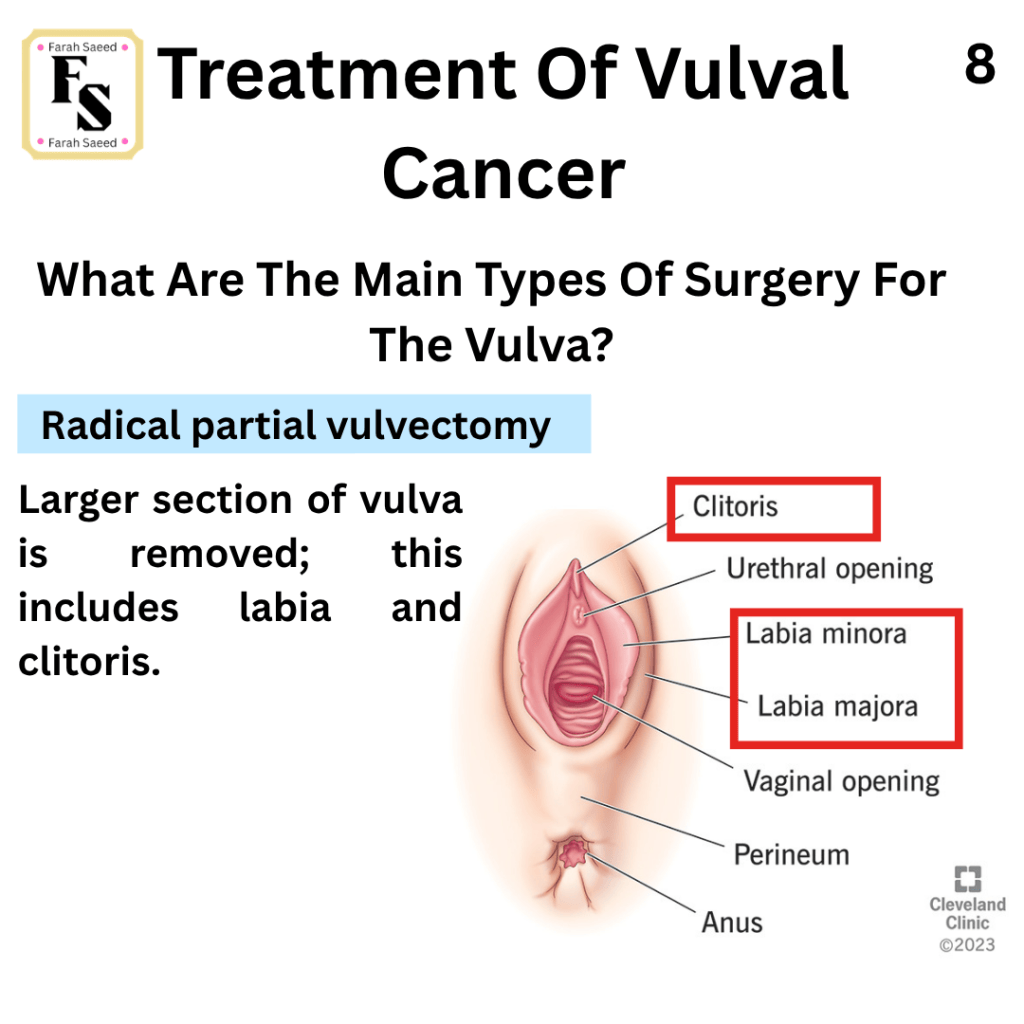
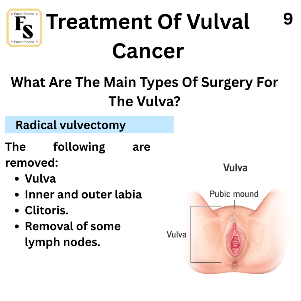
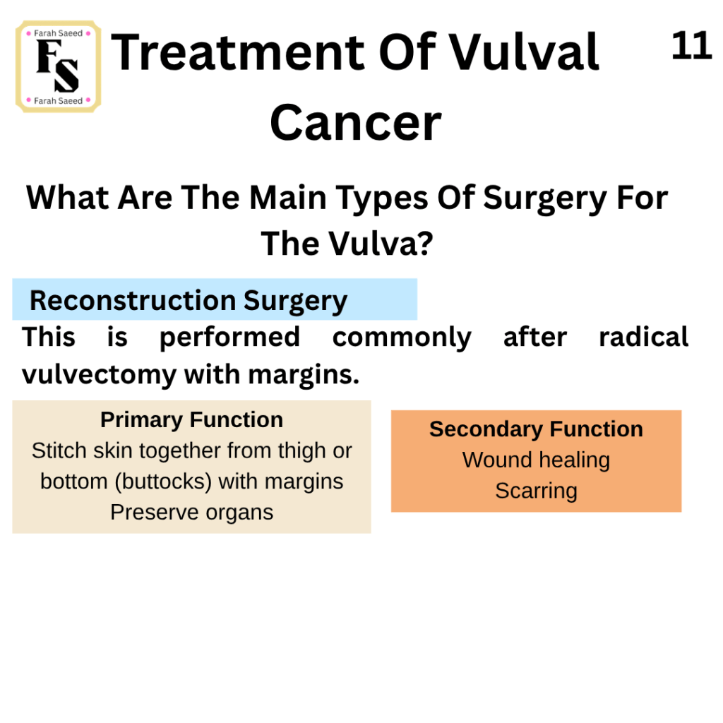
Additional Resources For Main Types Of Surgery
By Dr PK Mohan on Medical Education Briefs
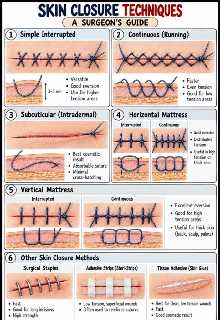
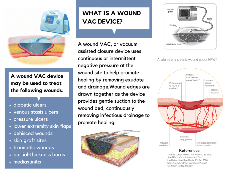
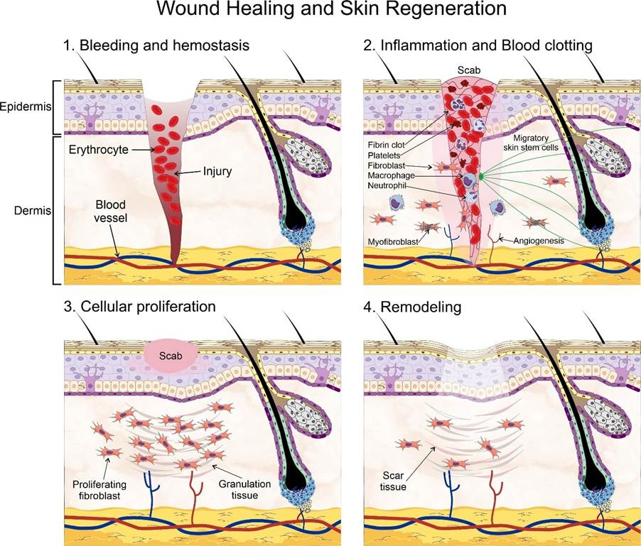
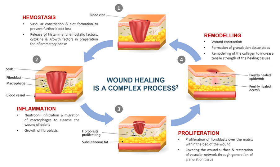
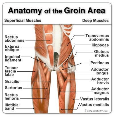
Robotic Surgery and Vulval Cancer
Vulvar Cancer Treatment by Robotic Gynec Oncologist – Cancerclinix
Treatment for Vulval Squamous Cell Carcinoma
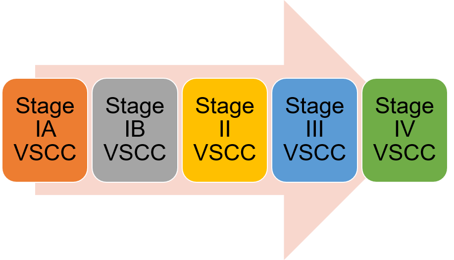
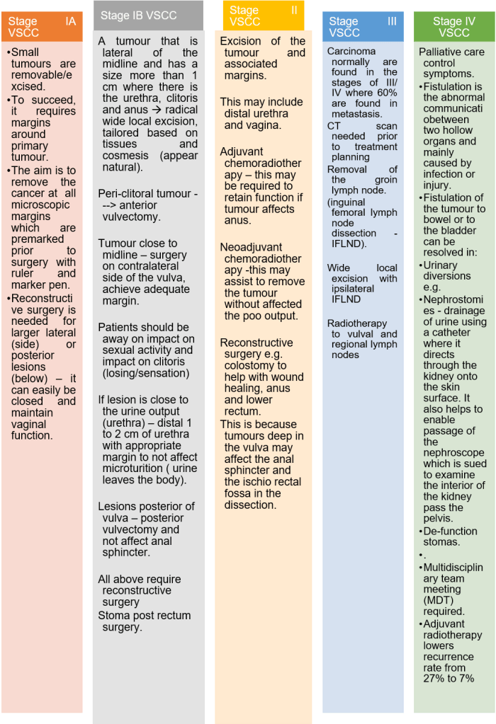
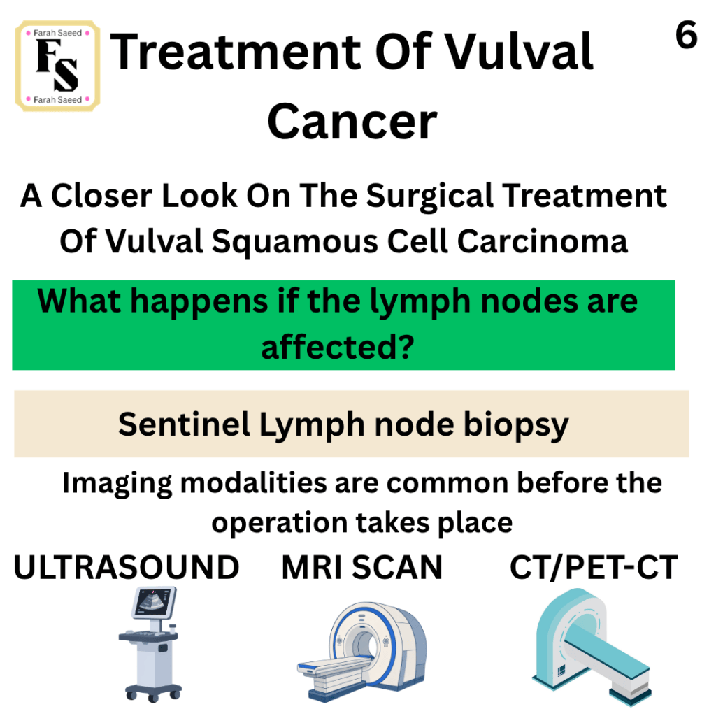
Squamous cell carcinoma of the vulva is the most common type of vulval cancer. Staging is an important process that can be defined by either radiological techniques, for instance, ultrasound, computed tomography (CT) and magnetic resonance imaging (MRI) or it can be achieved through surgical biopsy and histology. The purpose is to help identify the size of the tumour which will guide in the necessary prognosis and treatment.
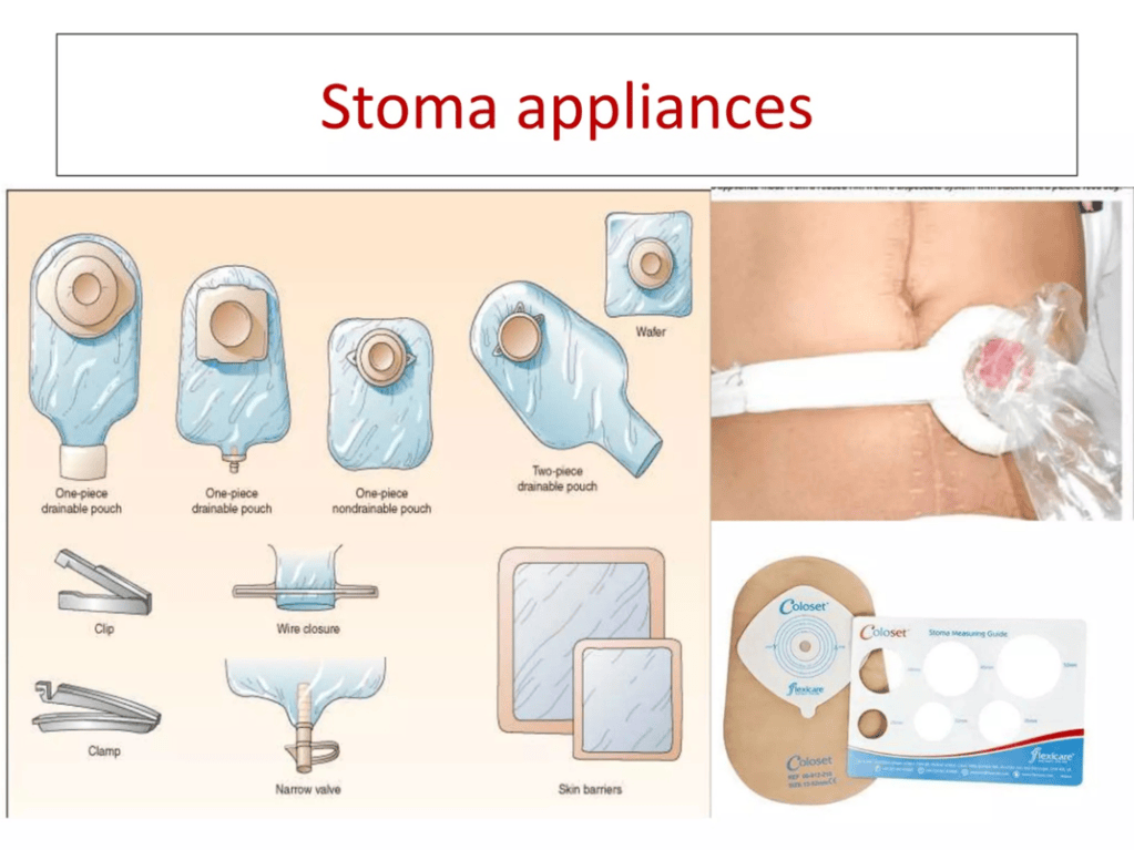
Types Of Stoma
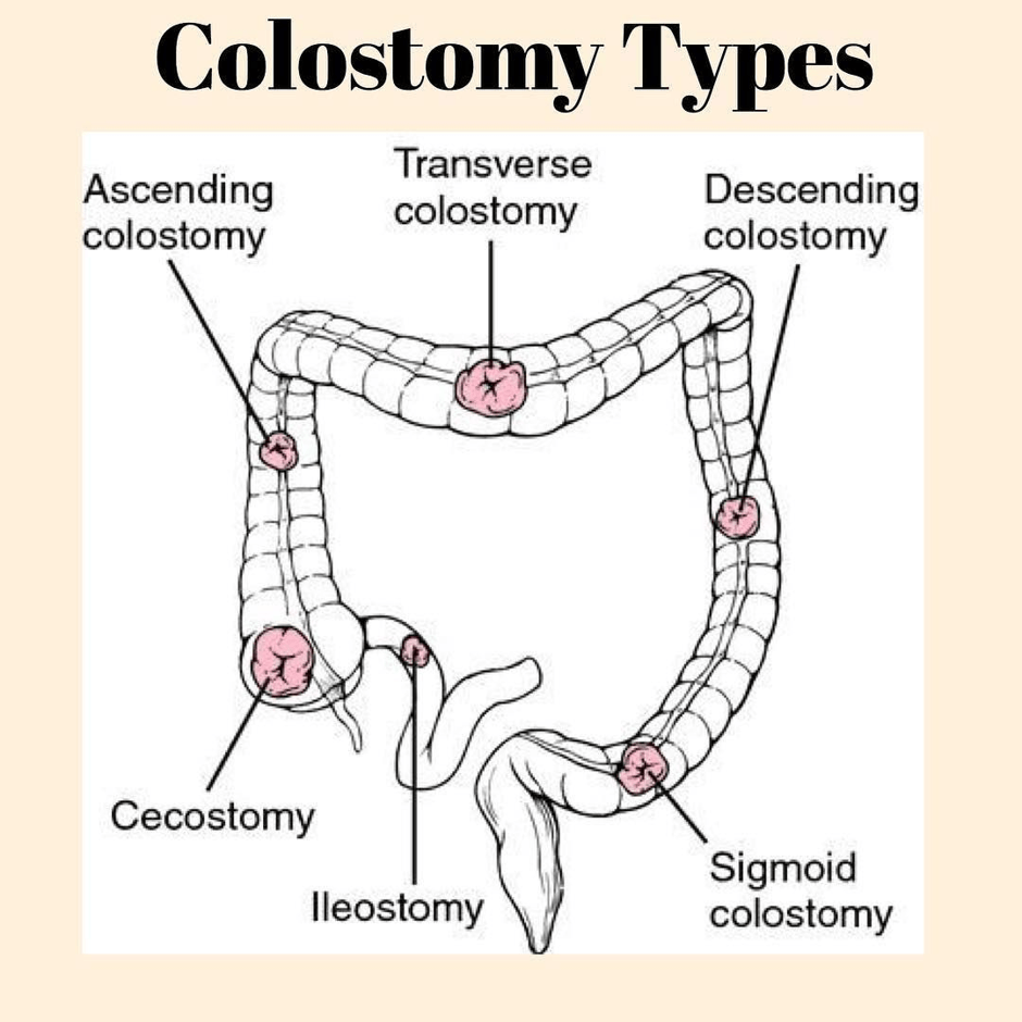
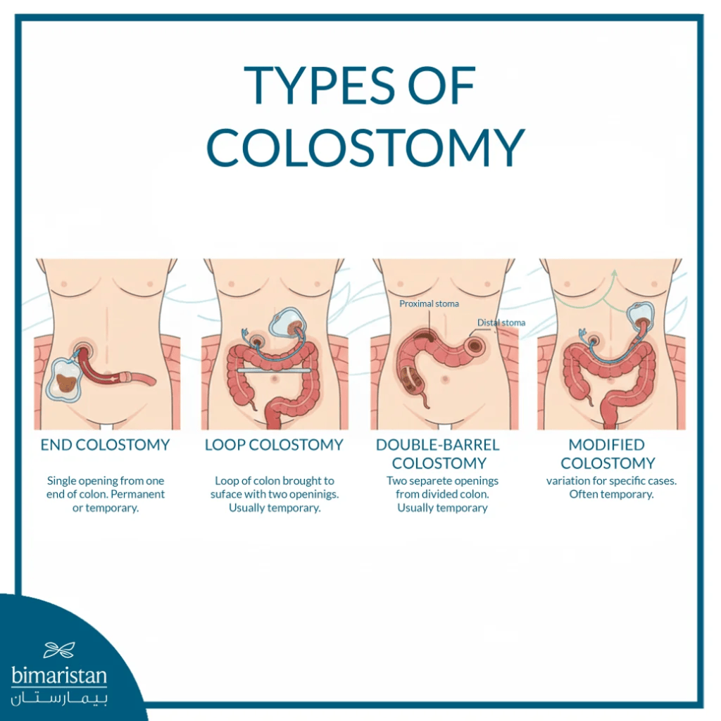
The purpose of stoma is for the colon to be taken throughout the abdominal wall to decompress the intestine.
Iliac colostomy onto the left lower abdomen.
Transverse colostomy on the upper abdomen.
Surgery For VSCC – A Closer Look
A gentle reminder: This is in accordance to the British Guidelines and contains key points. Please visit the guidelines directly for additional information.
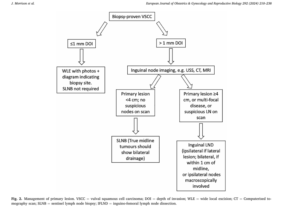
Early stages – VIN/VAIN
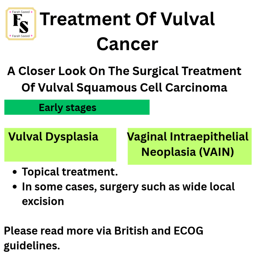
Additional Resources of VIN
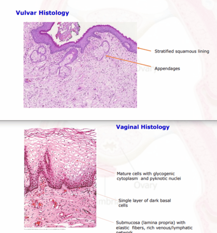
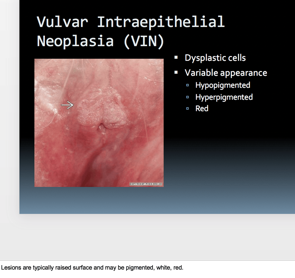
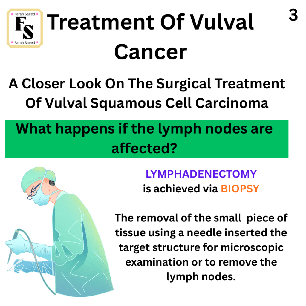
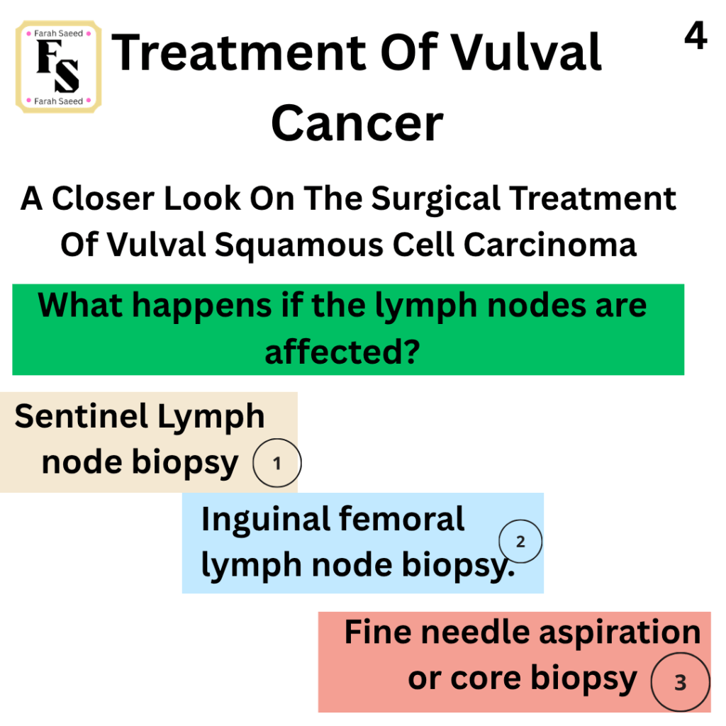
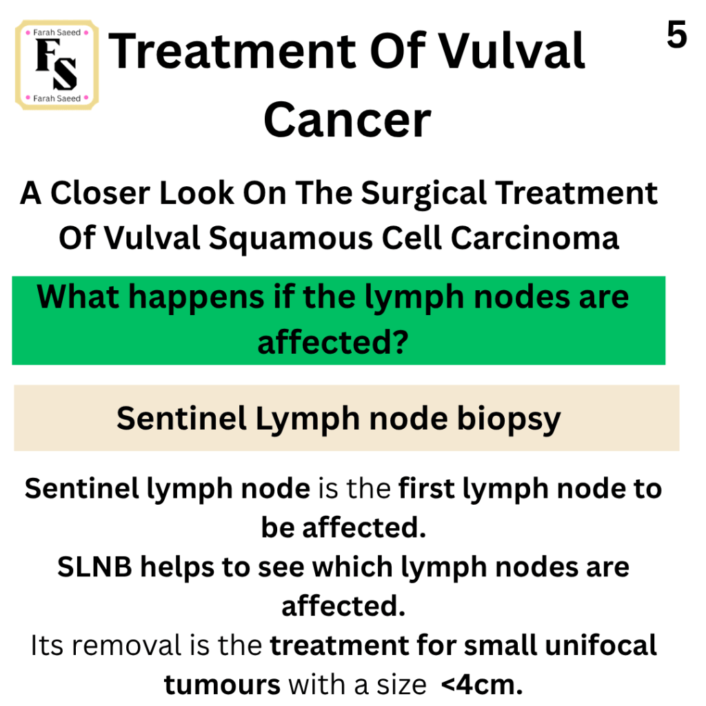
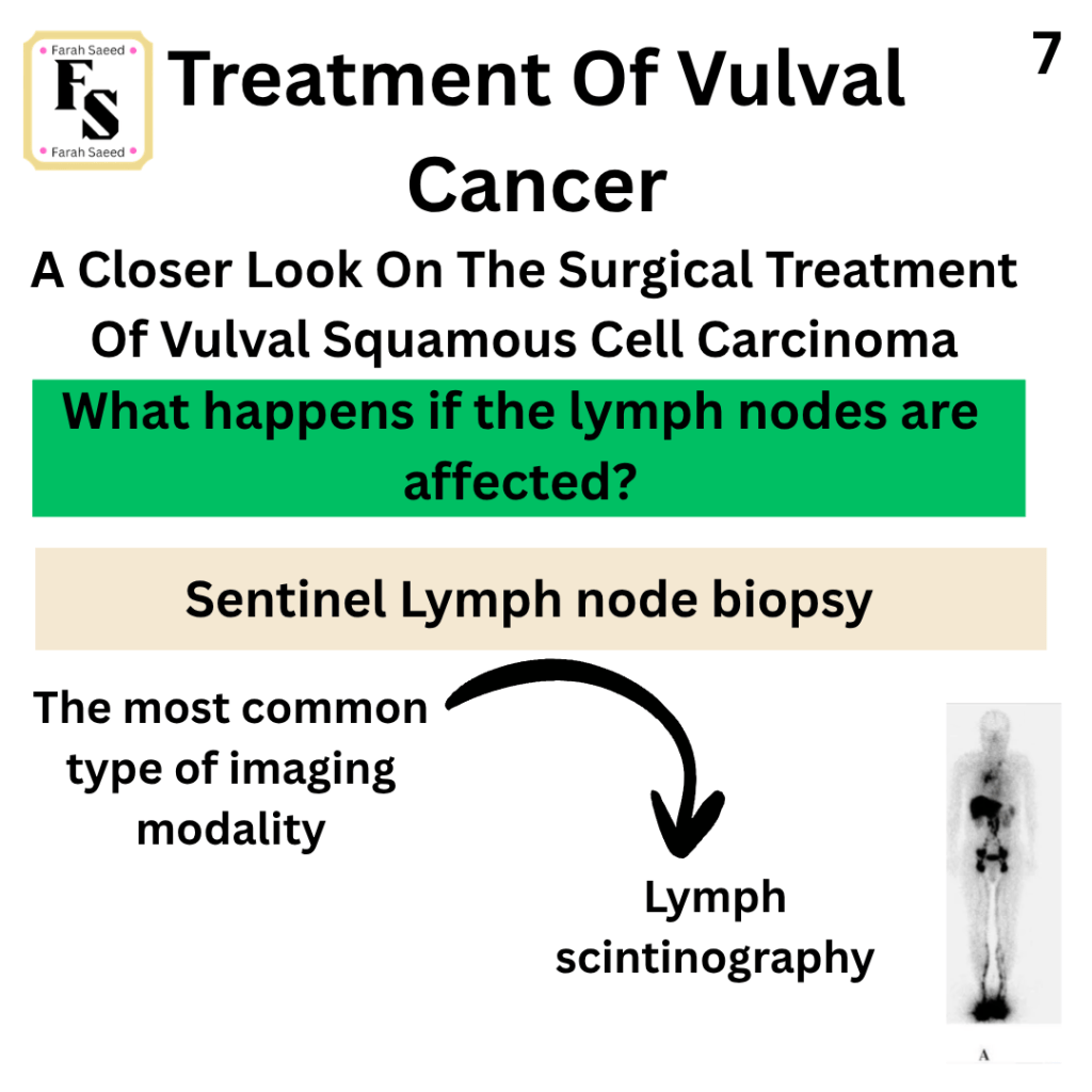
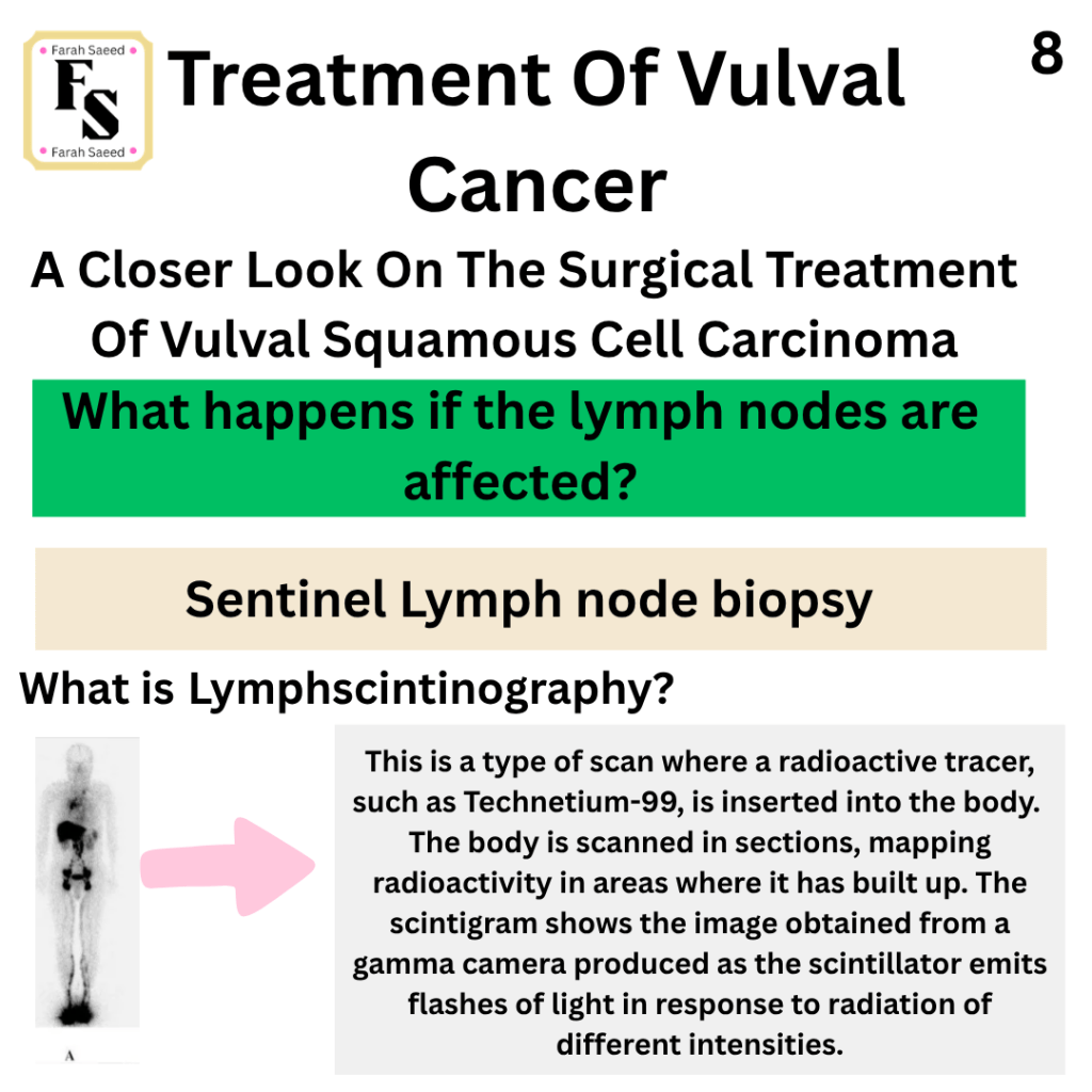
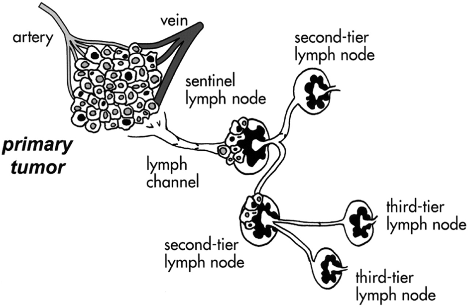
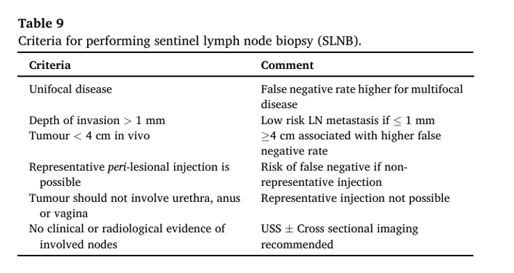
1) VSCC – lymphadenectomy – Sentinel Lymph Node Biopsy
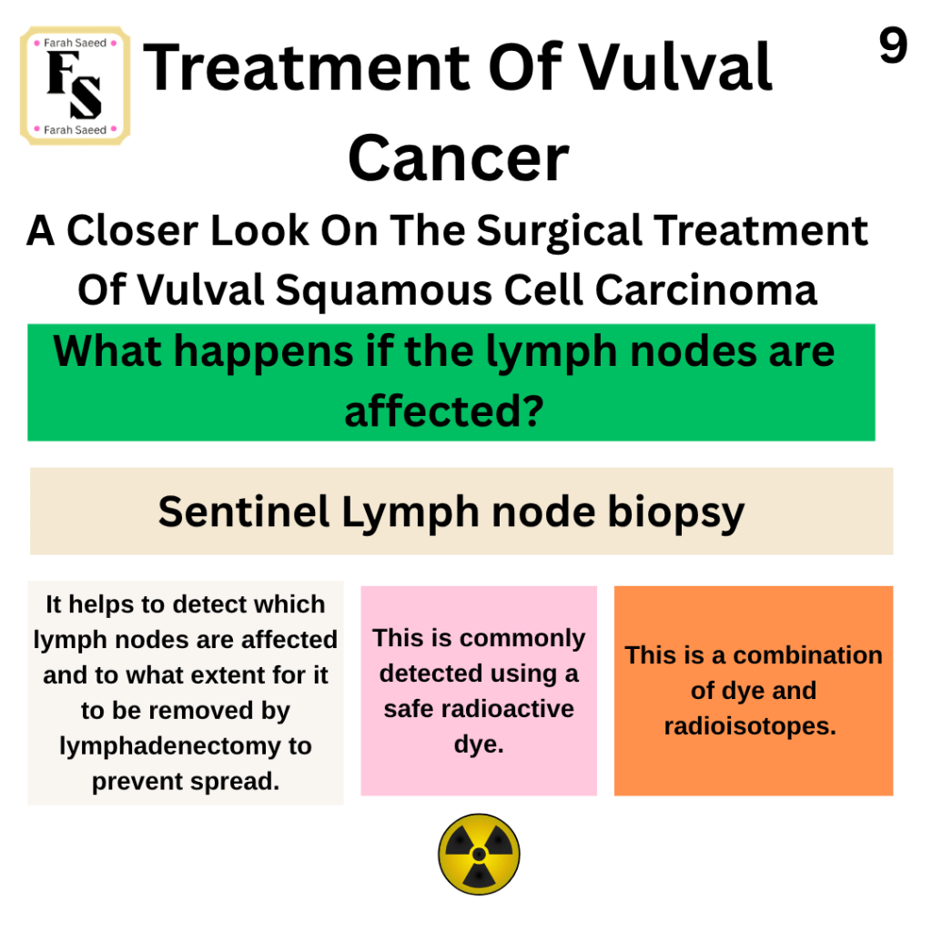
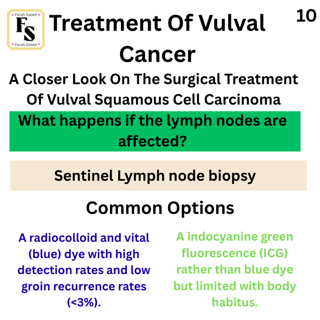
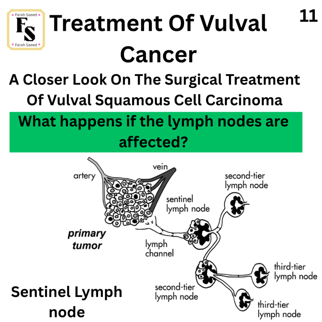
Supporting Images For SLNB
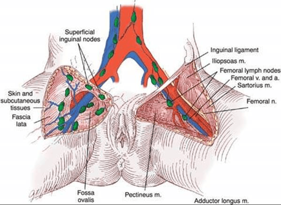
Bilateral drainage commonly applied for tumours that are midline.
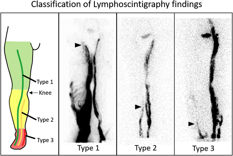
What are the lymphatic drainage in the human body?
Upper & Lower Limb Lymphatic Drainage: Inguinal and Axillary Lymph Nodes (Clinical Guide) By Human Anatomy Education Dr Akram Jaffar
Ipsilateral Versus Contralateral Lymphadectomy
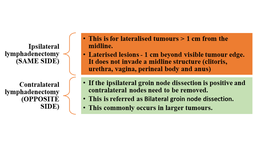
SAME CONCEPT WITH SLNB
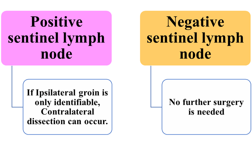
Royal College of Obstetricians and Gynaecologists (2014) Guidelines for the Diagnosis and Management of Vulval Carcinoma.
Ultrastaging may take place by sectioning at 200 micrometres and immunohistochemistry with epithelial marker AE1/AE3 to detect if there is any micro metastatic disease.
Further information, please see the guidelines
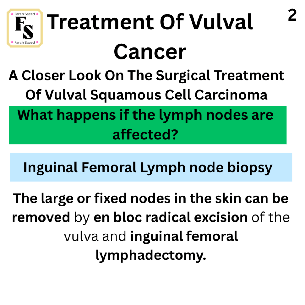
What If There Is Large Lymph Nodes Being Affected Greater Than 4cm (>4cm)?
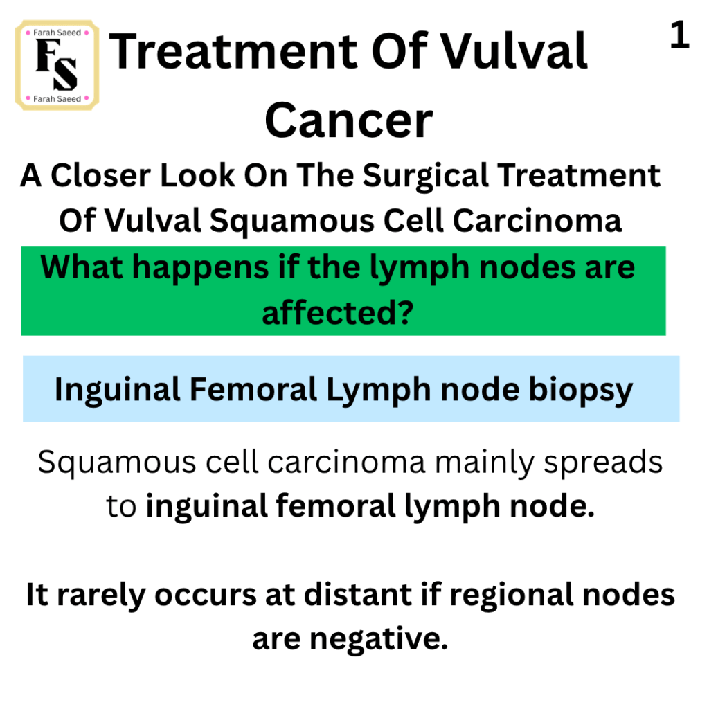
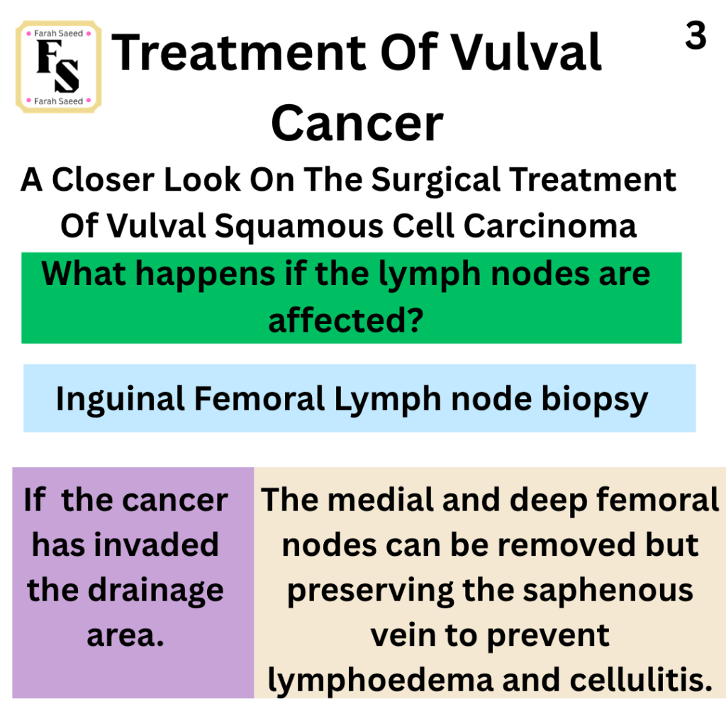
2) VSCC – lymphadenectomy – Inguinal Femoral Lymph Node Biopsy
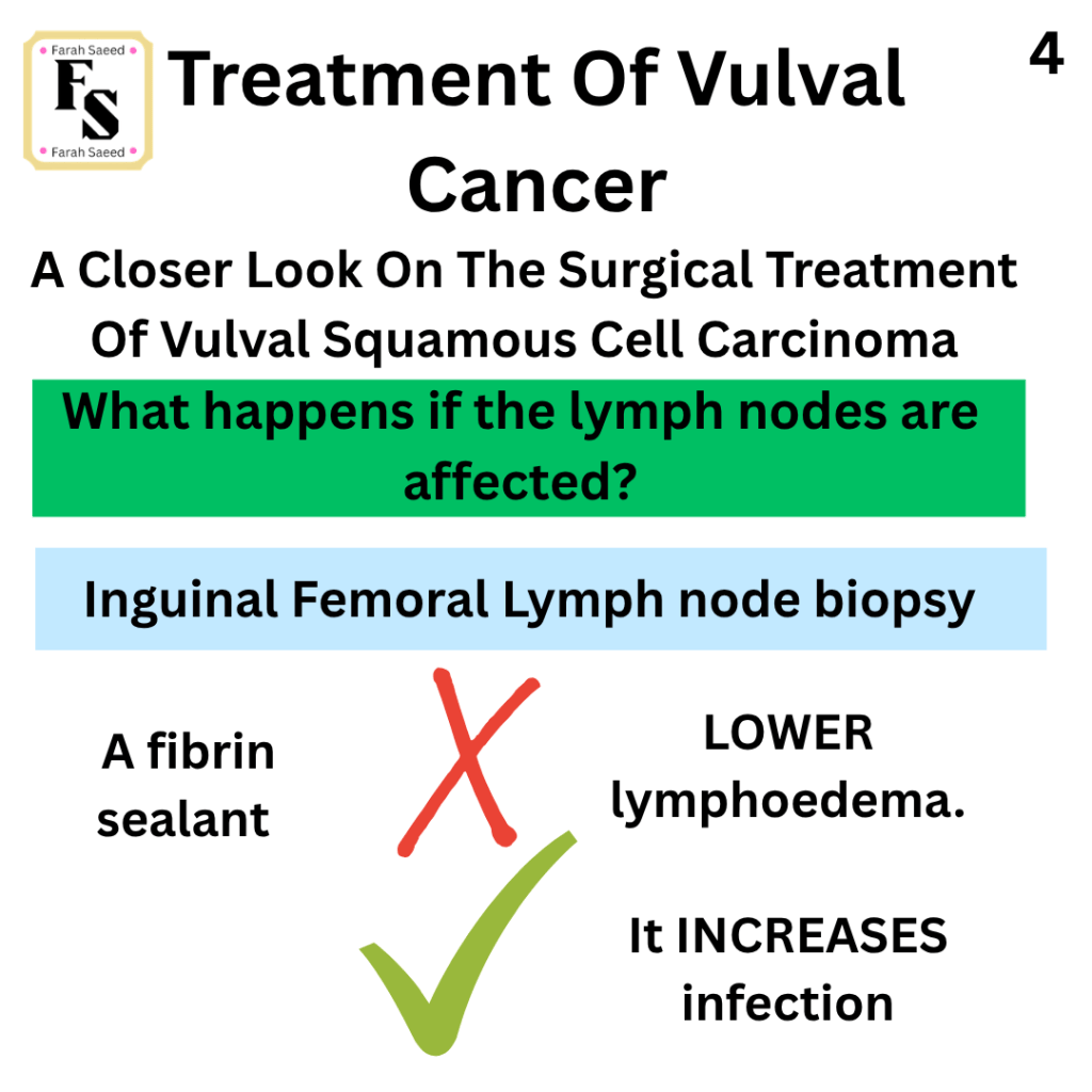
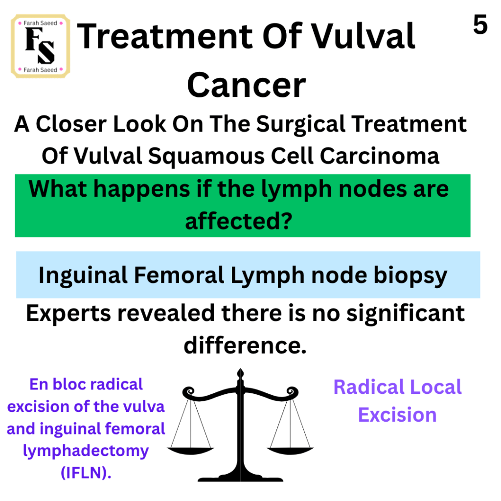
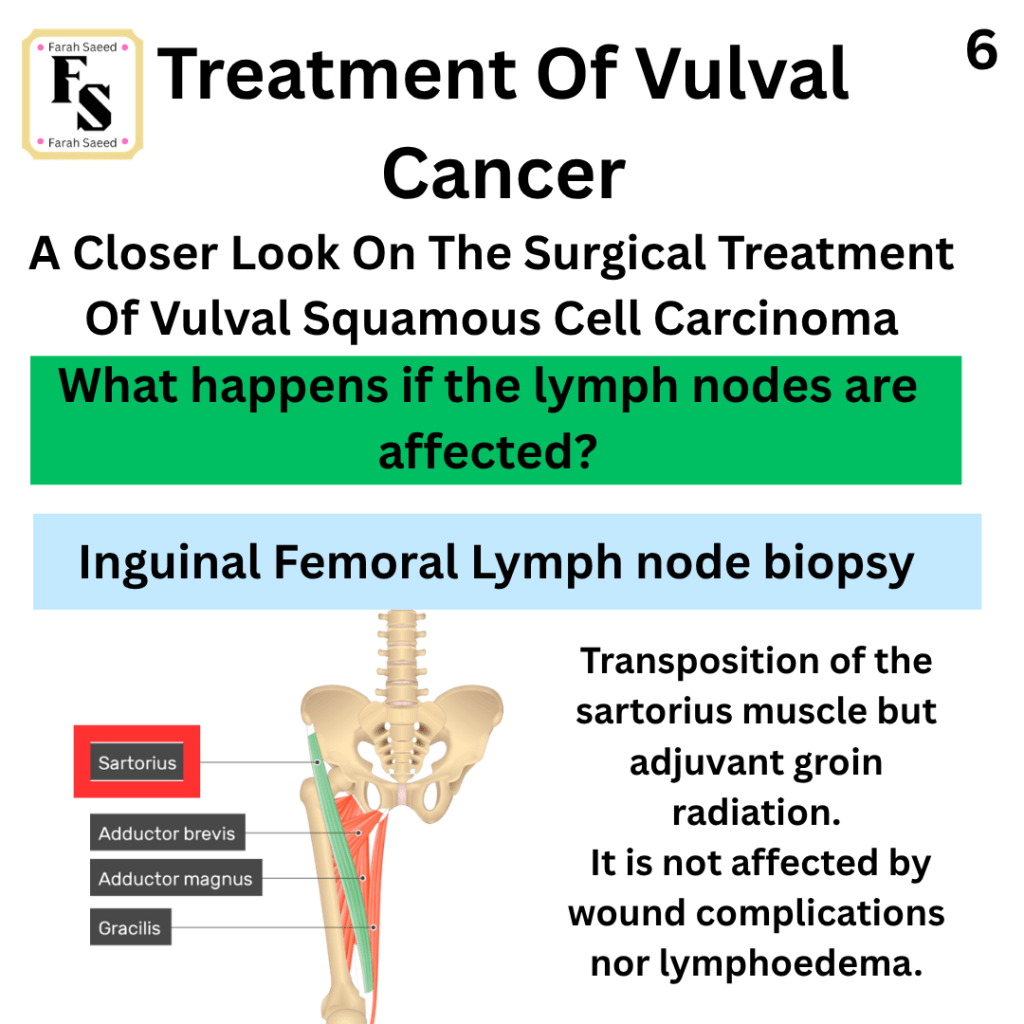
3) VSCC – lymphadenectomy – Fine Needle Aspiration Biopsy
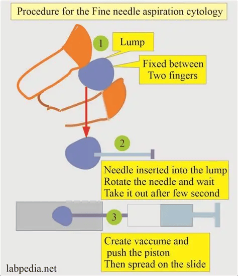
However, if there is suspected groin nodes on imaging, USS-guided fine needle aspiration (FNA) or core biopsy, where node positivity would change management.
Common Challenges In Lymph Node Biopsy and Lymphadenectomy
Cellulitis
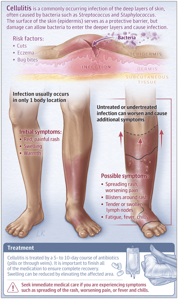
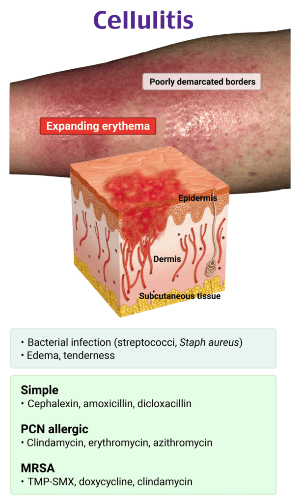
Cellulitis is the infection of the dermis of the skin caused by the bacterium Beta haemolytic streptococci. It commonly affected the lower legs. It is commonly associated with lymphangitis which is the inflammation of the lymphatic vessel where it appears as red streaks on the skin near the streptococcal infection and may result in lymphoedema. Another common cause of cellulitis is the inflammation of the lymph nodes where they appear swollen, painful and soft. They are commonly acute (sudden) and localised. Their normal role is to help combat infection.
Lymphoedema
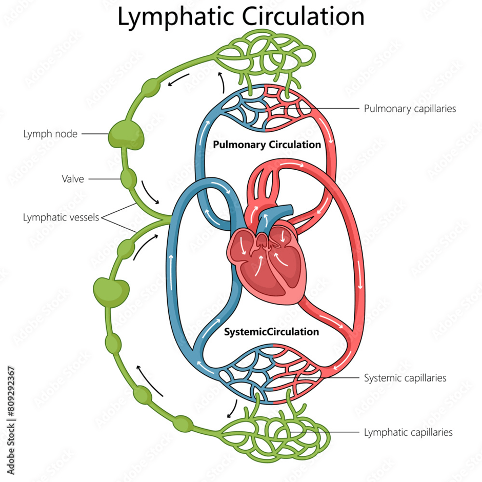
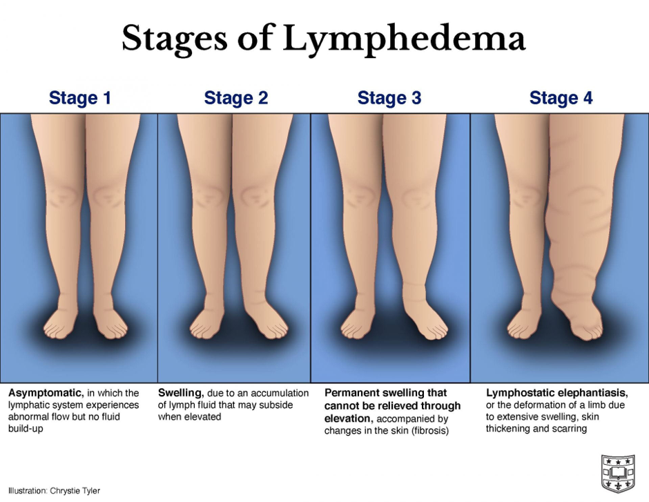
Lymphoedema is the build up of lymph in the tissue that commonly occurs due to inflammation, tumour, injury, parasites and other obstructions.
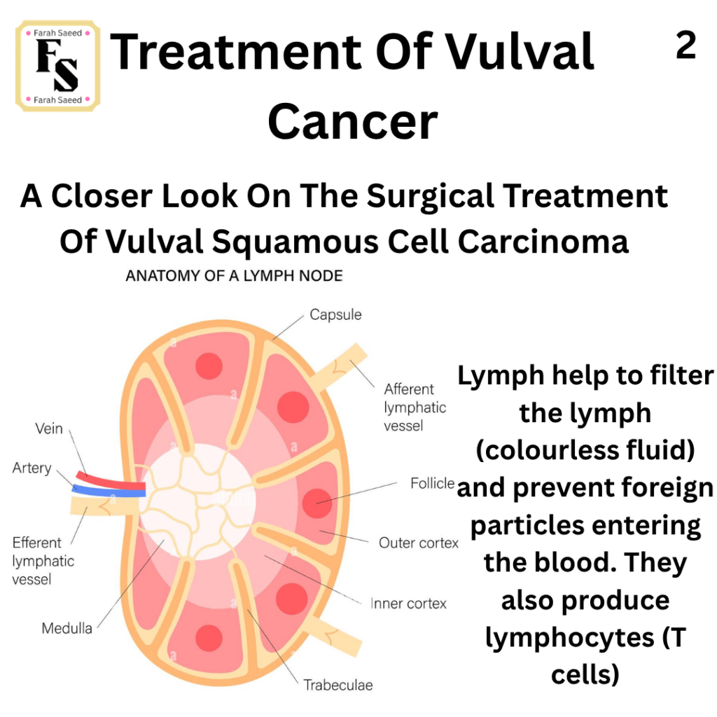
The lymph is a colourless fluid that bathes the tissues, drained by lymphatic vessels and is filtered through lymph nodes before returning back to the blood.
It commonly affects the lower the limbs, abdomen and pelvis after surgery like inguinal femoral lymph node dissection (IFLND) in 17 to 50% are affected but more so in patients with surgery and radiotherapy.
Removing less lymph nodes helps prevent lymphoedema.
Pre-operative measurements like height and weight for body mass index (BMI) is important as high levels of BMI, lack of exercise and pre-existing lymphoedema leads to more risk of lymphoedema.
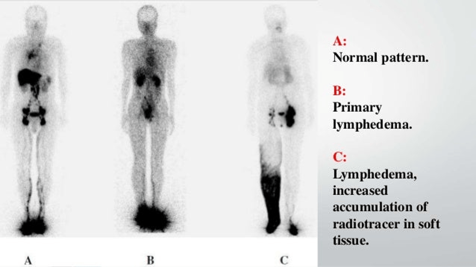
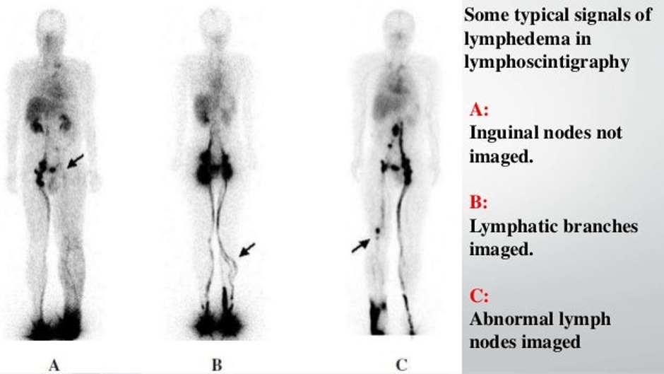
How Can You Overcome Lymphoedema?
Compression garments – this adds pressure on the area and help overcome the build up.
Manual lymphatic drainage
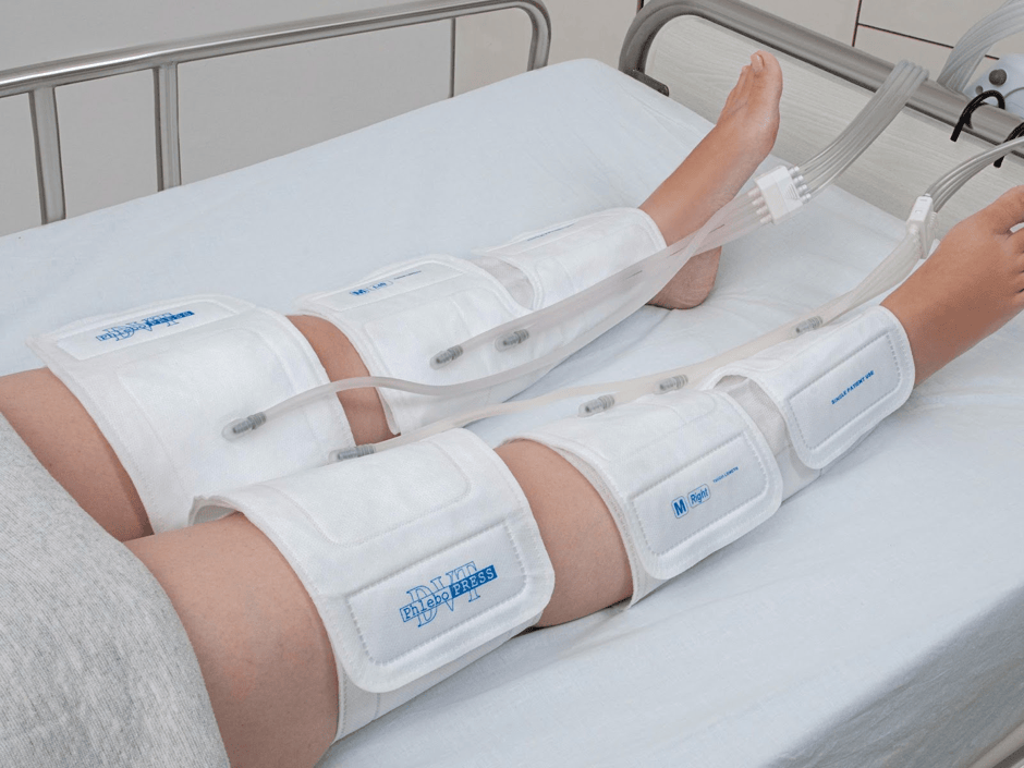
Pneumatic compression: Air-inflated sleeves that adds pressure to the limb, blood systemic circulation (improve venous return), lower swelling, prevent blood clots. It is commonly between 20 to 120 mmHg (blood pressure).
Patients before operation and post operation aim to maintain healthy lifestyle: weight, exercise and daily skin care.
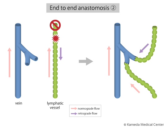
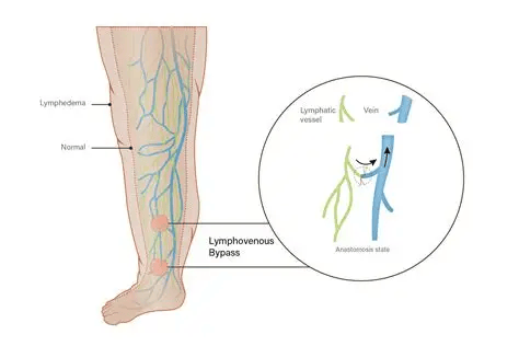
Lymphaticovenular anastomosis – the joining of the lymphatic vessel with the vein. This is commonly performed for early stage lymphoedema, especially if cellulitis has been repeated.
Drainage by suction helps after IFLND.
What Happens After Lymphadenectomy?
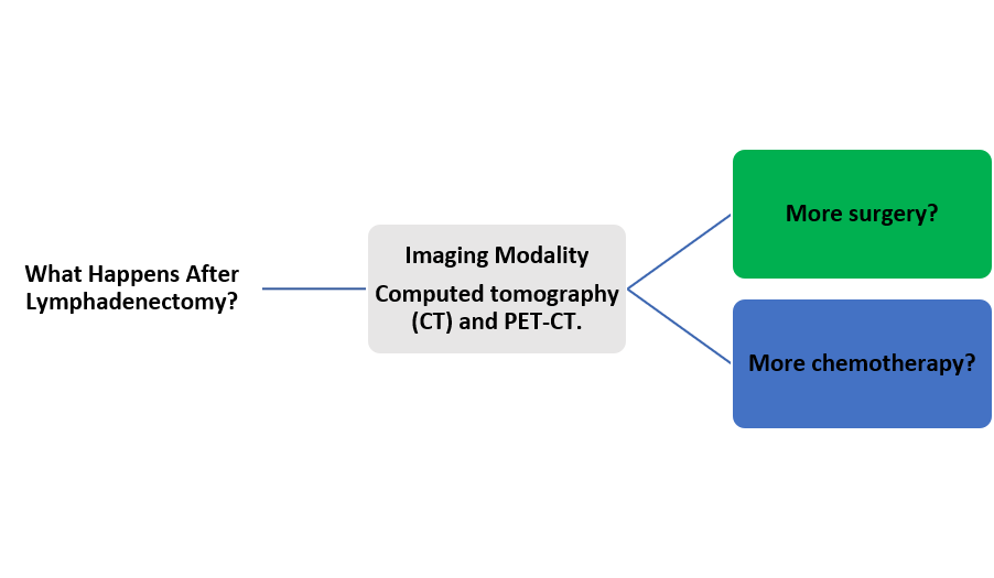
Please see guidelines for further details.
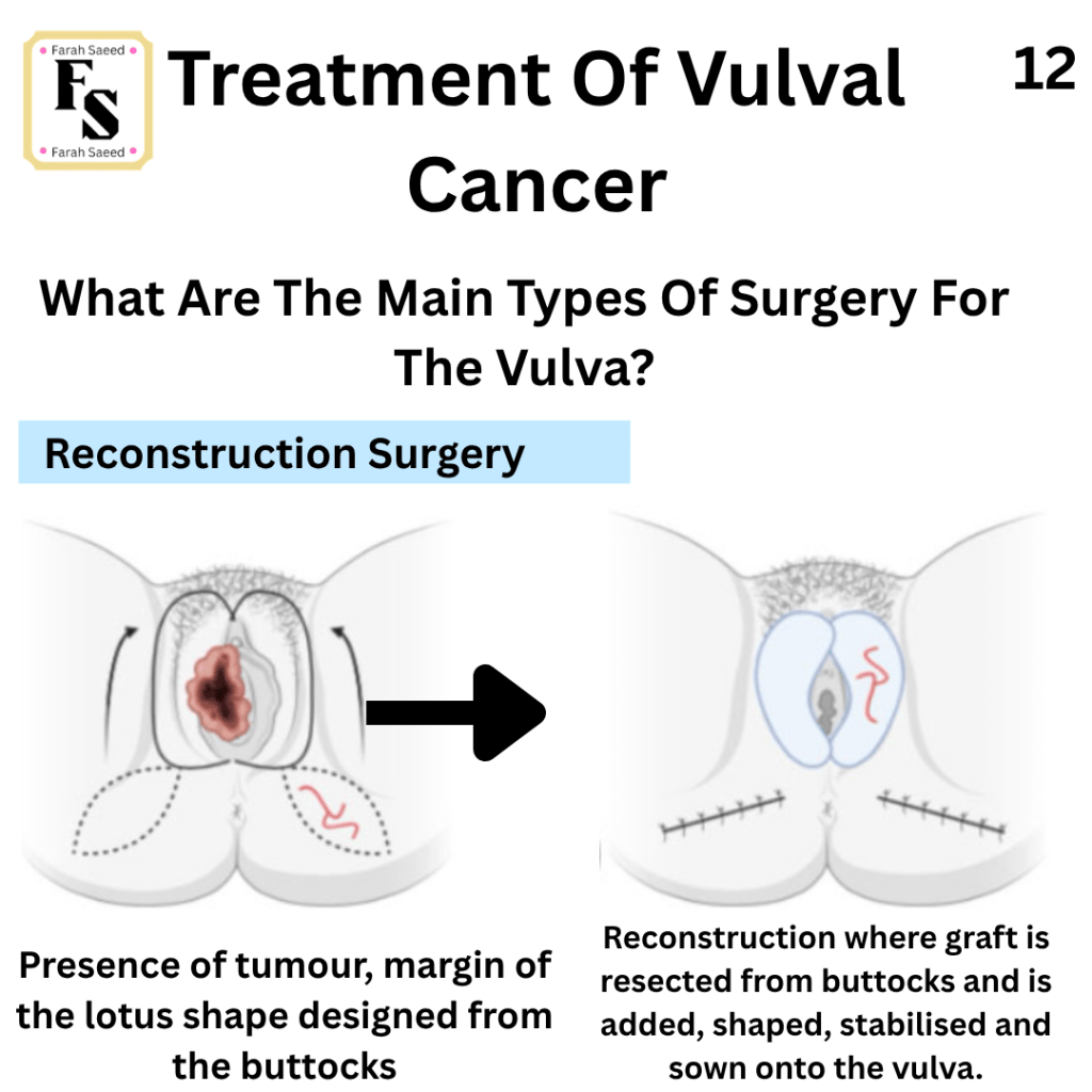
Reconstructive Surgery
It is a form of surgical art that requires to design and make structures to help:
- Complete curative surgery with margins.
- Maintain function of the organs
- Wound-healing
- Lower risk of scarring

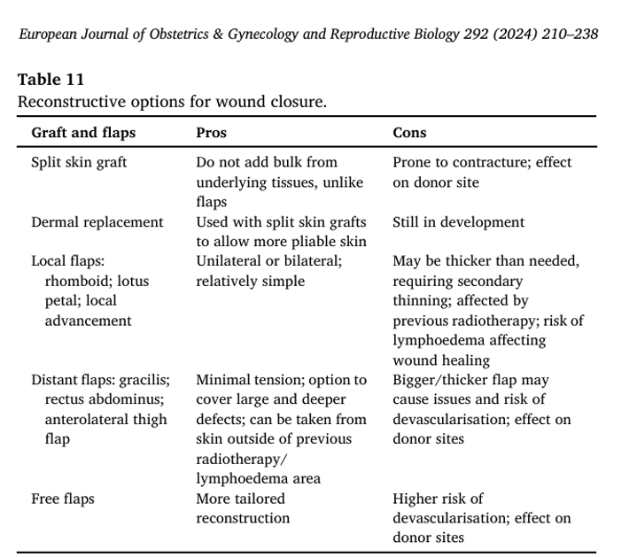
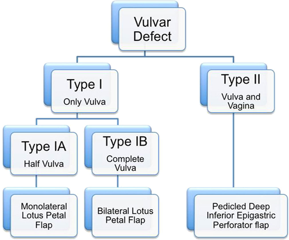
Types Of Reconstructive Options For Wound Closure
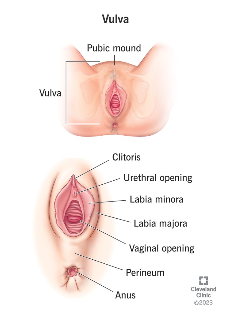

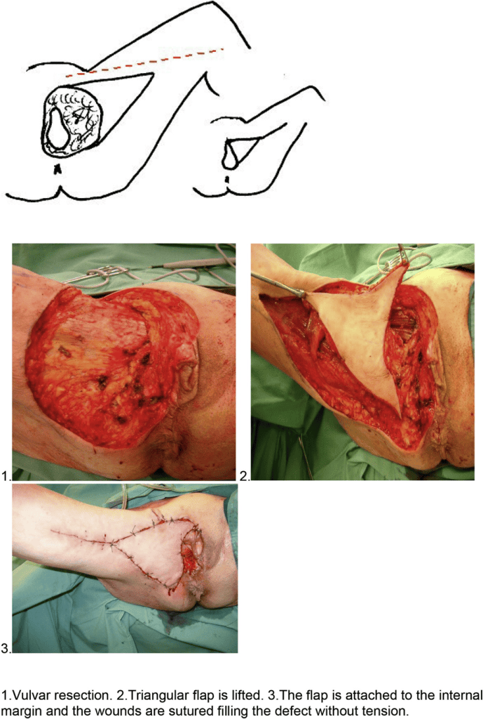
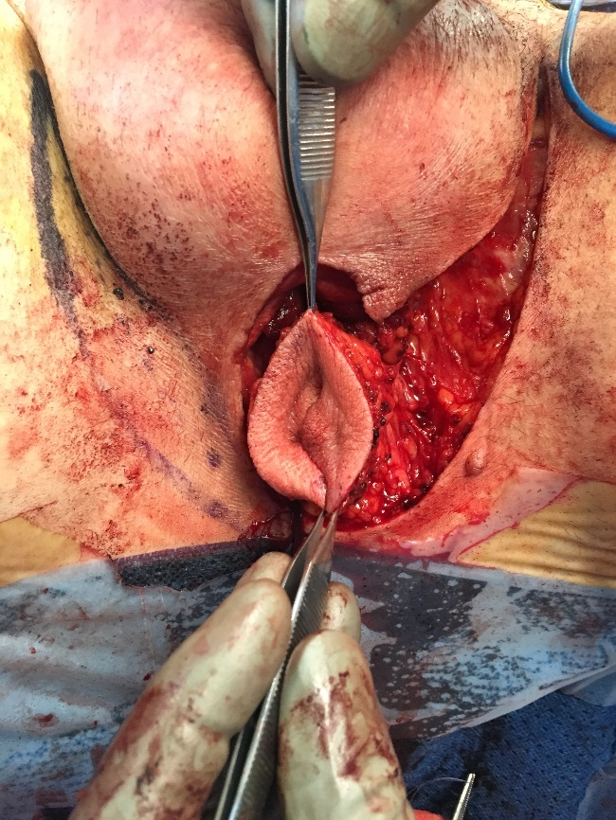
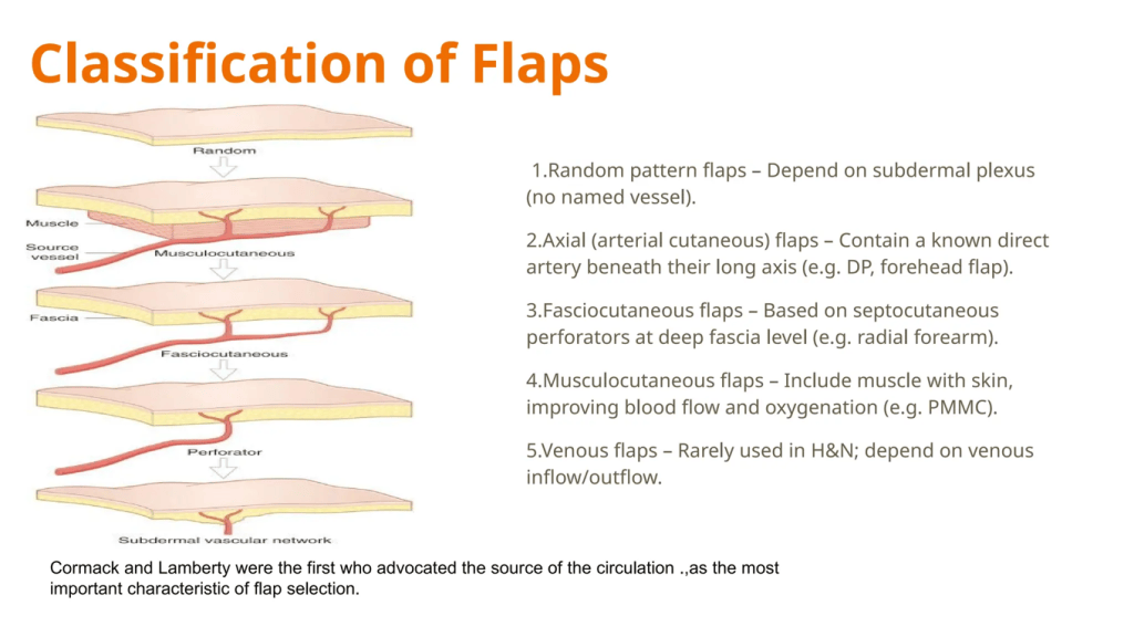
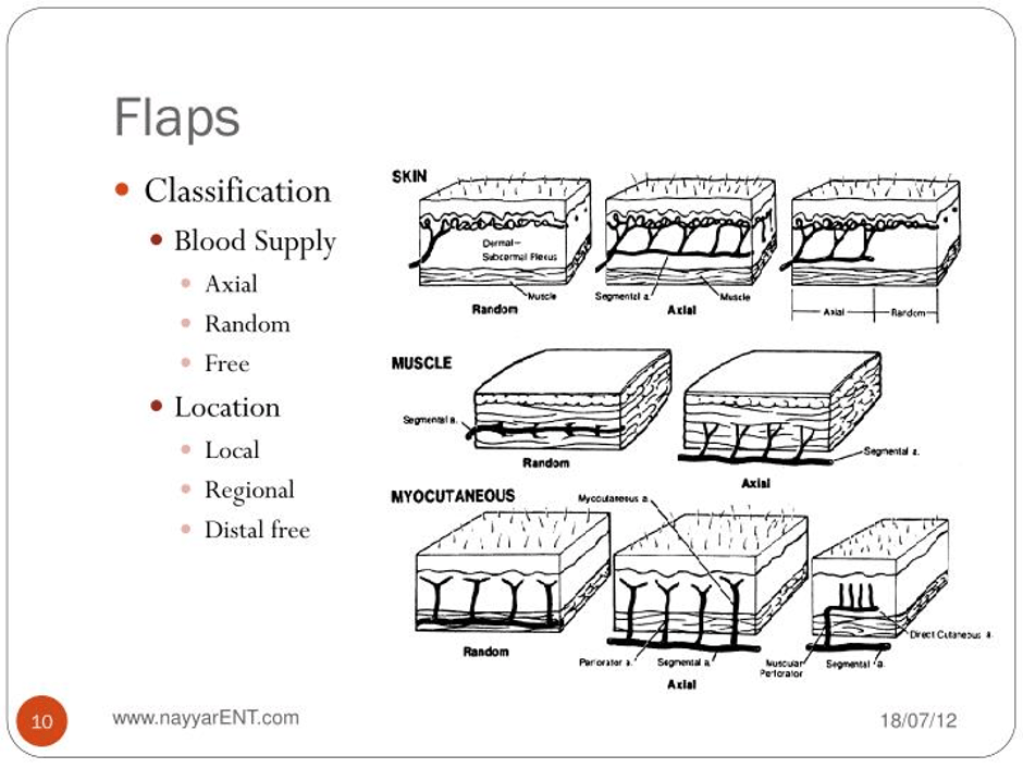
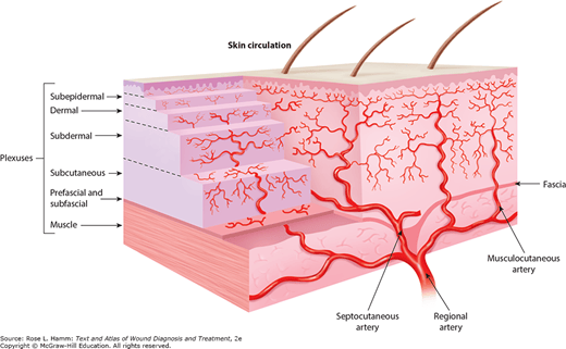
The Anal Margin And Vulval Cancer
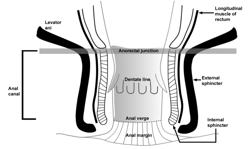
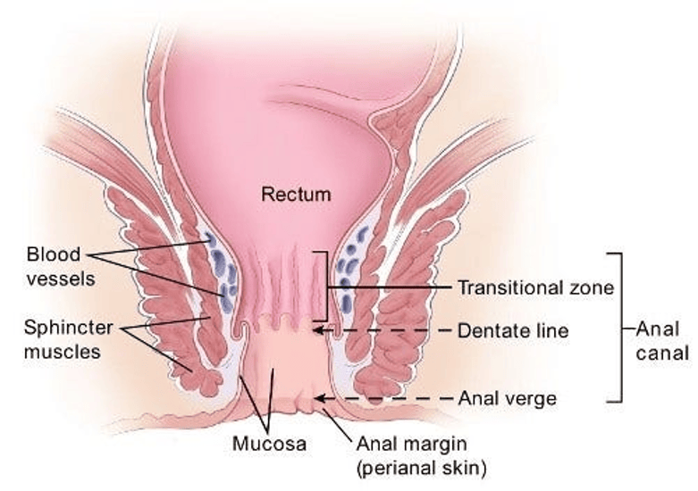
Both images illustrates the anatomy of the anus, the former is in black, grey and white mode whereas the other in colour mode.
The anal structure is the end of the large intestine and where stools/faeces/poo leave the body. The anus is relevant because the anal orifice/opening is part of the vulva. The pelvis is where the female reproductive system is situated alongside the gastrointestinal tract (rectum, sigmoid colon and coils of the ileum), urinary (ureters, bladder), fascia (coverings) and the peritoneum that connects with the organs. The levator ani is the muscle that lifts the anus. The anal canal is the termination point and is surrounded by anal sphincters that help release defecation. One is made of smooth muscle, and the other is skeletal.
The anal canal begins at the narrowing of the rectal ampulla at the anorectal junction, where there is a section where the anus connects with the rectum that stores stools/faeces/poo. More specifically, the rectum enters the puborectalis sling at the apex (tip) of the anal sphincter complex. It extends distally for approximately 4 cm and ends at the anal verge.
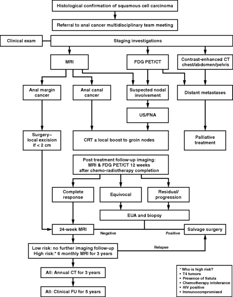
There is also a Dentate line, transitional zone, anal verge, and anal margin. The anal verge connects the canal to the outside skin at the anus, called the perianal skin, otherwise known as the anal margin, and is within a 5 cm radius of the anal verge. The anal margin contains pigmented skin with folds surrounding the anus. . If the anal margin is involved in the vulval disease or cancer, primary treatment with chemoradiation is given.
There are different types of cells found in the anus. The perianal skin is made of non-keratinised squamous cells and also sweat glands (e.g. apocrine) and hair follicles. However, they are not found in the lining of the lower anal canal. The lining of the canal consists of mucus glands and ducts to help lubricate the area. They are also found in the rectum. The transitional zone contains cuboid-like shapes. The dentate line is found in the middle of the anal canal and functions as the site where mucus is emptied into the anus from the glandular cells that produce it. It is visible macroscopically (naked eye) but not on MRI scan. This can be overcome by the use of transanal endoscopic ultrasound for supervision and localised tumours.
However, transanal endoscopic ultrasound has several disadvantages: operator dependency, inability to assess stenotic tumours (tumours arising from abnormal passage), and regional lymph nodes higher in the pelvis or groins are outside the FOV of endosonography, and supplementary MRI is needed for N (node) staging.
FDG PET/CT helps in the diagnosis and treatment of anal carcinoma, as 98% of anal cases are particularly because up to 98% of anal tumours are FDG-avid (positive). Perirectal lymph nodes are commonly irradiated. However, PET/CT has low sensitivity but can detect pelvic and inguinal lymph nodes.
The cells found above the anal verge are anal cancer, whereas cancers below the anal verge are referred to as cancers of the perianal skin. Anal margin tumours can be treated by surgical excision alone, whereas anal canal tumours have a worse prognosis and are treated with chemoradiotherapy.
Staging helps define anal margin tumours, which can be treated by local excision if small (< 2 cm), well differentiated, do not involve the sphincter, and have no nodal spread. Defunctioning colostomy is considered for patients with transmural vaginal involvement (at risk of development of an anorectal-vaginal fistula), those with fecal incontinence, and those with severe anal pain and obstructive symptoms. During staging, assessment of the cervix and vulva is important because of the common viral cause of vulval, cervical, and anal neoplasms.
Vulval Paget’s disease can affect skin anywhere in the body, especially the perianal vulva and chest. Vulval melanoma arises in melanin-containing cells, and a very small number of anal cancers are melanomas and are hard to see.
Therefore, according to the British guidelines, a temporary or permanent stoma with excision/removal of the necessary amount of anal margin is recommended. The role of chemoradiotherapy in the treatment of vulval cancers. Chemoradiotherapy decreases the size to preserve the anus. After radiotherapy, local flap reconstruction is done to the flap field, but this needs to be measured based on the length-to-breadth ratio. If it is difficult to assess, frozen sections should be done before flaps for reconstruction.
Limitations Of Post-Reconstructive Surgery
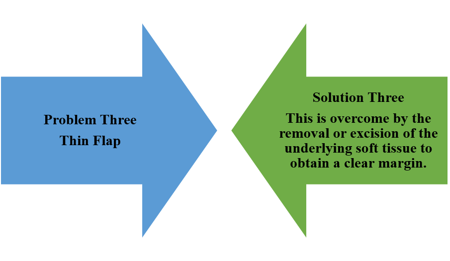
When flap has been reconstructed, there may be a few issues but every issue has a surgical solution.



Other Treatments For Vulval Cancer



References For Vulval Surgery
American Cancer Society (2026) Anal Cancer. Available at: https://www.cancer.org/cancer/types/anal-cancer.html (Accessed: 2nd October 2026)
British Association of Dermatologists (2026) Imiquimod cream. Available at: https://www.bad.org.uk/pils/imiquimod-cream (Accessed: 30th September 2026)
Cancer Research UK (2028) Chemotherapy for vulval cancer. Available at: https://www.cancerresearchuk.org/about-cancer/vulval-cancer/treatment/chemotherapy (Accessed: 30th September 2026)
Cleveland Clinic (2026) Volumetric Modulated Arc Therapy (VMAT). Available at: https://my.clevelandclinic.org/health/treatments/17626-volumetric-modulated-arc-therapy-vmat (Accessed: 30th September 2026)
Hoppe-Seyler, K., Bossler, F., Braun, J.A., Herrmann, A.L. and Hoppe-Seyler, F. (2018). The HPV E6/E7 Oncogenes: Key Factors for Viral Carcinogenesis and Therapeutic Targets. Trends in Microbiology, [online] 26(2), pp.158–168. doi:10.1016/j.tim.2017.07.007.
Kochhar, R., Plumb, A.A., Carrington, B.M. and Saunders, M. (2012). Imaging of Anal Carcinoma. American Journal of Roentgenology, 199(3), pp.W335-W344. doi:10.2214/ajr.11.8027.
Lobna Ouldamer, Z. Chraibi, Flavie Arbion, Barillot, I. and Body, G. (2013). Bartholin’s gland carcinoma: Epidemiology and therapeutic management. 22(2), pp.117–122. doi:10.1016/j.suronc.2013.02.004.
Management of Vulval Carcinoma. Available at: https://www.rcog.org.uk/media/dqwnw2a1/vulvalcancerguideline.pdf (Accessed: 1st October 2026)
Morrison, J., Baldwin, P., Hanna, L., Andreou, A., Buckley, L., Durrant, L., Edey, K., Faruqi, A., Fotopoulou, C., Ganesan, R., Hillaby, K. and Taylor, A. (2024). British Gynaecological Cancer Society (BGCS) vulval cancer guidelines: An update on recommendations for practice 2023. PubMed, 292, pp.210–238. Doi: 10.1016/j.ejogrb.2023.11.013.
National Cancer Institute (2026) Vulvar Cancer Treatment (PDQ®)–Health Professional Version. Available at: https://www.cancer.gov/types/vulvar/hp/vulvar-treatment-pdq (Accessed: 17th August 2026)
Royal College of Obstetricians and Gynaecologists (2014) Guidelines for the Diagnosis and Management of Vulval Carcinoma. Available at: https://www.rcog.org.uk/media/dqwnw2a1/vulvalcancerguideline.pdf (Accessed: 1st October 2026)
The Eve Appeal (2026) Vulval cancer Available at; https://eveappeal.org.uk/information-and-advice/gynaecological-cancers/vulval-cancer/ (Accessed: 7th August 2026)

