
Risk Factors Of Osteosarcoma Part A
A) Genetic Syndromes
There are many genetic syndromes that increase the risk and is associated with osteosarcoma. Here are some of them
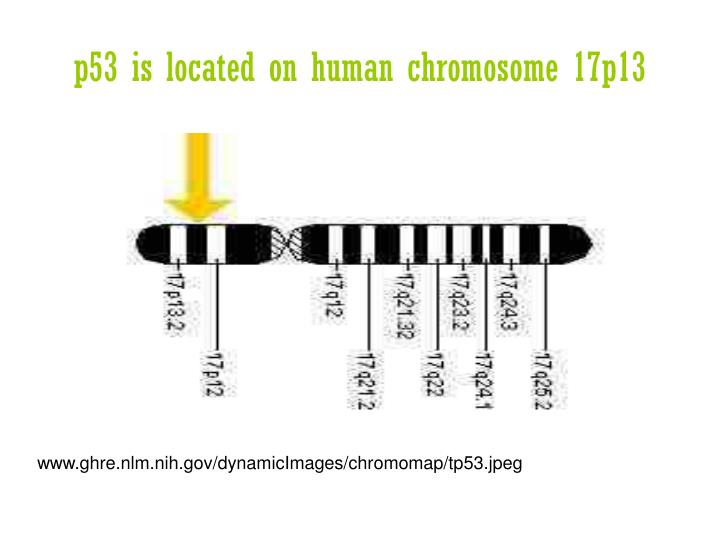
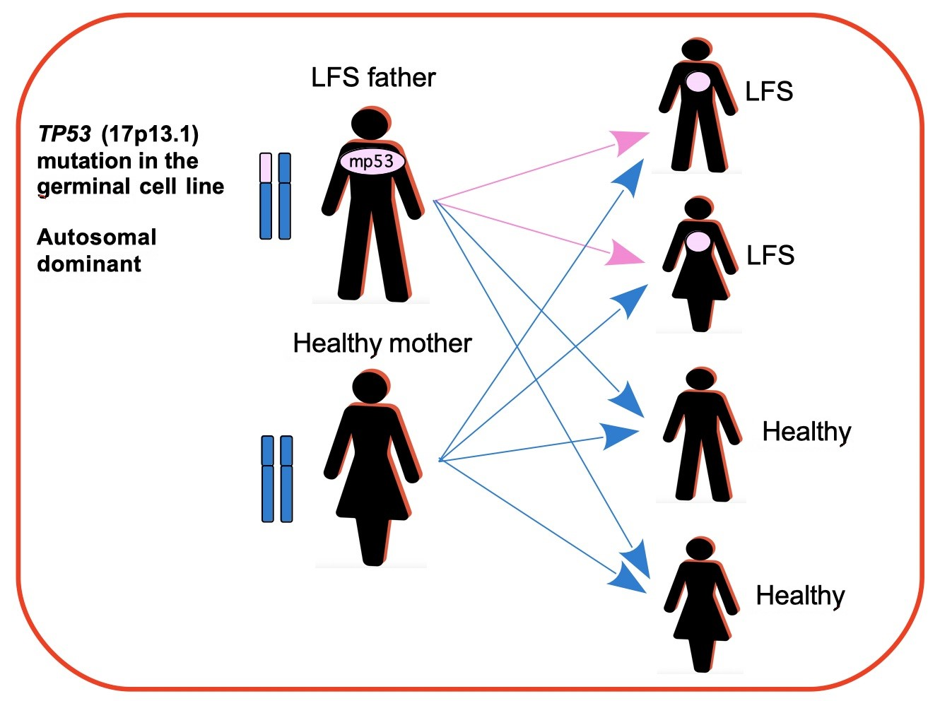
Li Fraumeni Syndrome
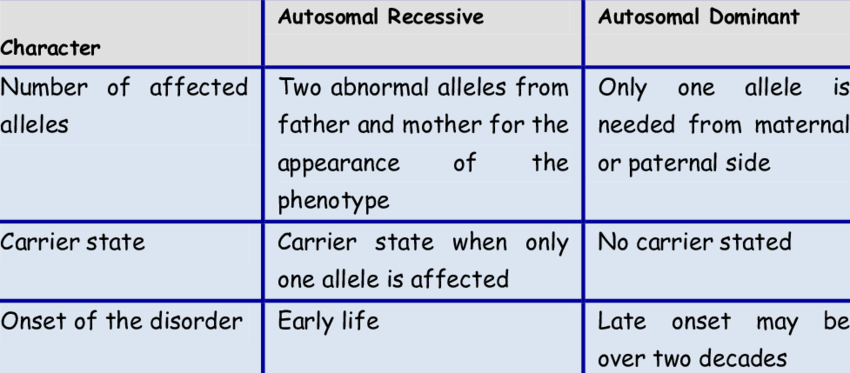
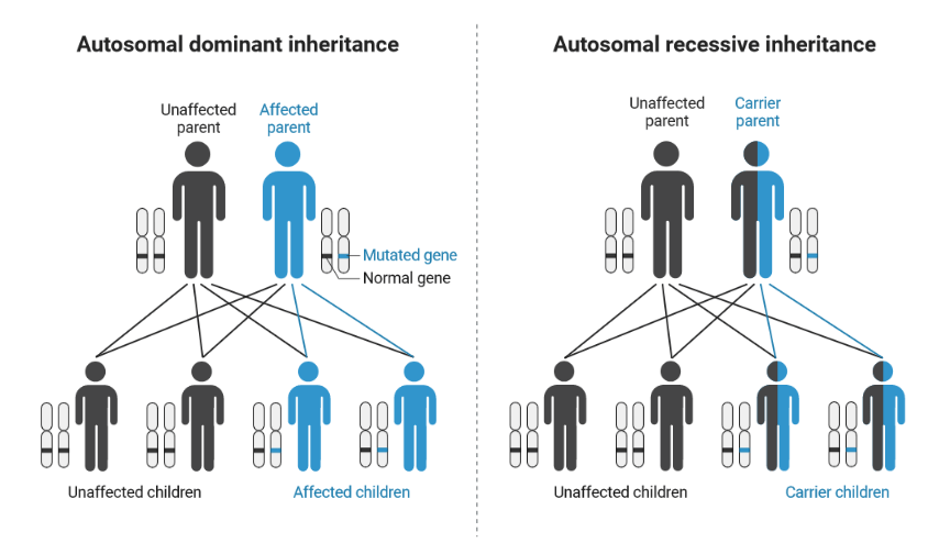
It is an autosomal dominant disorder.
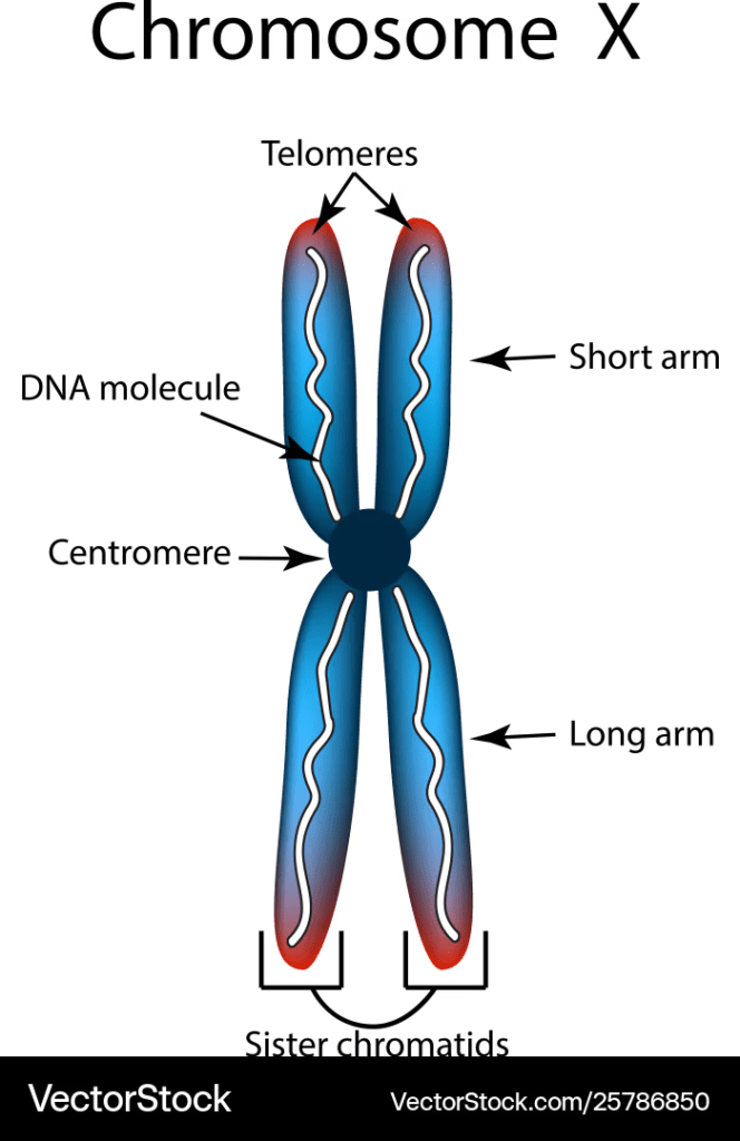
Autosomal refers to the 22 pairs of numbered chromosomes in humans that are not involved in determining biological sex. The 23rd pair determines the sex/gender of the subject.
Germline mutation in the TP53 tumour suppressor gene on chromosome 17p13.1 causing loss of function.
Germline –> Sex cells (egg in females) and (sperm in males).
Tumour suppressor –> It stops tumours from growing.
Classical LFS diagnosed before 45 years old who had a relative of first degree before 45 or a second-degree relative with any cancer before age 45 or sarcoma at any age.
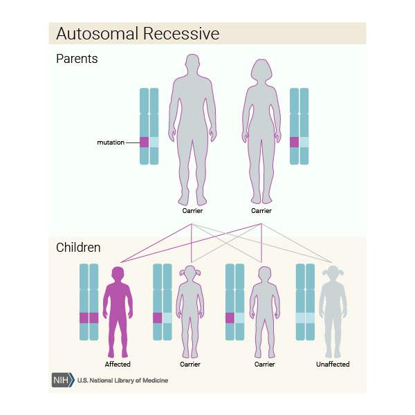
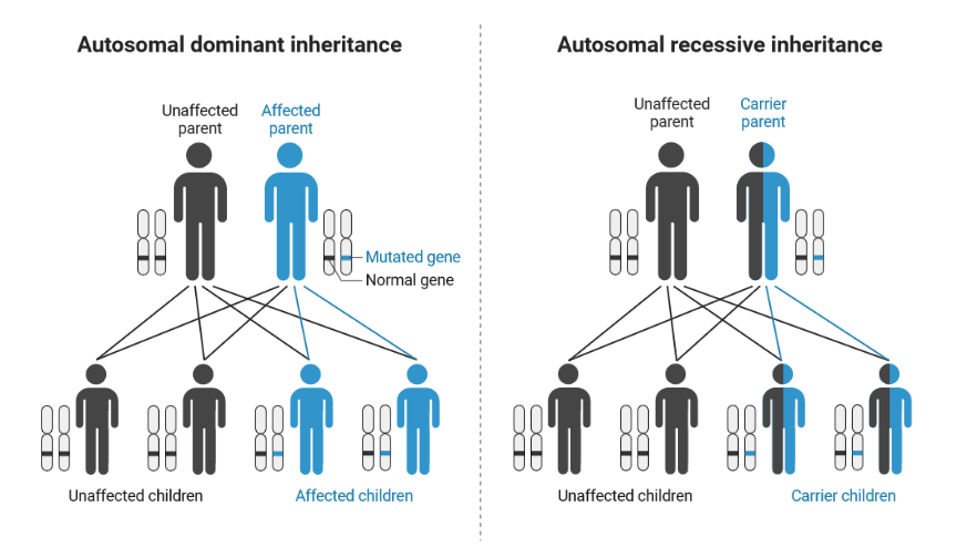
What is the difference between autosomal dominant and autosomal recessive?
Age at risk of cancer:
50% by 30 years of age and 90% by age 60 years.
5% of osteosarcoma cases are under the age of 30
The metaphysis of long bones most commonly affected area.
It has pleomorphic-shaped tumour cells (different forms) and is subdivided into osteoblastic, chondroblastic, and fibroblastic osteosarcoma depending on the variable amounts of osteoid matrix produced by the tumour.
Familial Retinoblastoma
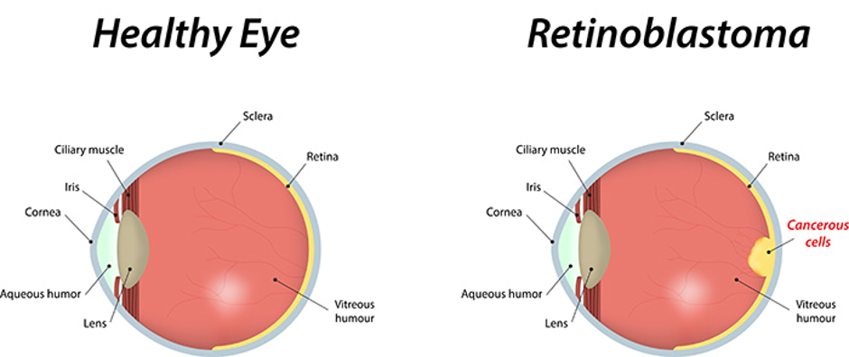
It is a rare autosomal dominant disorder.
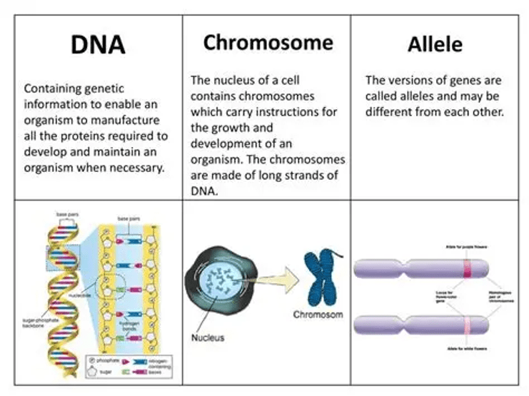
It is caused by biallelic mutation of the retinoblastoma gene RB1 (70% of cases)
It is present on chromosome 13q14.2.
It affects the retina, especially children under two.
The retina is a light-sensitive area.
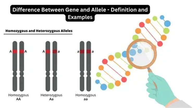
What is biallelic?
The Knudson’s Two-Hit Hypothesis
In 1971, Dr Alfred G. Knudson introduced the two-hit hypothesis, where cancer arises by two mutational events.
There are two types of retinoblastomas: hereditary and sporadic.
At first, the inherited form is where the mutation of the RB gene is inherited from one allele in a germ cell (sex cells) from either the mother or father. This is called hereditary. The second is when the mutation arises in a body cell (somatic cell).
In sporadic retinoblastoma, both mutational events arise in the somatic cells.
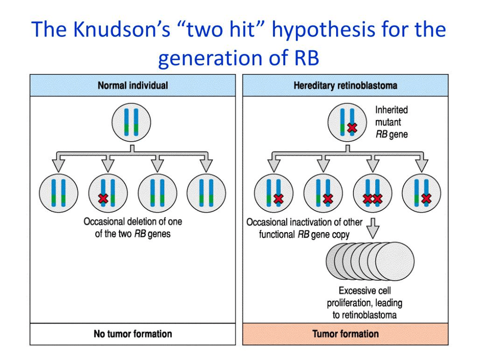
Patients with hereditary retinoblastoma have a higher survival rate but they are at risk of developing bone and soft tissue sarcoma especially osteosarcoma.
A combination of genetic factors, exposure to radiation and chemotherapy causes retinoblastoma but highest is related to radiotherapy where there is a 400-fold increase of risk.
The common areas affected areas in 75 patients who were diagnosed with radiotherapy-induced hereditary retinoblastoma:
- Skull and face (61%)
- Lower limb (29%)
- Trunk (7%)
- Unknown locations (3.8%)
(Kleinerman et al., 2005; Hameed and Mandelker, 2019).
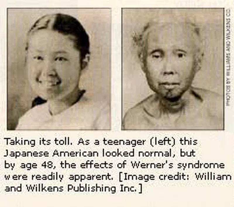
Werner Syndrome
It is autosomal recessive disorder where the WRN gene is mutated/changed.
The normal role of WRN gene.
The WRN gene encodes the Werner protein and is involved in DNA replication and repair.
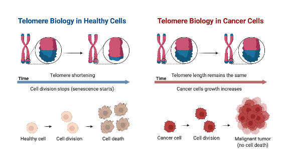
The WRN maintains the ends of chromosomes (telomeres) which helps to maintain DNA.
DNA undergoes copying before cell division and transfers informed in genes that help make protein (transcription).
It requires the following enzymes:
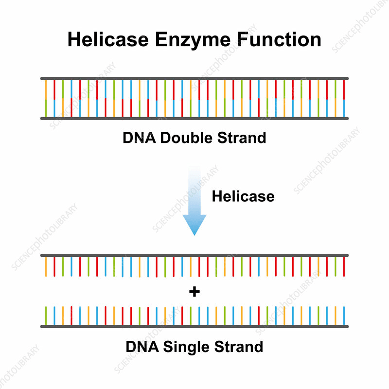
- RecQ helicase: This is located on chromosome 8p11.1 or 8p12. Helicase unwindS and separates the two strands of DNA.
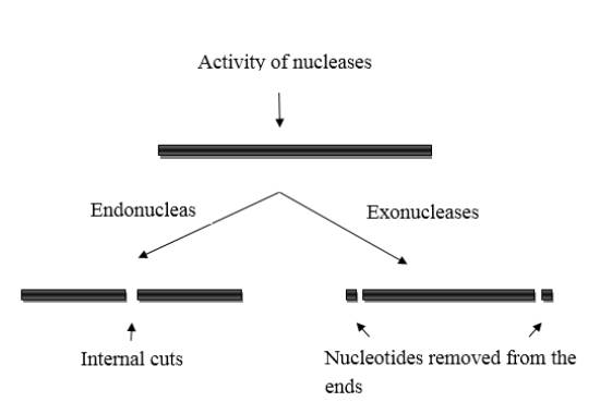
- Exonuclease: This enzyme cut the ends to remove DNA building blocks (nucleotides) of damaged DNA.
There are several examples of exonucleases:
Bal31: This removes nucleotides from both strand of the DNA.
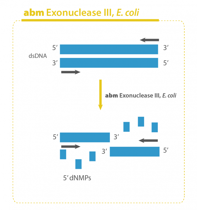
The E.coli exonuclease III: This enzyme from the bacteria removed nucleotide from the 3′ end of the DNA molecule.
Lambda exonuclease: This enzyme is from bacteriophage (bacterial viruses) that infects E.coli and it removes nucleotides from the 5′ end of the DNA molecule.
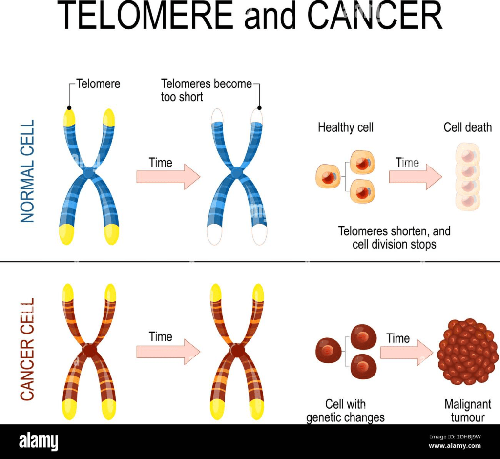
Mutation in the WRN gene
A number of symptoms may appear:
- Premature ageing (progeria) that starts at teenage years e.g. 14
- Loss of bone density leading to osteoporosis
- Muscle wasting (atrophy)
- Hair loss
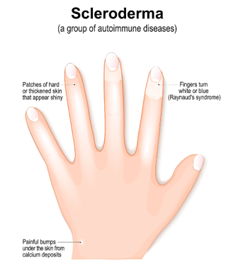
- Disease progression: vision (bilateral cataracts), skin changes (scleroderma due to abnormal production of collagen caused by abnormal immune system), diabetes and heart issues.
The link between osteosarcoma and Werner’s syndrome
Osteosarcomas occur at a later age in Werner syndrome patients (ages ranging from 35-57).
They tend to present at atypical sites such as foot, ankle and patella (kneecap) to more common sites such as long bones (Langer, Cunniff and Kucine, 2023).
Rothmund -Thomson syndrome type 2
RTS is an autosomal recessive disorder that commonly affects babies.
There are two types:
Type 1 is caused by a mutation in ANAPC1 gene
Type 2 caused by a biallelic mutation in chromosome 8q24.3 of the RECQLA gene. There can also be mutation in APC/C gene. Type 2 is associated with osteosarcoma.
The effects of mutation in RECQLA gene
Cell cycle delays.
RECQLA recruits cTIP to cause double-strand breaks.
The effects of mutation in APC/C gene
Mutation in APC/C degrades ctIP (CtBP-interacting protein) steadily to prevent hyper double-stranded break and maintain genomic instability.
There are several signs and symptoms of RTS
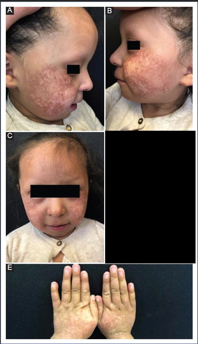
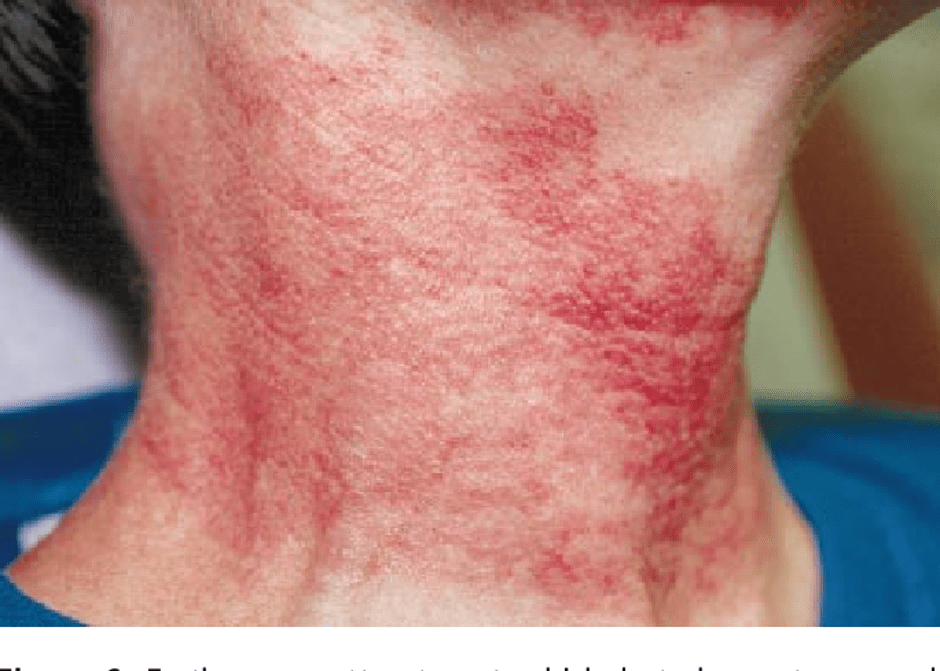
- Rash (usually on the cheeks)
- Changes to the bones, hair and teeth.
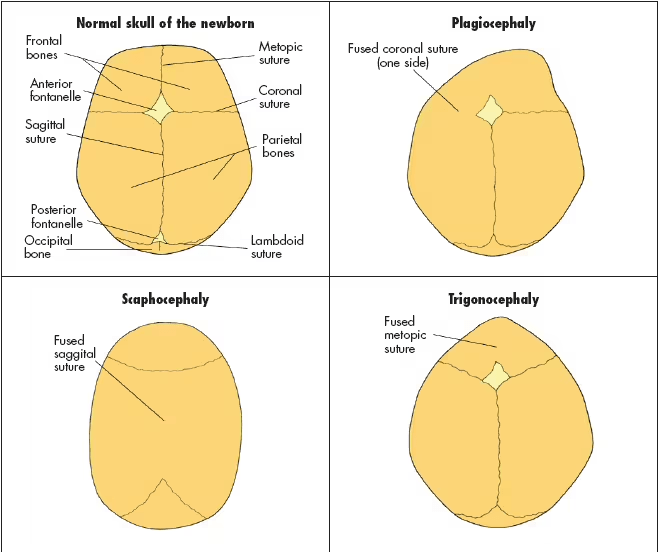
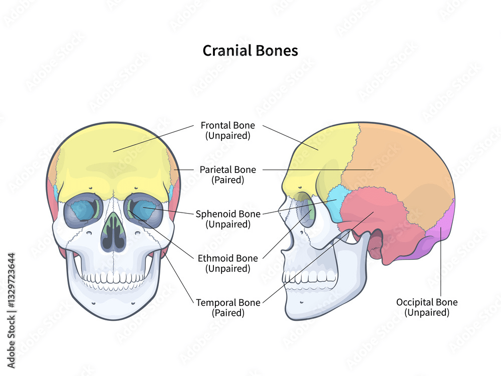
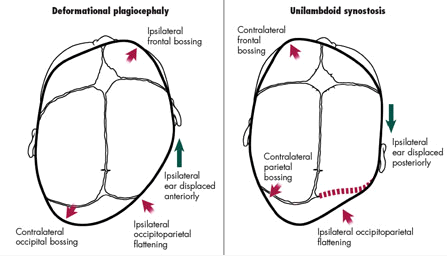
- Frontal bossing – enlarged forehead
- Short stature
- Radial defects
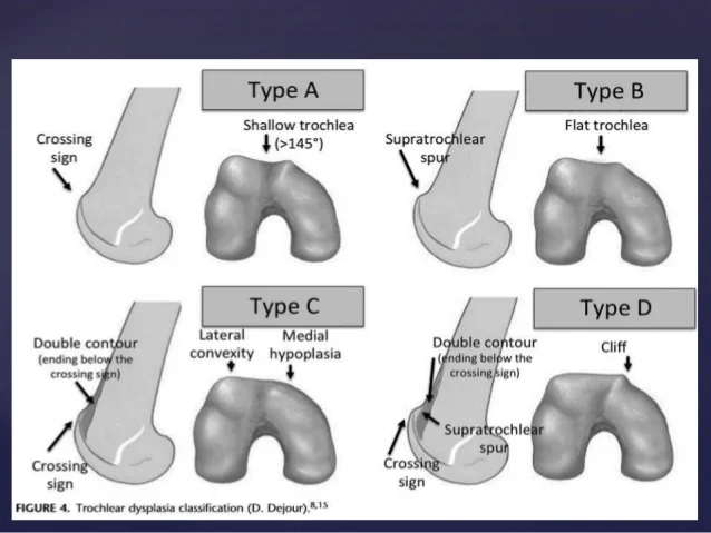
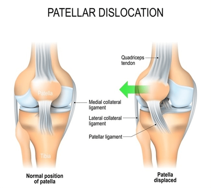
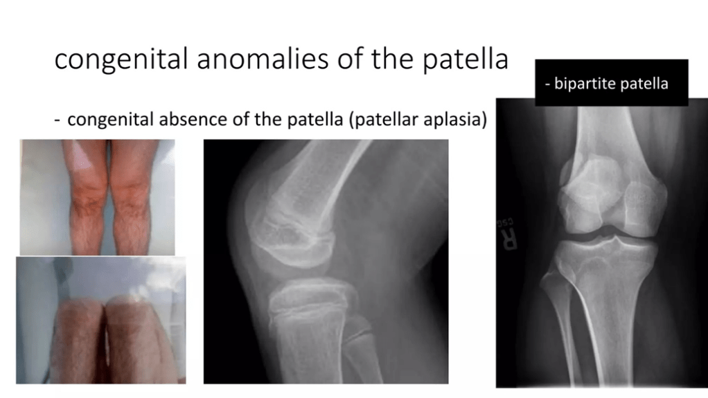
- Hypoplastic patellae – underdeveloped or abnormal kneecap.
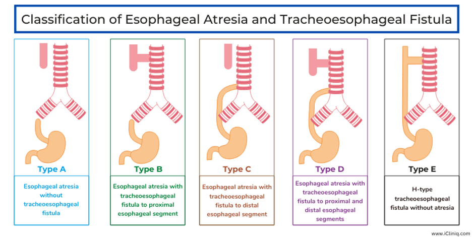
- Oesophageal or pyloric atresia – Oesophageal atresia is a rare birth defect where the oesophagus does not connect to the stomach, preventing food from reaching the stomach.
- Poikiloderma congenital or poikiloderma of Rothmund-Thomson
- Small saddle nose
- Protruding jaw
(Langer, Cunniff and Kucine, 2023)
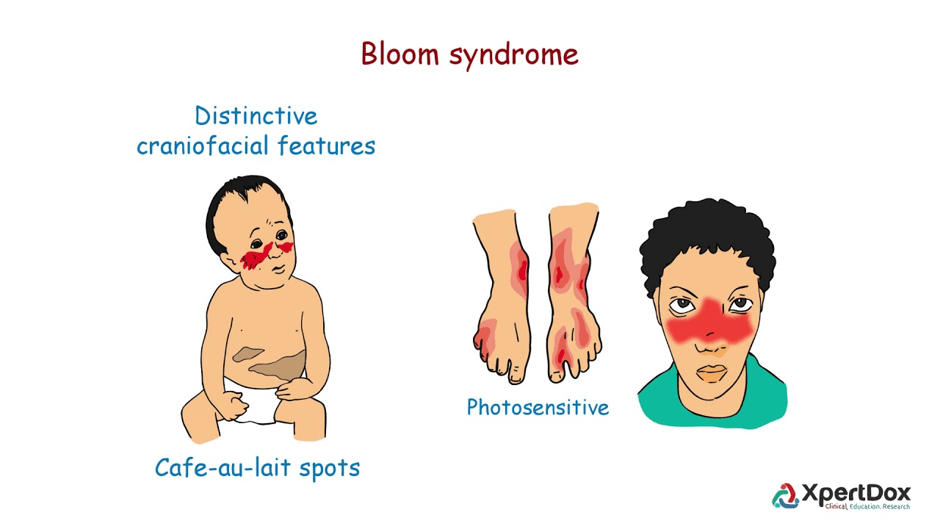
Bloom’s syndrome
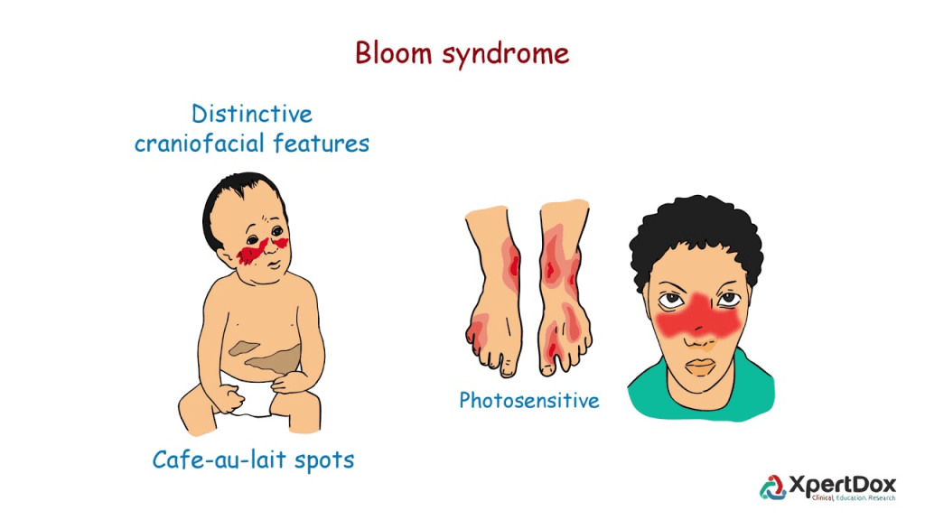
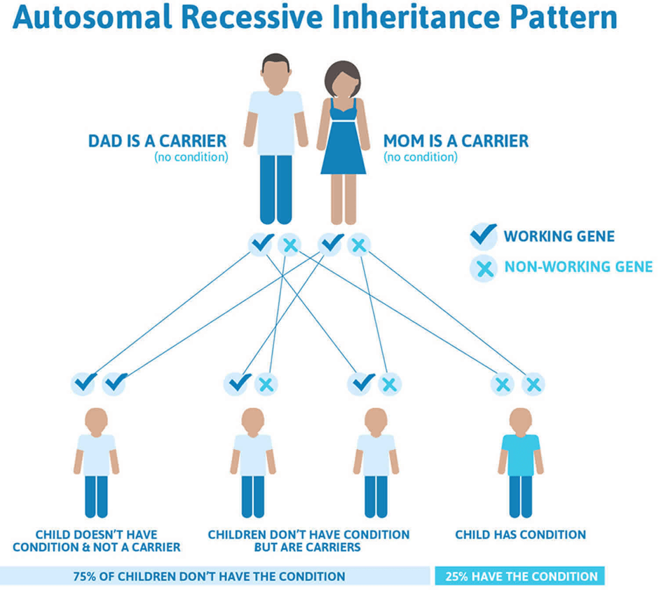
It is an autosomal recessive disorder.
There is a Biallelic mutations on Chromosome 15 (15q26.1) in the BLM gene.
BLM gene encodes for a RecQ helicase.
If both parents are heterozygous (carriers) of the BLM gene but are not affected, there is a 25% chance of having a child with BLM pathogenic gene and affected. 50% being a carrier and 25% no BLM pathogenic neither carrier nor affected. Prenatal and preimplantation genetic testing are affected.
Signs and symptoms
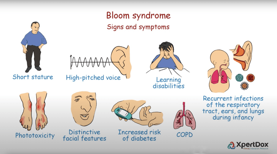
- Repeated infections due to immune abnormalities
- Blistering hands and lips
- short stature
- Sun-sensitive erythematous rash on face with a butterfly shape.
- Sparse subcutaneous fat
- Growth deficiency affects height, weight and head circumference throughout life. However, body proportions are normal.
- Resistant to insulin (type 2 diabetes)
- Normal intellectual ability.
- Fertile but early menopause
- Men tend to be infertile.
- Chronic obstructive pulmonary disease (COPD). Acondition where the lungs are intoxicated with smoking or airborne pollution and cause difficulty breathing and obstructs the lungs.
- Hypothyroidism (underactive thyroid gland /low production of thyroxine hormone) affecting metabolism
(Langer, Cunniff and Kucine, 2023)
RAPADILINO syndrome
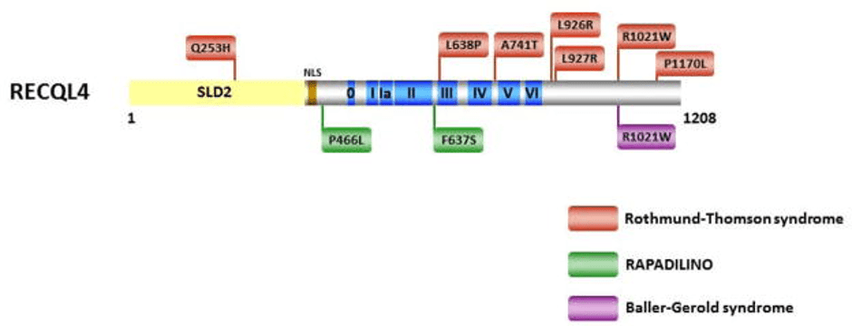
This is another genetic syndrome caused by the mutation of the RECQL4 gene.
It is an autosomal recessive condition.
The association with RAPADILINO and Osteosarcoma
This can be seen on the signs how it affects the growth of the bone and soft tissue of the human skeleton.
The RAPADILINO is mneumonic for its signs
RA Radial dysplasia
Dysplasia – abnormal development of lining (epithelium)
PA Patella aplasia or hypoplasia/ left high arched palate
Hypoplasia – underdevelopment of cells and tissues and organs.
Aplasia –
Aplasia refers to the incomplete or absent development of an organ, tissue, or body part, often evident at birth. This is different from agenesis. Agenesis is the complete absence of an organ where there is no structure at all.
DI Diarrhoea and dislocated joints
LI little size and limb malformations
NO long, slender Nose and Normal intelligence.
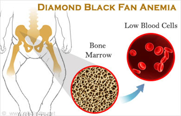
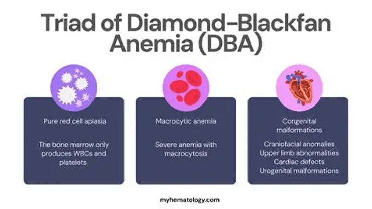
Diamond-Blackfan anaemia (DBA syndrome)
It is an autosomal dominant disorder.
45% of cases have been passed down through heredity (parents).
Other cases may occur randomly (sporadic) when cells are dividing.
Other causes of genetic mutations of DBA are:
Viruses
UV radiation from sunlight
Other environmental factors.
What are the effects of DBA?
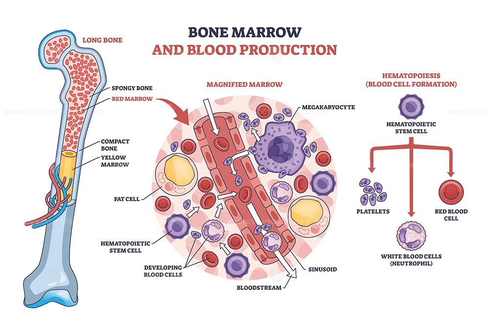
The bone marrow unable to produce red blood cells.
This is partially caused by inefficient or disrupted metabolism.
Metabolism is a series of chemicals to make energy and remove toxins.
Each chemical reaction are maintained by hormones and enzymes.
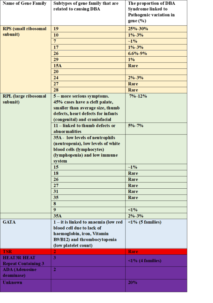
What are the genes affected by Diamond Black fan anaemia?
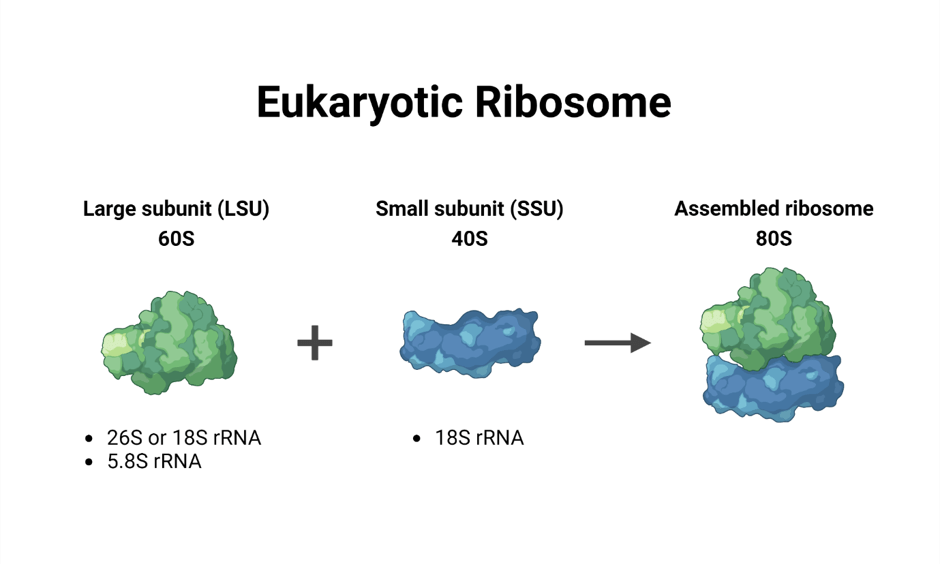
The ribosomal subunit
The ribosomes produce proteins.
They are structurally divided into two subunits: large and small
The size of proteins in plants, animals and fungi (eukaryotic) is different compared to bacteria (prokaryotes)
The large ribosomal subunit in eukaryotes is 60S. This is referred to as RPL.
The small ribosomal subunit in eukaryotes is 40S. This is referred to as RPS.
Deficiency in maturation of 60S causes the buildup and rapid maturation of 40S.
GATA1
It encodes the GATA1 protein.
GATA is needed for the development of many cells (blood, heart, and skin).
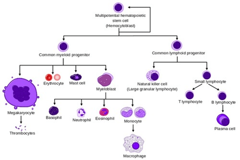
GATA 1, 2, and 3 are specifically needed for normal production of blood cells (haematopoiesis).
GATA1 is an X-linked gene that encodes a DNA-binding protein needed for the growth and development of erythroid cells and immune cells (megakaryocytes, mast cells, eosinophils, and basophils).
TSR2 ribosomal maturation factor
The TSR2 gene encodes a protein that represses or stops the transcription of NF-kappaB (NF-κB), which is important for transcription, inflammation, cell cycle control, cell death (apoptosis), survival, and production of cytokine proteins involved in the immune system.
HEATR3 Gene – HEAT Repeat Containing 3
The gene encodes a protein that is involved in the transport of the 60S ribosomal protein subunit RPL5.
It helps in the production of ribosomes. Ribosomes are organelles that produce proteins.
It also helped to assemble the 5S ribonucleoprotein particle (5S RNP).
It is involved in the NOD2-mediated NF-kappaB signaling for inflammation.
It helps to mature red blood cells (erythrocytes).
ADA2 (Adenosine deaminase)
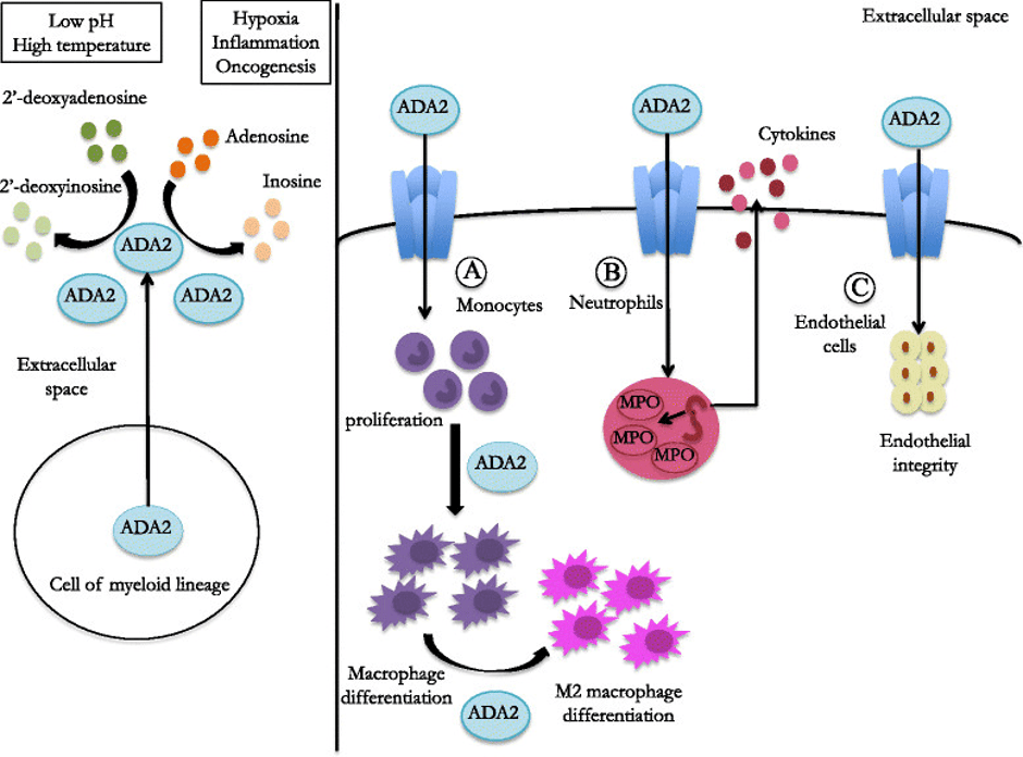
The ADA2 gene encodes a protein enzyme called adenosine deaminase 2.
This gene is needed for the growth and development of many immune cells involved in inflammation, where to increase or decrease.
For instance, macrophages promote inflammation.
What are the pathogenic variants affected by Diamond-Blackfan anaemia?
There are three forms:
- Autosomal recessive
- Autosomal dominant
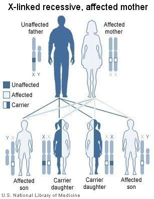
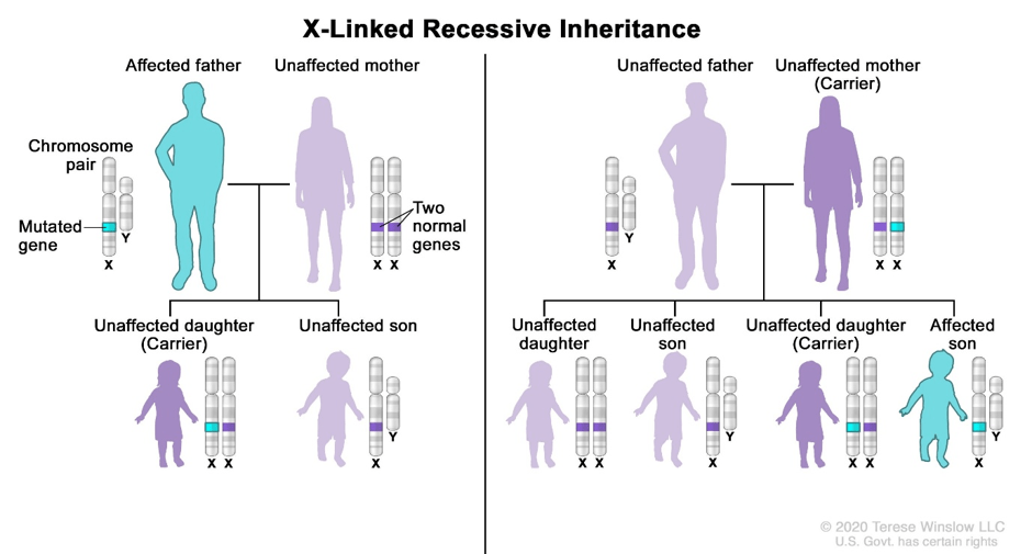
- X-linked
A closer depth
Autosomal recessive DBA
Example: HEATR
There is a biallelic pathogenic variant.
There is a 25% chance that each sibling of an affected patient is at risk of being affected. 50% a carrier without symptoms, and 25% are neither carriers nor affected.
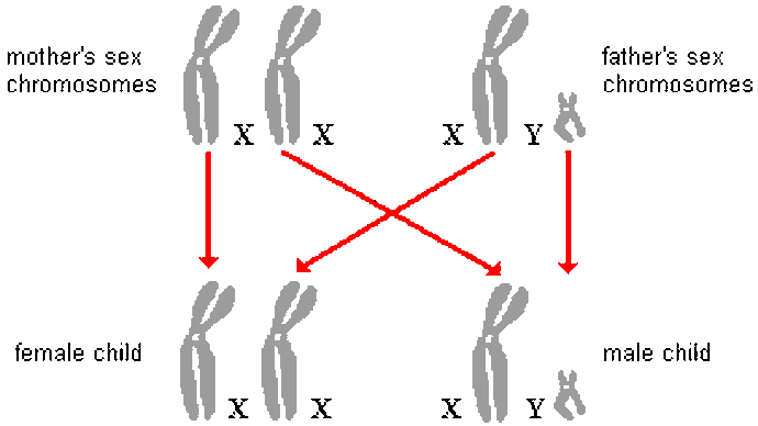
Hemizygous – a gene on a single copy that is normally an X-linked gene in males. Males have an XY chromosome. X from the mother and Y from the father.
Genetic testing can be done to identify any at-risk relatives for the HEATR3 gene.
Autosomal dominant DBA
It represents 40 to 45% of cases of all DBA syndromes.
55 to 60% of the autosomal dominant DBA cases are de novo.
De novo refers to having the condition for the first time in a family member.
X-linked DBA
Example: GATA1 or TSR2 gene.
For example:
The mother has a GATA1 or TSR2 mutation.
There is a 50% chance the offspring will have it.
Males have an XY chromosome.
Females have an XX chromosome.
The condition is on an X chromosome.
Therefore, females could have the condition or could be a carrier (heterozygote).
Males who have the mother’s X chromosome will have the condition because men carry only one X chromosome.
The affected male can transmit the mutation to their daughters but not to his sons.
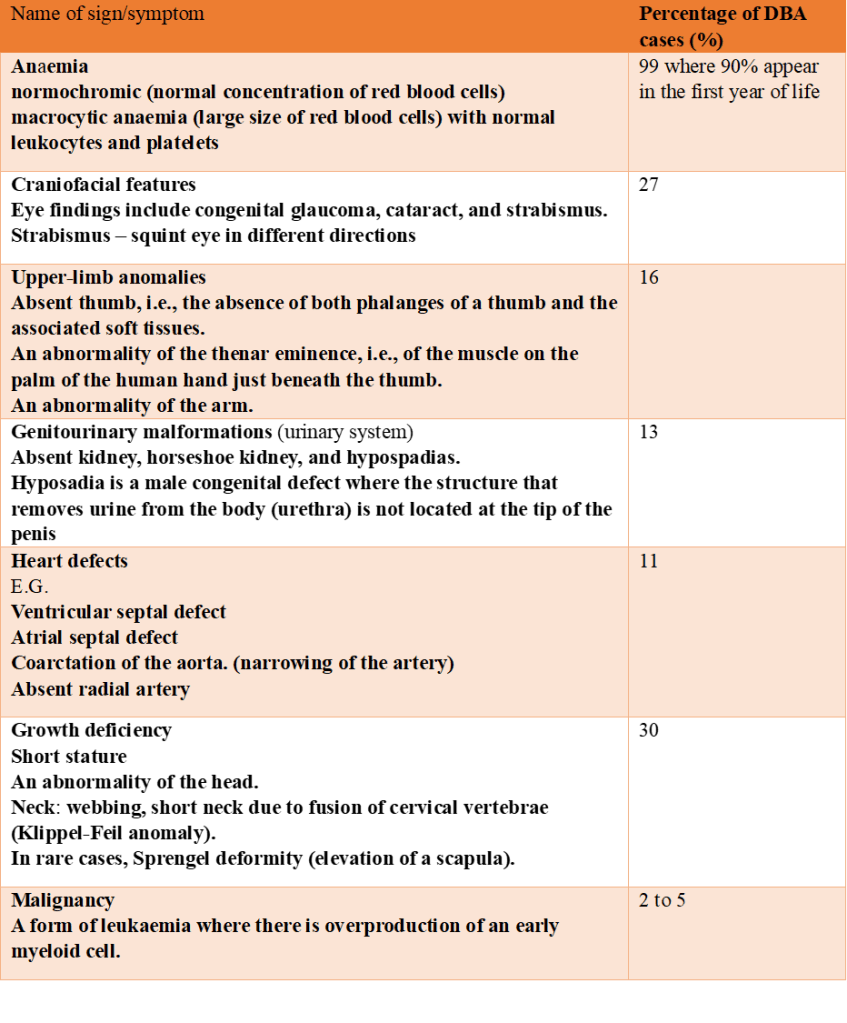
Signs and symptoms of DBA syndrome
Symptoms arise from birth.
Congenital malformations in up to 50% of affected individuals.
(Genetic and Rare Diseases Information Center, 2026; Cheng et al., 2019; Crispino and Horwitz, 2017; MedlinePlus, 2018; Gene Cards, 2026a; Gene Cards,2026b; Sieff, 2025)
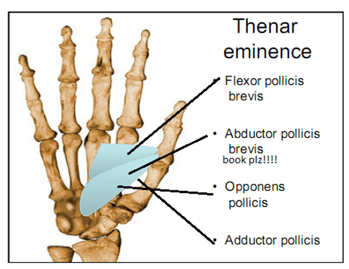
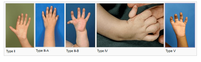
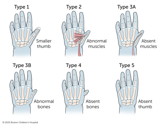
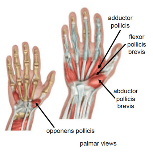
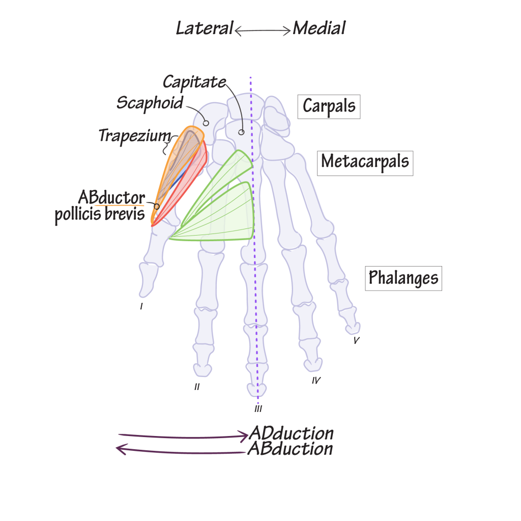
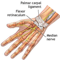
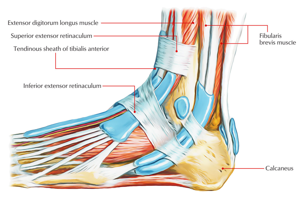
Abductor pollicis brevis: “brevis” means short or limited. The origin of abductor pollicis brevis is situated at the flexor retinaculum and the tubercles of the scaphoid and trapezium (carpals). The retinaculum is a band of dense fibrous connective tissue containing collagen and it provides tensile strength for the tendons and nerves as they cross the joints to ensure smooth efficient movement whilst withstanding pressure. This prevents displacement.
The insertion of the abductor pollicis brevis is the lateral side of the base and proximal phalange of the thumb. It abducts (moves away) the thumbs and helps opposes it.
The flexor pollicis brevis has the same origin and insertion of abductor pollicus brevis but has an opposing action of flexing the thumb. Flexor movement is bending towards.
Adductor pollicis: Oblique head: bases of 2nd and 3rd metacarpals, capitate, adjacent carpals. tranvese head. Anterior surface body and 3rd metacarpals. Insertion is the medial side of the base of the proximal phalange of the thumb. Thumb is Adducted (brings towards) thumb toward the middle digit.
Opponens pollicis: The origin is the Flexor retinaculum and tubercles of scaphoid and trapezium. Insertion involves the lateral side of the 1st metacarpal. The action involves draws the first metacarpal bone laterally to oppose thumb towards the center of palm and rotates medially.
References
Ajeawung, N.F., Nguyen, T.T.M., Lu, L., Kucharski, T.J., Rousseau, J., Molidperee, S., Atienza, J., Gamache, I., Jin, W., Plon, S.E., Lee, B.H., Teodoro, J.G., Wang, L.L. and Campeau, P.M. (2019). Mutations in ANAPC1, Encoding a Scaffold Subunit of the Anaphase-Promoting Complex, Cause Rothmund-Thomson Syndrome Type 1. The American Journal of Human Genetics, 105(3), pp.625–630. doi:https://doi.org/10.1016/j.ajhg.2019.06.011.
Alexiev, B. (2021) Bone & joints Osteoclastic giant cell rich tumors Giant cell tumor of bone, NOS, Available at: https://www.pathologyoutlines.com/topic/bonegiantcelltumor.html (4th April 2026t
Alexiev, B. and Jennings, L. (2026) Bone & joints Undifferentiated spindle and small round cell sarcomas of soft tissue and bone NFATC2 rearranged sarcoma. Available at: https://www.pathologyoutlines.com/topic/boneroundcellsarcomaewsr1.html (Accessed: 26th April 2026)
Alexiev, B. and Laskin, W. (2022) Bone & joints. Other tumors of bone. Adamantinoma. Available at: https://www.pathologyoutlines.com/topic/boneadamantinoma.html (Accessed: 5th May 2026)
Bădilă, A.E., Rădulescu, D.M., Niculescu, A.-G., Grumezescu, A.M., Rădulescu, M. and Rădulescu, A.R. (2021). Recent Advances in the Treatment of Bone Metastases and Primary Bone Tumors: An Up-to-Date Review. Cancers, [online] 13(16), p.4229. Doi: https://doi.org/10.3390/cancers13164229.
Biermann, J.S., Hirbe, A., Ahlawat, S., Bernthal, N.M., Binitie, O., Boles, S., Brigman, B., Callan, A.K., Cipriano, C., Cranmer, L.D., Davis, J., Donnelly, E., Ferguson, M., Graham, A., Groundland, J., Hess, M., Hiniker, S.M., Hoover-Regan, M.L., Hornick, J.L. and Jonard, B. (2025). Bone Cancer, Version 2.2025, NCCN Clinical Practice Guidelines in Oncology. Journal of the National Comprehensive Cancer Network, [online] 23(4). does: https://doi.org/10.6004/jnccn.2025.0017.
Cancer Research UK (2025a) Types of bone cancer. Available at: https://www.cancerresearchuk.org/about-cancer/bone-cancer/types (Accessed 3rd April 2026)
Cheng, Z., Mugler, C.F., Keskin, A., Hodapp, S., Chan, L.Y.-L., Weis, K., Mertins, P., Regev, A., Jovanovic, M. and Brar, G.A. (2019). Small and Large Ribosomal Subunit Deficiencies Lead to Distinct Gene Expression Signatures that Reflect Cellular Growth Rate. Molecular Cell, [online] 73(1), pp.36-47.e10. doi:https://doi.org/10.1016/j.molcel.2018.10.032.
Cleveland Clinic (2022) Rothmund-Thomson Syndrome (RTS). Available at: https://my.clevelandclinic.org/health/diseases/24154-rothmund-thomson-syndrome-rts (Accessed: 21st April 2026)
Cleveland Clinic (2024) Werner Syndrome Available at: https://my.clevelandclinic.org/health/diseases/werner-syndrome (Accessed: 21st April 2026)
Caorsi, R., Penco, F., Schena, F. and Gattorno, M. (2016). Monogenic polyarteritis: the lesson of ADA2 deficiency. Pediatric Rheumatology, 14(1). doi:10.1186/s12969-016-0111-7.
Cowan, P., Launico, M. and Kahai, P. (2024) Anatomy, Bones. Available at: https://www.ncbi.nlm.nih.gov/books/NBK537199/ (Accessed: 9th April 2026)
Crispino, J.D. and Horwitz, M.S. (2017). GATA factor mutations in hematologic disease. Blood, [online] 129(15), pp.2103–2110. doi:https://doi.org/10.1182/blood-2016-09-687889.
Gene Cards (2026a) TSR2 Gene – TSR2 Ribosome Maturation Factor. Available at: https://www.genecards.org/cgi-bin/carddisp.pl?gene=TSR2 (Accessed: 22nd April 2026)
Gene Cards (2026b) HEATR3 Gene – HEAT Repeat Containing 3. Available at: https://www.genecards.org/cgi-bin/carddisp.pl?gene=HEATR3 (Accessed: 22nd April 2026)
Genecards (2026c) H3-3A Gene – H3.3 Histone A. Available at: https://www.genecards.org/cgi-bin/carddisp.pl?gene=H3-3A#summaries (Accessed: 9th April 2026)
Genetic and Rare Diseases Information Center (2026) Diamond-Blackfan anemia. Available at: https://rarediseases.info.nih.gov/diseases/6274/diamond-blackfan-anemia (Accessed: 21st April 2026)
Gerrand, C., Amary, F., Anwar, H.A., Brennan, B., Dileo, P., Kalkat, M.S., McCabe, M.G., McCullough, A.L., Parry, M.C., Patel, A., Seddon, B.M., Sherriff, J.M., Tirabosco, R. and Strauss, S.J. (2024). UK guidelines for the management of bone sarcomas. British Journal of Cancer. doi:https://doi.org/10.1038/s41416-024-02868-4.
Hameed, M. and Mandelker, D. (2018). Tumor Syndromes Predisposing to Osteosarcoma. Advances in anatomic pathology, [online] 25(4), pp.217–222. doi:https://doi.org/10.1097/PAP.0000000000000190.
Hosseini, H., Heydari, S., Kiavash Hushmandi, Salman Daneshi and Rasoul Raesi (2025). Bone tumors: a systematic review of prevalence, risk determinants, and survival patterns. BMC Cancer, 25(1). doi:https://doi.org/10.1186/s12885-025-13720-0.
Kleinerman, R.A., Tucker, M.A., Tarone, R.E., Abramson, D.H., Seddon, J.M., Stovall, M., Li, F.P. and Fraumeni, J.F. (2005). Risk of New Cancers After Radiotherapy in Long-Term Survivors of Retinoblastoma: An Extended Follow-Up. Journal of Clinical Oncology, 23(10), pp.2272–2279. doi:https://doi.org/10.1200/jco.2005.05.054.
Langer, K., Cunniff, C. and Kucine, N. (2023) Bloom Syndrome. Available at: https://www.ncbi.nlm.nih.gov/sites/books/NBK1398/ (Accessed: 21st April 2026)
MedlinePlus (2018) Adenosine deaminase 2 deficiency. Available at: https://medlineplus.gov/genetics/condition/adenosine-deaminase-2-deficiency/#causes (Accessed: 22nd April 2026)
Piazza, Y. and Kaseb, H. (2026) Bone & joints Other tumors of bone Undifferentiated pleomorphic sarcoma. Available at: https://www.pathologyoutlines.com/topic/boneMFH.html (Accessed: 26th April 2026)
Pietrangelo, A. (2019) What is fibrosarcoma and how is it treated? Available at: https://www.healthline.com/health/fibrosarcoma (Accessed 3rd April 2026)
Pullan, J.E., and Lotfollahzadeh, S. (2024) Primary Bone Cancer Available at: https://www.ncbi.nlm.nih.gov/books/NBK560830/ (Accessed: 9th April 2026)
Sieff, C. (2025) DBA Syndrome Available at: https://www.ncbi.nlm.nih.gov/books/NBK7047/ (Accessed: 22nd April 2026)
WHO Editorial Board. WHO classification of bone tumours. In WHO Classification of Tumours Editorial Board. Soft tissue and bone tumours. Bovee J, Flanagan AM, Lazar AJ, Nielsen GP and Yoshida A (eds) pp 338. International Agency for Research on Cancer (2020)
Updated July 2026 Next Review July 2028


Leave a Reply