Question: How Can We Tell The Difference Between Enchondroma And Low-grade Chondrosarcoma?
What is low-grade chondrosarcoma?
Type of tumour: malignant, slow-growing cartilage tumour.
Location: Long bones, flat bones (pelvis) and ribs.
Symptoms: Deep continuous dull pain.
Risk factors: Increased age, and there is a 40% risk of patients with Ollier and Mafucci syndrome developing chondrosarcoma, which depends on age. Patients with endochondroma in the long bones and the pelvis (axial) have an increased risk of developing chondrosarcoma. Thus, regular screening is needed to detect malignant transformation.
What is an enchondroma?
- Type of tumour: Benign cartilage tumour
- Location: Mostly found in the long bones (proximal humerus, distal femur, or proximal tibia metaphyses),
- Age of onset: 10 to 40 years.
- Symptoms: Patients have no symptoms and are diagnosed via X-rays in the small bones of the hands and feet.
- Size of tumour: Enchondromas are typically smaller than 5 cm, and 80% of enchondromas in the long bones are smaller than 2 cm.
- Risk factors:
- The risk is small if it affects a small area, but if it is caused by rare genetic conditions, the risk increases, causing malignant multiple enchondromas.
- Examples of genetic conditions are Ollier’s disease and Mafucci syndrome.
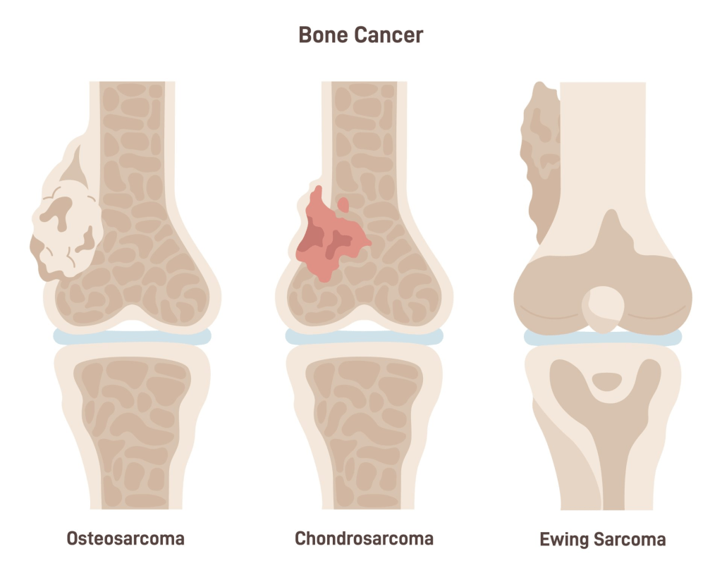
What is the difference between Ollier’s disease and Maffucci syndrome?
The extent to which it is distributed in the body.
Ollier’s disease, otherwise known as dyschondroplasia, has unequal distribution of benign cartilaginous tumours in the body. It is caused by malformed ossification of cartilages. The tumours lead to stunted growth and deformation.
Maffucci syndrome causes enchondroma and soft tissue vascular growths called hemangioma. This causes a greater risk and is linked to the size and number of lesions, especially in the pelvis and long bones.
Classification of endochondroma and low-grade chondrosarcoma
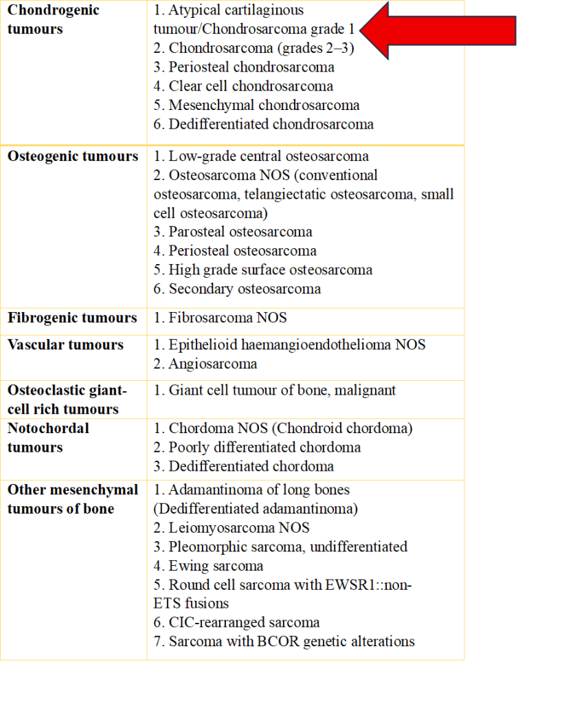
Endochondroma and low-grade chondrosarcoma are classified as part of atypical cartilaginous tumours (ACT) in the APPENDICULAR skeleton, particularly long bones, by the World Health Organisation in 2013 and 2020.
They are intermediate chondroid tumours because of their rarity of metastasis but are locally aggressive and have a better clinical outcome than the same tumour in the axial skeleton (<2%).
Since the World Health Organisation (2020), the same lesions of Endochondroma and low-grade chondrosarcoma are also classified as grade 1 chondrosarcoma in the AXIAL skeleton (scapula, pelvis, ribs, spine, sternum (breastbone) and skull) because they have a poorer outcome.
If there are multiple lesions or in the pelvis, it can cause chondrosarcoma in solitary osteochondroma and enchondroma.
Table 1 Classification of malignant primary bone tumours (WHO, 2020)
Diagnosis of enchondroma and chondrosarcoma
Both enchondroma and low-grade chondrosarcoma vary in the outcomes of diagnosis and treatment.
The tests that can be done are in the form of X-ray, Magnetic Resonance Imaging (MRI), Computed Tomography (CT), and the study of tissues (histology). A sample of this is extracted via biopsy of the concerned area to be visualised under the microscope by a histopathologist.
The role of CT in the diagnosis of enchondroma and ACT
It is not a routine imaging test for enchondroma and ACT.
However, if the MRI/X-ray does not help with the diagnosis, it is used to detect the following:
- Subtle matrix mineralization.
- To measure the amount of bone destruction in axial lesions, e.g., skull or pelvis, and the extent of the tumour if it has reached the joint.
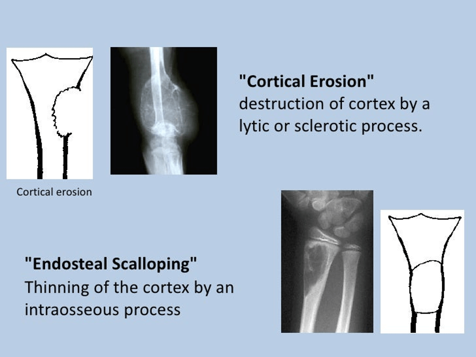
- It can help detect whether there is expansion and cortical destruction.
- It helps to detect other pathological fractures.
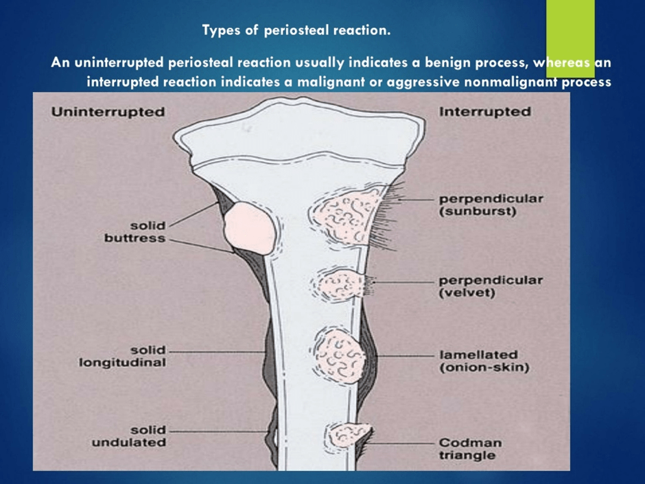
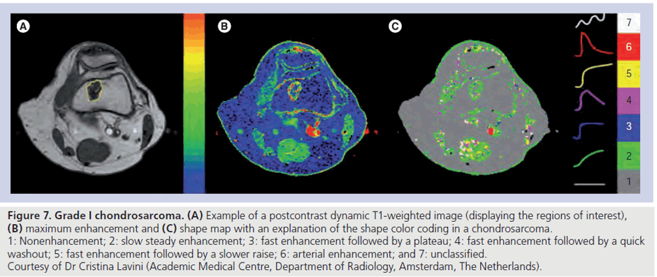
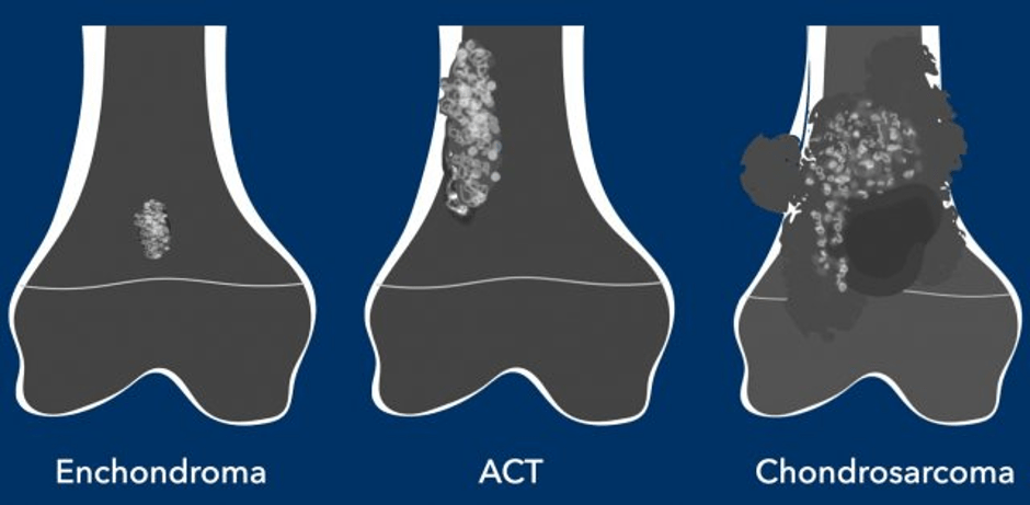
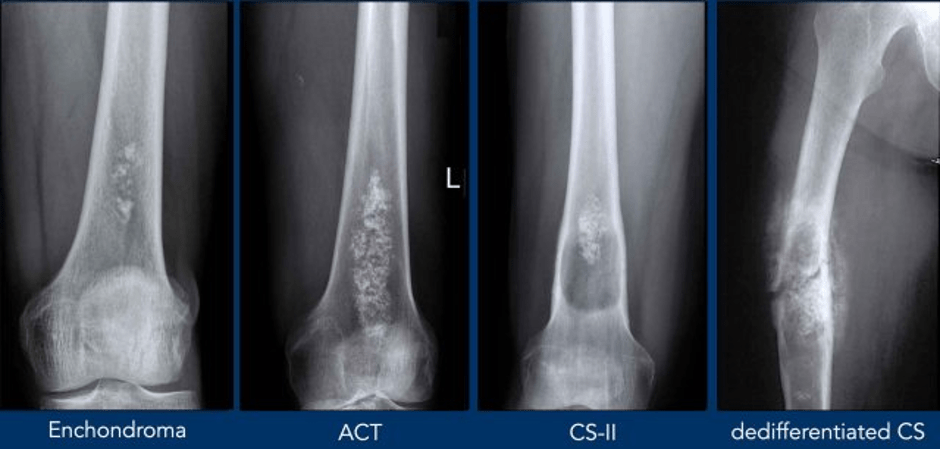
Features to distinguish enchondroma and chondrosarcoma
Supporting Images And Resources
Treatment differences between enchondroma and chondrosarcoma
Enchondromas are benign and do not require follow-up.
High-grade chondrosarcomas are treated with wide resection, since they are insensitive to chemotherapy and radiation.
Over time, treatment of ACT has shifted towards the conservative side, i.e. watchful waiting or “wait and scan” by MRI instead of curettage.
Recommended source
https://radiologyassistant.nl/musculoskeletal/bone-tumors/chondrotumors-1
References
Acibadem Health Point (2026) Chondrosarcoma Vs Enchondroma: Key Differences Available at: https://www.acibademhealthpoint.com/chondrosarcoma-vs-enchondroma-key-differences/ (Accessed: 23rd April 2026)
Bădilă, A.E., Rădulescu, D.M., Niculescu, A.-G., Grumezescu, A.M., Rădulescu, M. and Rădulescu, A.R. (2021). Recent Advances in the Treatment of Bone Metastases and Primary Bone Tumors: An Up-to-Date Review. Cancers, [online] 13(16), p.4229. Doi: https://doi.org/10.3390/cancers13164229.
Biermann, J.S., Hirbe, A., Ahlawat, S., Bernthal, N.M., Binitie, O., Boles, S., Brigman, B., Callan, A.K., Cipriano, C., Cranmer, L.D., Davis, J., Donnelly, E., Ferguson, M., Graham, A., Groundland, J., Hess, M., Hiniker, S.M., Hoover-Regan, M.L., Hornick, J.L. and Jonard, B. (2025). Bone Cancer, Version 2.2025, NCCN Clinical Practice Guidelines in Oncology. Journal of the National Comprehensive Cancer Network, [online] 23(4). does: https://doi.org/10.6004/jnccn.2025.0017.
Biology Insights (2025) Enchondroma vs. Chondrosarcoma: Key Differences Explained. Available at: https://biologyinsights.com/enchondroma-vs-chondrosarcoma-key-differences-explained/ (Accessed: 23rd April 2026)
Cancer Research UK (2025a) Types of bone cancer. Available at: https://www.cancerresearchuk.org/about-cancer/bone-cancer/types (Accessed 3rd April 2026)
Cowan, P., Launico, M. and Kahai, P. (2024) Anatomy, Bones. Available at: https://www.ncbi.nlm.nih.gov/books/NBK537199/ (Accessed: 9th April 2026)
Gazendam, A., Popovic, S., Parasu, N., & Ghert, M. (2023). Chondrosarcoma: A Clinical Review. Journal of Clinical Medicine, 12(7), 2506. https://doi.org/10.3390/jcm12072506
Gerrand, C., Amary, F., Anwar, H.A., Brennan, B., Dileo, P., Kalkat, M.S., McCabe, M.G., McCullough, A.L., Parry, M.C., Patel, A., Seddon, B.M., Sherriff, J.M., Tirabosco, R. and Strauss, S.J. (2024). UK guidelines for the management of bone sarcomas. British Journal of Cancer. doi:https://doi.org/10.1038/s41416-024-02868-4.
Guney, A. and Qasem, S. (2024) Bone and joints Chondrosarcoma. Available at: https://www.pathologyoutlines.com/topic/bonechondrosarcoma.html (Accessed: 20th July 2026)
Hosseini, H., Heydari, S., Kiavash Hushmandi, Salman Daneshi and Rasoul Raesi (2025). Bone tumors: a systematic review of prevalence, risk determinants, and survival patterns. BMC Cancer, 25(1). doi:https://doi.org/10.1186/s12885-025-13720-0.
Pullan, J.E., and Lotfollahzadeh, S. (2024) Primary Bone Cancer Available at: https://www.ncbi.nlm.nih.gov/books/NBK560830/ (Accessed: 9th April 2026)
Thakkar, J.B., Deshpande, S.V. and Janu, A. (2019) Enchondroma Vs Low grade Chondrosarcoma : Differentiating features and role of imaging in management. Available at: https://epos.myesr.org/poster/esr/ecr2019/C-2823/Background (Accessed: 23rd April 2026)
van Langevelde, K. and Robin Smithuis, R. (2023) Cartilage tumors with special attention to Atypical Cartilaginous Tumors. Available at: https://radiologyassistant.nl/musculoskeletal/bone-tumors/chondrotumors-1 (Accessed: 23rd April 2026)
Verdegaal, S.H.M, Bovée, J.V.M.G., Pansuriya, T.C., Grimer, R.J., Ozger, H., Jutte, P.C., San Julian, M., Biau, D.J., Ingrid C.M. Geest, Leithner, A., Streitbürger, A., Klenke, F.M., Gouin, F.G., Campanacci, D.A., Marec-Berard, P., Hogendoorn, P.C.W., Brand, R. and Taminiau, A.H.M. (2011). Incidence, Predictive Factors, and Prognosis of Chondrosarcoma in Patients with Ollier Disease and Maffucci Syndrome: An International Multicenter Study of 161 Patients. The Oncologist, 16(12), pp.1771–1779. doi:10.1634/theoncologist.2011-0200.
WHO Editorial Board. WHO classification of bone tumours. In WHO Classification of Tumours Editorial Board. Soft tissue and bone tumours. Bovee J, Flanagan AM, Lazar AJ, Nielsen GP and Yoshida A (eds) pp 338. International Agency for Research on Cancer (2020)
Updated July 2026 Next Review July 2028


Leave a comment