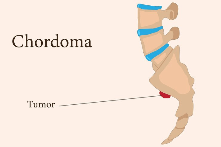
Overview of Chordoma
Malignancy: very rare malignant
Type of bone tumour: primary
Grade: slow-growing/low grade
Location: It affects the vertebral bodies.
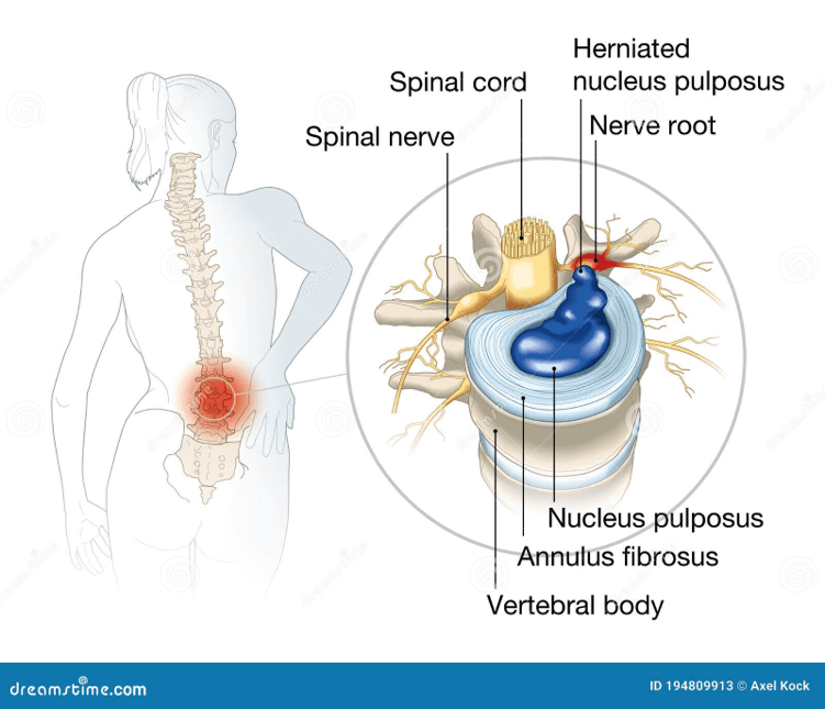
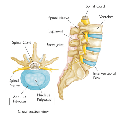
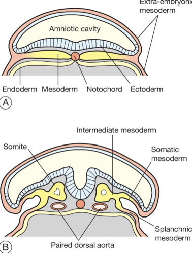
The vertebral bodies are the bony area of the spine rather than the jelly disc, though they contain vestigial notochord within the nucleus pulposus.
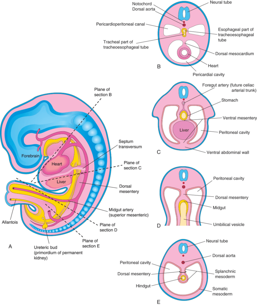
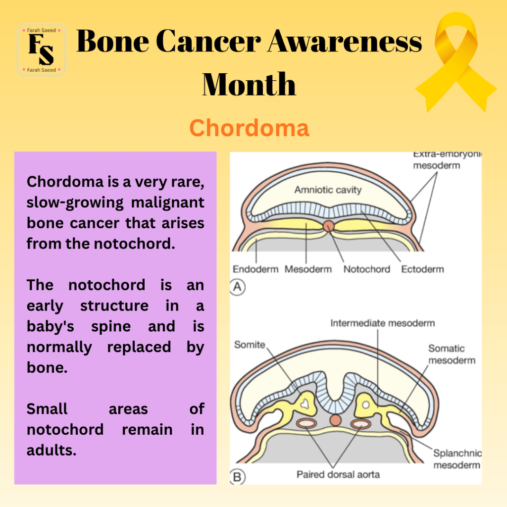
The notochord is the early stage of the spine in a baby, which normally gets replaced by bone.
Small areas of the notochord remain in adults.
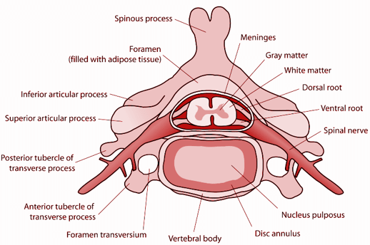
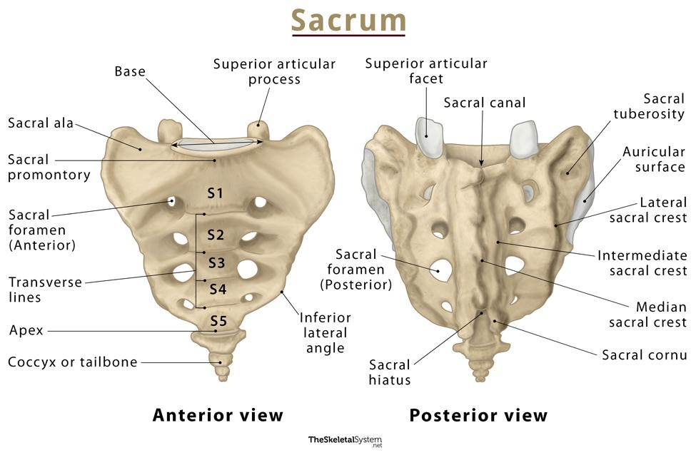
50% grow in the lower end of the spinal vertebrae (sacrum)
Other locations of the chordoma.
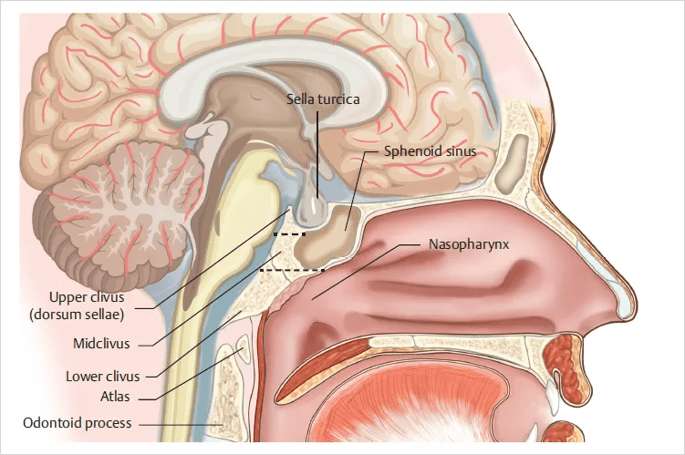
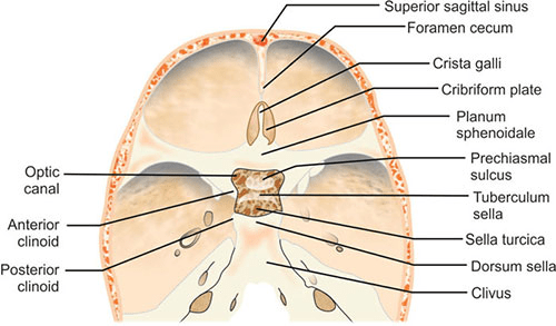
It may occur in the skull (30%), like the clivus.
The clivus is a bony structure at the base of the skull that supports the brainstem and connects the skull with the spine.
Mobile spine and other parts of the bone (20%).
Extraskeletal tumours are rare.
Onset: Mean age is 60 years.
Appearance:
Chordomas are infiltrative.
They destroy the bony portion of the spine and invade surrounding neural spaces.
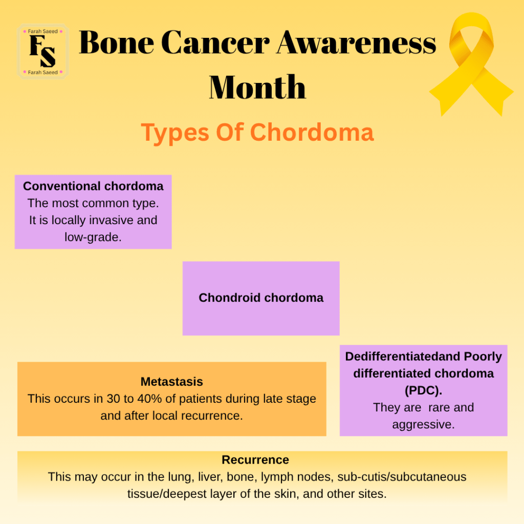
Types Of Chordoma
Risk Factors Of Chordoma
A) Genetics:
Example One of the genes that increases the risk of Chordoma: Brachyury
Brachyury (T) is a transcription factor of the T-box family.
It binds to target genes in the nucleus, facilitates development, and in some cases disease.
A mutated gene or its duplication of Brachyury is linked with the rare bone cancer (chordoma)
Overexpression of the mutated gene is linked to epithelial tumours that line the gastrointestinal tract (oesophagus, stomach, small intestine), urogenital (kidney, bladder, testis), respiratory (lungs), and the female gynaecological system (uterus, ovaries).
Recent evidence indicates tumour-promoting and tumour suppressor properties. TBX3 promotes tumour via epithelial-mesenchymal transition (EMT) during metastasis.
Overexpression of Brachyury causes resistance to anti-cancer therapy and confers stem cell traits on cancer cells.
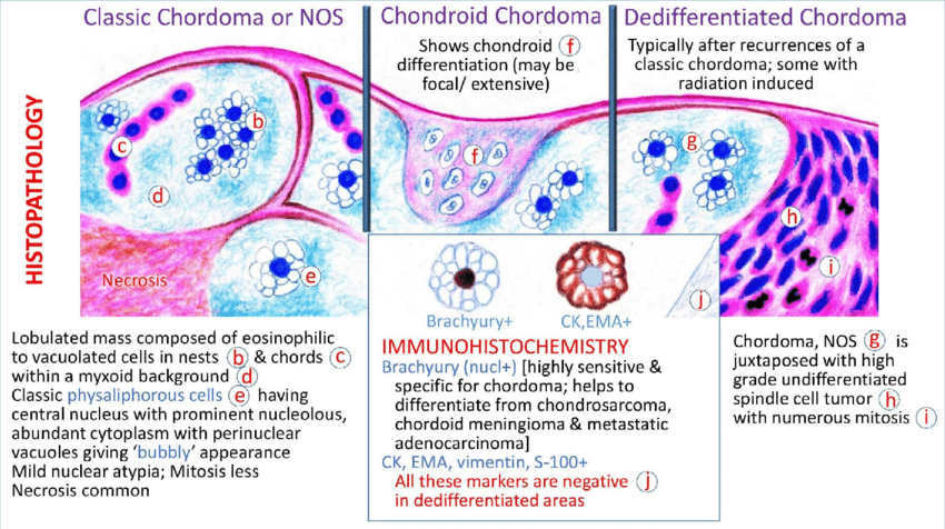
This can be detected using high levels of mesenchymal markers (fibronectin, N-cadherin, vimentin) and loss of epithelial markers (E-cadherin, plakoglobin) through immunohistochemistry testing.
(Wansleben et al., 2014)
Other risk factors of chordoma
B) Gender
More in males than females
C) Ethnicity:
Not applicable – no influence on ethnicity.
Appearance Under Microscope
Cell Morphology:
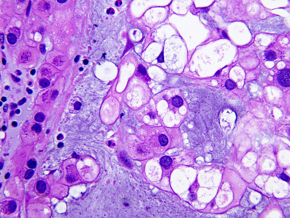
Under the microscope, they commonly have physaliphorous cells (vacuolated or soap-bubbled cells). It is also found in adenocarcinomas and chondrosarcomas. This can be distinguished using immunohistochemistry markers.
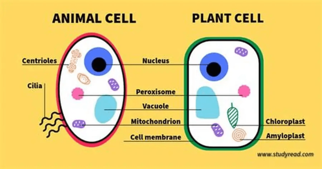
Vacuoles are membrane-bound sacs in the cytoplasm of the cells.
The membrane is selectively permeable and is called a tonoplast.
The role of the tonoplast is to control movement of substances in and out of the vacuole and depends on the size.
Inside the vacuole, there are a variety of nutrients, ions, water, enzymes and in some cases waste products. They vary based on the type of cell, size, number, and physiological state.
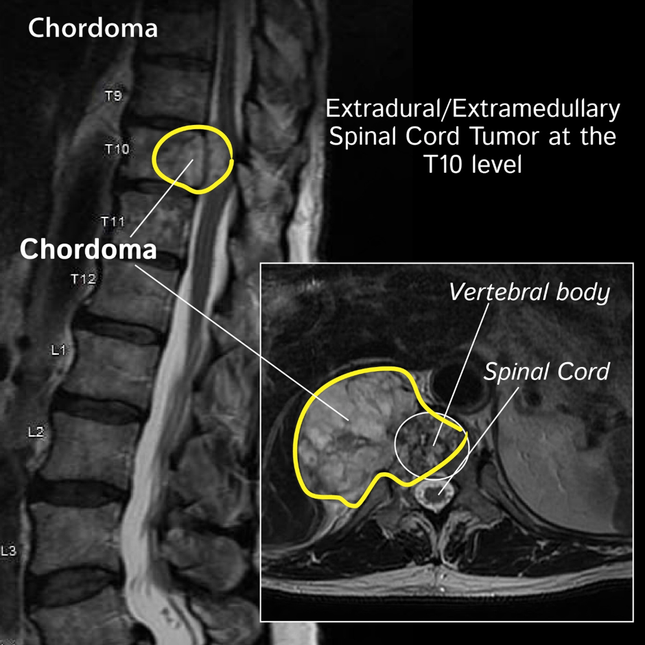
Infiltrative chordoma in position T10 affecting the vertebral body Dikti (n.d.)
References
Elsamadicy, A. A., Sayeed, S., Sherman, J. J. Z., Serrato, P., Ghanekar, S. D., Lo, S.-F. L., & Sciubba, D. M. (2025). The Association of Gender in the Management and Prognosis of Vertebral and Sacral Chordoma: A SEER Analysis. Journal of Clinical Medicine, 14(5), 1737. https://doi.org/10.3390/jcm14051737
Bădilă, A.E., Rădulescu, D.M., Niculescu, A.-G., Grumezescu, A.M., Rădulescu, M. and Rădulescu, A.R. (2021). Recent Advances in the Treatment of Bone Metastases and Primary Bone Tumors: An Up-to-Date Review. Cancers, [online] 13(16), p.4229. Doi: https://doi.org/10.3390/cancers13164229.
Biermann, J.S., Hirbe, A., Ahlawat, S., Bernthal, N.M., Binitie, O., Boles, S., Brigman, B., Callan, A.K., Cipriano, C., Cranmer, L.D., Davis, J., Donnelly, E., Ferguson, M., Graham, A., Groundland, J., Hess, M., Hiniker, S.M., Hoover-Regan, M.L., Hornick, J.L. and Jonard, B. (2025). Bone Cancer, Version 2.2025, NCCN Clinical Practice Guidelines in Oncology. Journal of the National Comprehensive Cancer Network, [online] 23(4). does: https://doi.org/10.6004/jnccn.2025.0017.
Cancer Research UK (2025a) Types of bone cancer. Available at: https://www.cancerresearchuk.org/about-cancer/bone-cancer/types (Accessed 3rd April 2026)
Cima, L., Das, A., Venkatesh Dhanasekaran, Mirabassi, N. and Pagliuca, F. (2021). The “Make Surgical Pathology Easy” project: learning Pathology through tailored digital infographics – the case for renovation of an old teaching method. Pathologica, 113(4), pp.252–261. doi:10.32074/1591-951x-269.
Cowan, P., Launico, M. and Kahai, P. (2024) Anatomy, Bones. Available at: https://www.ncbi.nlm.nih.gov/books/NBK537199/ (Accessed: 9th April 2026)
Ditki (n.d) Chordoma Available at: https://ditki.com/course/pathology/glossary/pathophysiologic-disorder/chordoma (Accessed: 28th April 2026)
Frontal cortex (2009) Clival chordoma, H&E stain x 400. Available at: https://frontalcortex.com/?page=oll&topic=24&qid=1909 (Accessed: 28th April 2026)
Gerrand, C., Amary, F., Anwar, H.A., Brennan, B., Dileo, P., Kalkat, M.S., McCabe, M.G., McCullough, A.L., Parry, M.C., Patel, A., Seddon, B.M., Sherriff, J.M., Tirabosco, R. and Strauss, S.J. (2024). UK guidelines for the management of bone sarcomas. British Journal of Cancer. doi:https://doi.org/10.1038/s41416-024-02868-4.
Hosseini, H., Heydari, S., Kiavash Hushmandi, Salman Daneshi and Rasoul Raesi (2025). Bone tumors: a systematic review of prevalence, risk determinants, and survival patterns. BMC Cancer, 25(1). doi:https://doi.org/10.1186/s12885-025-13720-0.
Pullan, J.E., and Lotfollahzadeh, S. (2024) Primary Bone Cancer Available at: https://www.ncbi.nlm.nih.gov/books/NBK560830/ (Accessed: 9th April 2026)
Sieff, C. (2025) DBA Syndrome Available at: https://www.ncbi.nlm.nih.gov/books/NBK7047/ (Accessed: 22nd April 2026)
Thakkar, J.B., Deshpande, S.V. and Janu, A. (2019) Enchondroma Vs Low grade Chondrosarcoma : Differentiating features and role of imaging in management. Available at:https://epos.myesr.org/poster/esr/ecr2019/C-2823/Background (Accessed: 23rd April 2026)
Tumor Surgery (2026) Adamantinoma: A Rare Bone Tumor of the Tibia Explained. Available at: https://www.tumorsurgery.org/2025/12/16/adamantinoma-rare-bone-tumor-tibia/ (Accessed: 4th April 2026)
van Langevelde, K. and Robin Smithuis, R. (2023) Cartilage tumors with special attention to Atypical Cartilaginous Tumors. Available at: https://radiologyassistant.nl/musculoskeletal/bone-tumors/chondrotumors-1 (Accessed: 23rd April 2026)
Verdegaal, S.H.M, Bovée, J.V.M.G., Pansuriya, T.C., Grimer, R.J., Ozger, H., Jutte, P.C., San Julian, M., Biau, D.J., Ingrid C.M. Geest, Leithner, A., Streitbürger, A., Klenke, F.M., Gouin, F.G., Campanacci, D.A., Marec-Berard, P., Hogendoorn, P.C.W., Brand, R. and Taminiau, A.H.M. (2011). Incidence, Predictive Factors, and Prognosis of Chondrosarcoma in Patients with Ollier Disease and Maffucci Syndrome: An International Multicenter Study of 161 Patients. The Oncologist, 16(12), pp.1771–1779. doi:10.1634/theoncologist.2011-0200.
Wansleben, S., Peres, J., Hare, S., Goding, C.R. and Prince, S. (2014). T-box transcription factors in cancer biology. Biochimica et Biophysica Acta (BBA) – Reviews on Cancer, [online] 1846(2), pp.380–391. doi:https://doi.org/10.1016/j.bbcan.2014.08.004.
WHO Editorial Board. WHO classification of bone tumours. In WHO Classification of Tumours Editorial Board. Soft tissue and bone tumours. Bovee J, Flanagan AM, Lazar AJ, Nielsen GP and Yoshida A (eds) pp 338. International Agency for Research on Cancer (2020)
Updated July 2026 Next Review July 2028


Leave a comment