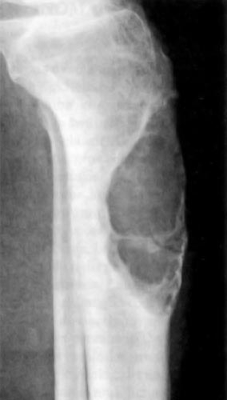
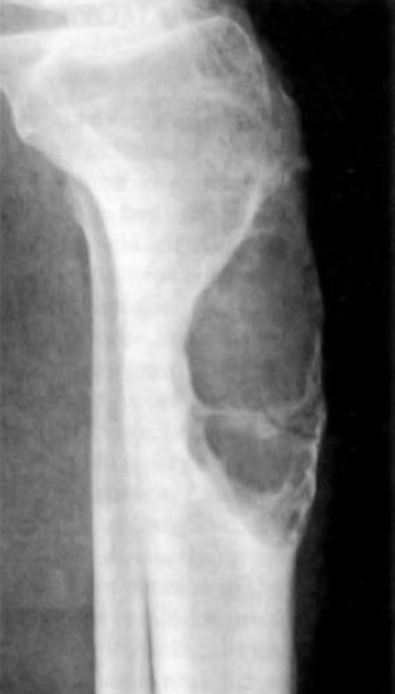
Overview of Adamantinoma
Malignancy: Rare, slow-growing, malignant tumour
Type of bone tumour: primary
0.3 to 1% of all primary bone tumours
Grade: Low grade but can be treated surgically.
Location:
It originates from epithelial cells trapped in bone (intracortical) during development in mesenchymal (osteofibrous dysplasia-like) stroma.
Lower leg: Diaphysis of Tibia (middle of shin bone) and in the medial and distal area and Fibula (calf bone)
Mouth: Mandible (Jawbone)
Upper limbs: Humerus, Ulna and Radius (Forearms)
Carpals (Hands)
Tarsals (Feet)
Onset:
Young adults (20 to 40 years of age)
Very rare in children.
Risk Factors Of Adamantinoma
A) Genetics:
Exact cause unclear.
Possible linked to genetic mutations:
- KMT2D (MLL2).
KMT2D encodes Histone-lysine N-methyltransferase 2D. This enzyme catalyses the transfer of the methyl (CH3) group from S-adenosyl-L-methionine to the epsilon-amino group of Lys-4 of histone H3 (H3K4). The most common type is H3K4me1 methylation that takes place at active sites of chromatin where transcription and DNA repair take place.
- EPHB4-MARCH10 somatic gene fusion.
A somatic cell is a body cell, whereas a fusion gene arises from an aberrant combination of two distinct genes. EPHB4 gene encodes a receptor tyrosine kinase protein called EPHB4. The EPHB4 gene is associated with the EphrinB2 ligand. This leads to a signalling cascade that facilitates key cellular events: embryogenic angiogenesis (production of blood vessels to provide nutrients and energy), adhesion, migration, and proliferation in blood and lymphatic endothelial cells.
On the other hand, MARCH10 is part of the MARCH family of membrane-bound E3 ubiquitin ligases. Their role is to add ubiquitin to target-specific amino acid residues called lysine in their target/substrate proteins. This helps to signal their vesicular transport between membranes, especially the plasma membrane that surrounds the cell.
- Overexpression of the DLK1 gene.
DLK1 is an abbreviation of delta-like non-canonical Notch ligand 1 (DLK1).
DLK1 encodes a transmembrane protein that consists of growth factor repeats, for instance, epidermal growth factor, which helps to regulate growth. It also helps to differentiate into basic fat cells (adipocytes).
- Extra chromosomal copies of 7, 8, 12, 19, and 21.
Other risk factors of Adamantinoma
B) Gender
Most patients at risk are males
C) Ethnicity:
Unknown
Appearance Under Microscope
Cell Morphology:
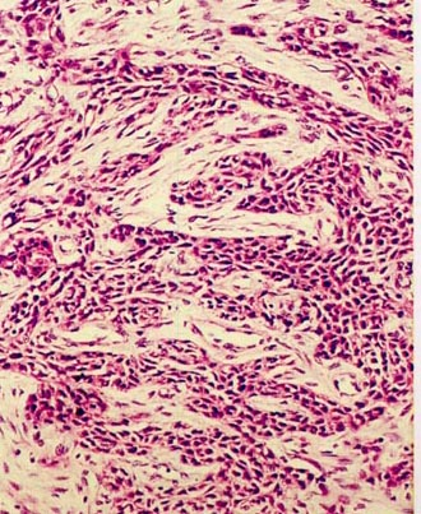
The cells in adamantinoma vary in size and mitotic activity, presence of keratin protein, and other features.
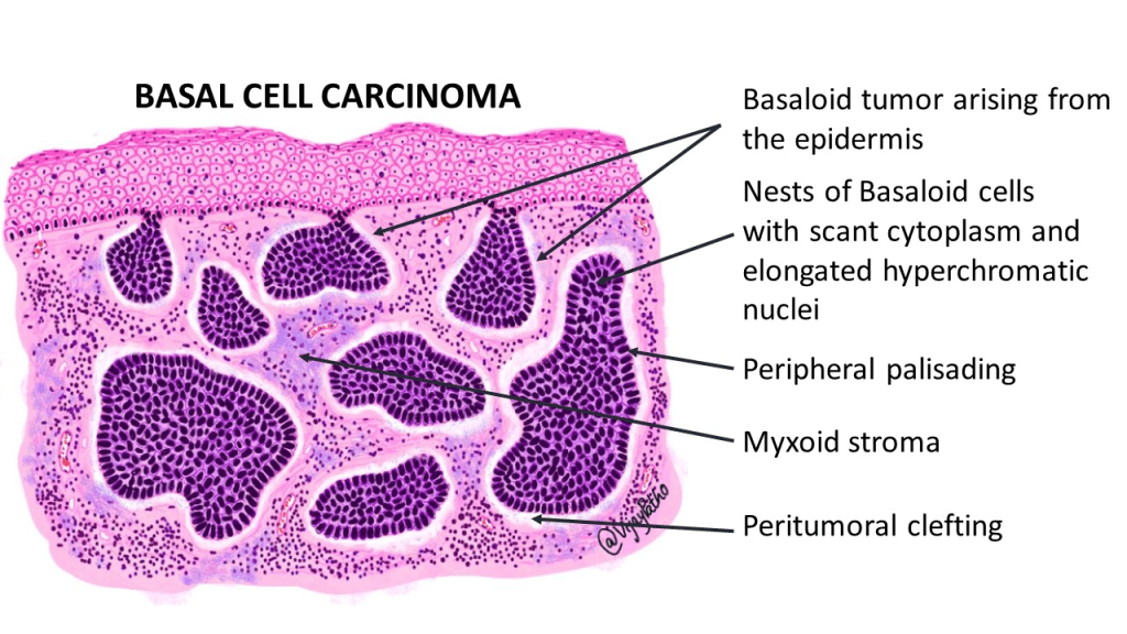
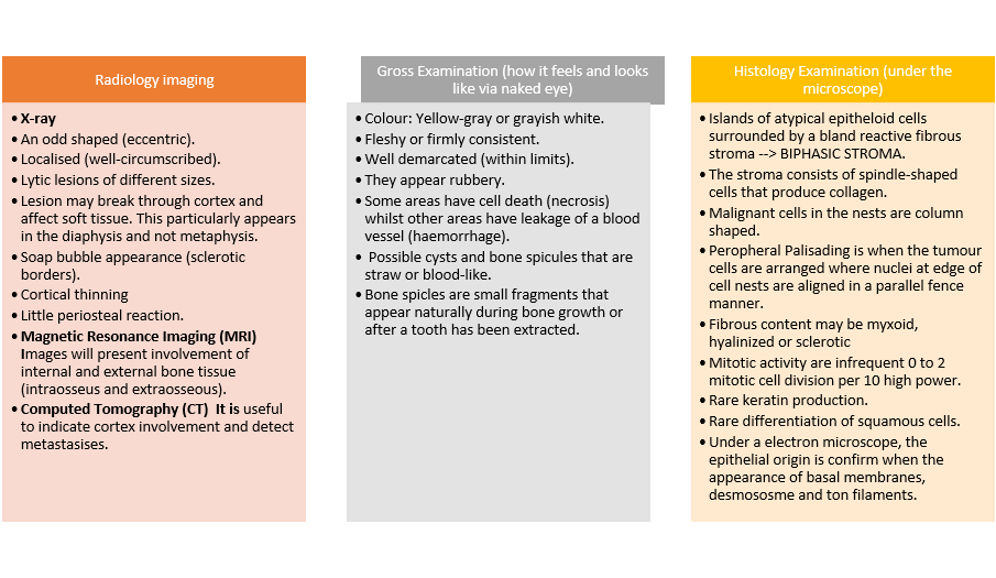
Classical adamantinoma: Mildly atypical, epithelial, solid basoloid nests, peripheral palisading but less common. There are spindled cell bundles, tubular structures, and keratinized squamous nests.
Osteofibrous dysplasia-like adamantinoma: Small scattered clusters of epithelial cells. They are less epithelial than classic adamantinoma. A positive test with keratin immunostaining. Multinucleated giant cells.
It is important to state that osteofibrous dysplasia shares several overlapping features with adamantinoma. It is a fibro-osseous proliferative lesion that affects the tibia and fibula of the long bones. It is a self-limited process and occurs during the first two decades of life. On the other hand, adamantinoma occurs later in life above the age of 20.
Dedifferentiated adamantinoma: They have negative keratin immunostaining. They are highly active in dividing cells by mitosis (mitotically active), with different forms and shapes (highly pleomorphic), and deposits of osteoid and chondroid tissue. In differentiated adamantinoma of the tibia, which usually occurs at an earlier age and has a low metastatic potential.
Recurrence
After incomplete excision of the tumour via surgery: Recurrence can occur beyond >20 years i.e late and occurs in about 30% of cases.
Lung metastasis: 10 to 20% of cases.
Supporting Videos And Images
“Adamantinoma – Everything You Need To Know” – Dr. Nabil Ebraheim
Adamantioma by Dr Vikram Deshpande
References
Bădilă, A.E., Rădulescu, D.M., Niculescu, A.-G., Grumezescu, A.M., Rădulescu, M. and Rădulescu, A.R. (2021). Recent Advances in the Treatment of Bone Metastases and Primary Bone Tumors: An Up-to-Date Review. Cancers, [online] 13(16), p.4229. Doi: https://doi.org/10.3390/cancers13164229.
Biermann, J.S., Hirbe, A., Ahlawat, S., Bernthal, N.M., Binitie, O., Boles, S., Brigman, B., Callan, A.K., Cipriano, C., Cranmer, L.D., Davis, J., Donnelly, E., Ferguson, M., Graham, A., Groundland, J., Hess, M., Hiniker, S.M., Hoover-Regan, M.L., Hornick, J.L. and Jonard, B. (2025). Bone Cancer, Version 2.2025, NCCN Clinical Practice Guidelines in Oncology. Journal of the National Comprehensive Cancer Network, [online] 23(4). does: https://doi.org/10.6004/jnccn.2025.0017.
Bone Tumour.org (2026) Adamantinoma Available at: https://www.bonetumor.org/tumors-fibrous-tissue/adamantinoma (Accessed: 4th May 2026)
Cancer Research UK (2025a) Types of bone cancer. Available at: https://www.cancerresearchuk.org/about-cancer/bone-cancer/types (Accessed 3rd April 2026)
Cowan, P., Launico, M. and Kahai, P. (2024) Anatomy, Bones. Available at: https://www.ncbi.nlm.nih.gov/books/NBK537199/ (Accessed: 9th April 2026)
Gerrand, C., Amary, F., Anwar, H.A., Brennan, B., Dileo, P., Kalkat, M.S., McCabe, M.G., McCullough, A.L., Parry, M.C., Patel, A., Seddon, B.M., Sherriff, J.M., Tirabosco, R. and Strauss, S.J. (2024). UK guidelines for the management of bone sarcomas. British Journal of Cancer. doi:https://doi.org/10.1038/s41416-024-02868-4.
GeneCards (n.d.) MARCHF10 gene. Available at: https://www.genecards.org/card/MARCHF10 (Accessed: 24th July 2026)
Hosseini, H., Heydari, S., Kiavash Hushmandi, Salman Daneshi and Rasoul Raesi (2025). Bone tumors: a systematic review of prevalence, risk determinants, and survival patterns. BMC Cancer, 25(1). doi:https://doi.org/10.1186/s12885-025-13720-0.
Martin-Almedina, S., Ogmen, K., Sackey, E., Grigoriadis, D., Karapouliou, C., Nadarajah, N., Ebbing, C., Lord, J., Mellis, R., Kortuem, F., Dinulos, M.B., Polun, C., Bale, S., Atton, G., Robinson, A., Reigstad, H., Houge, G., von der Wense, A., Becker, W.-H., Jeffery, S., Mortimer, P.S., Gordon, K., Josephs, K.S., Robart, S., Kilby, M.D., Vallee, S., Gorski, J.L., Hempel, M., Berland, S., Mansour, S. and Ostergaard, P. (2021). Janus-faced EPHB4-associated disorders: novel pathogenic variants and unreported intrafamilial overlapping phenotypes. Genetics in Medicine, 23(7), pp.1315–1324. doi:10.1038/s41436-021-01136-7.
National Cancer Institute (2019) Adamantinoma Available at: https://www.cancer.gov/pediatric-adult-rare-tumor/rare-tumors/rare-bone-tumors/adamantinoma (Accessed: 4th April 2026)
National Library of Medicine (2026) DLK1 delta-like non-canonical Notch ligand 1 [ Homo sapiens (human) Available at: https://www.ncbi.nlm.nih.gov/gene/?term=ENSG00000185559 (Accessed: 24th July 2026)
Pullan, J.E., and Lotfollahzadeh, S. (2024) Primary Bone Cancer Available at: https://www.ncbi.nlm.nih.gov/books/NBK560830/ (Accessed: 9th April 2026)
Tumor Surgery (2026) Adamantinoma: A Rare Bone Tumor of the Tibia Explained. Available at: https://www.tumorsurgery.org/2025/12/16/adamantinoma-rare-bone-tumor-tibia/ (Accessed: 4th April 2026)
UniProt (2026) O14686 · KMT2D_HUMAN. Available at: https://www.uniprot.org/uniprotkb/O14686/entry (Accessed: 24th July 2026)
WHO Editorial Board. WHO classification of bone tumours. In WHO Classification of Tumours Editorial Board. Soft tissue and bone tumours. Bovee J, Flanagan AM, Lazar AJ, Nielsen GP and Yoshida A (eds) pp 338. International Agency for Research on Cancer (2020)
Updated July 2026 Next Review July 2028


Leave a comment