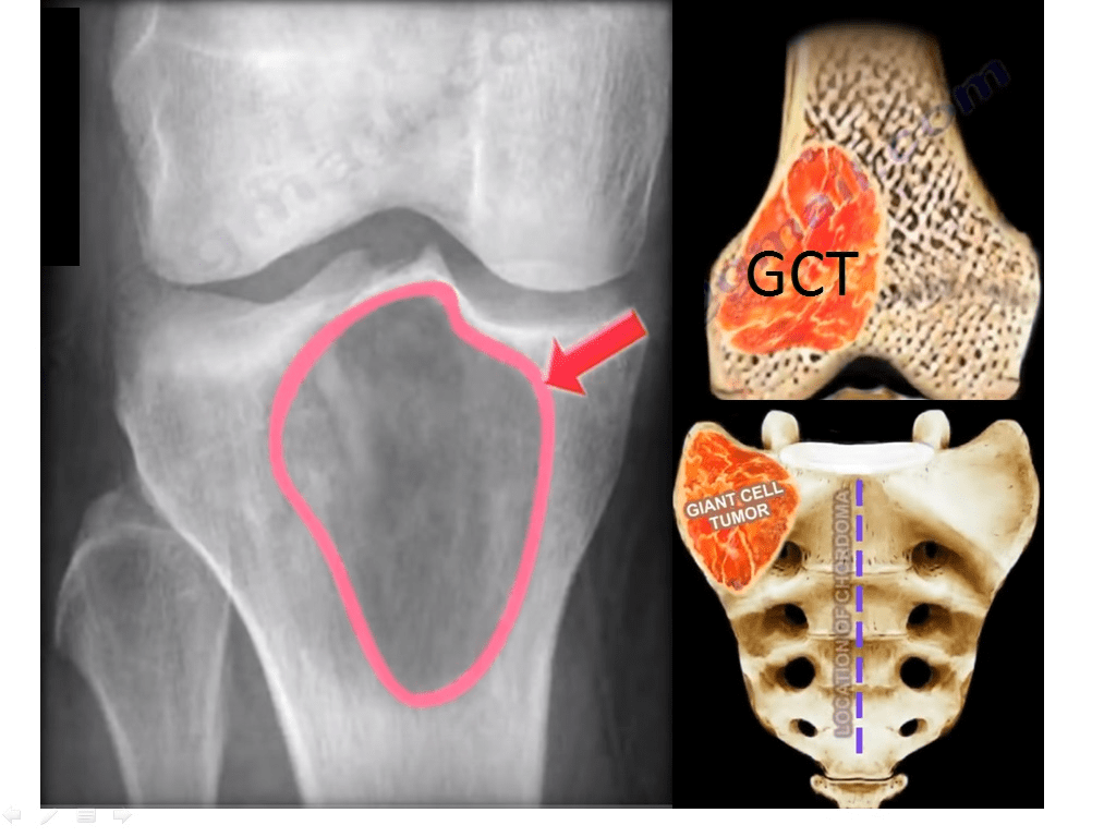
Overview of Giant Cell Tumour Of The Bone
Malignancy: Benign; aggressive; rarely metastasizes.
20% of benign skeletal tumours.
Type of bone tumour: Primary
5% of all primary tumours
Grade: Varies
Location: Multiple
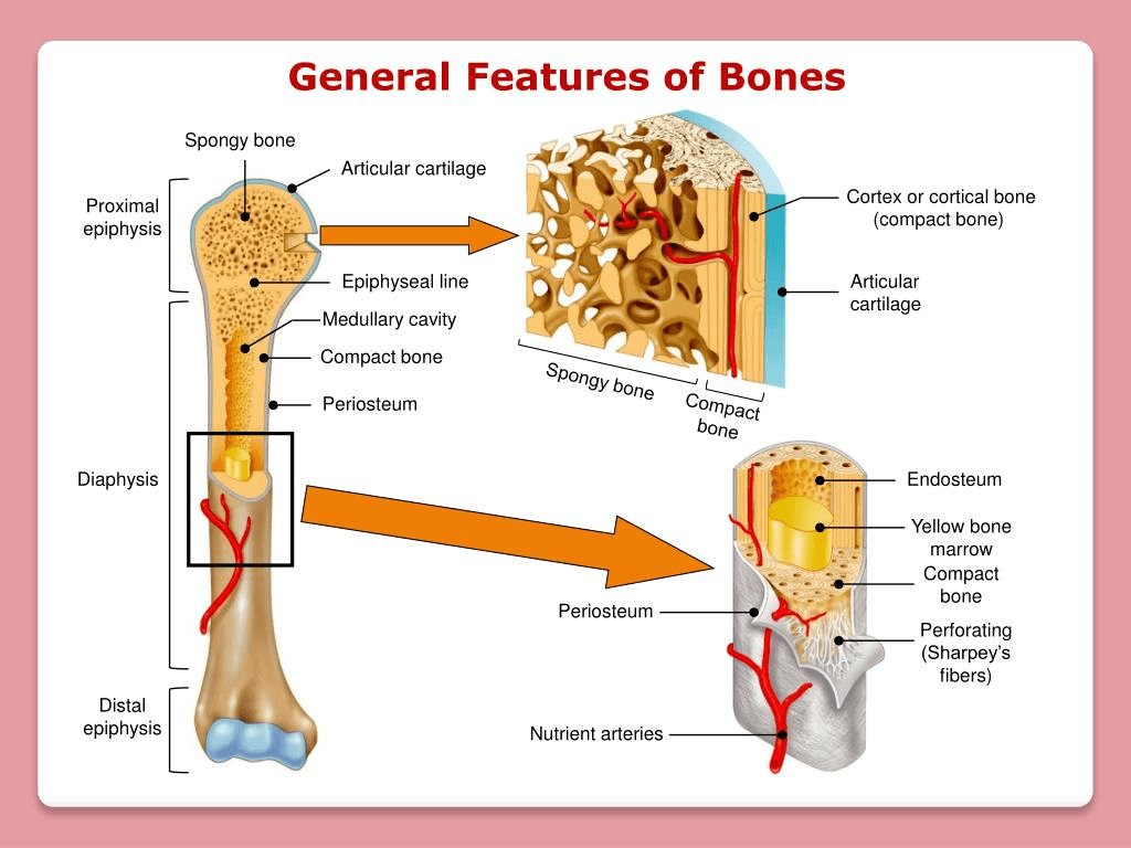
Most lesions (75% to 90%) form on the extension of metaphysis into epiphysis of long bones.
For instance, the frequent areas: distal femur (thigh bone), proximal tibia (lower limb/shin bone) and distal radius (arm bone).
Knee bones (50% to 65%) and is characterised by pain, swelling, joint loss, loss of ability to bear weight, and limited movement depending on tumour size.
Other sources suggest the most common area is around the knee.
Axial spine – sacrum but rarely in vertebral body or posterior areas.
Tumours of the lower back spine cause frequent radiation of legs, bladder, rectum and sexual dysfunction.
Uncommon in metatarsal, hands and feet.
Low prevalence in soft tissue but are at risk of spreading and being fatal.
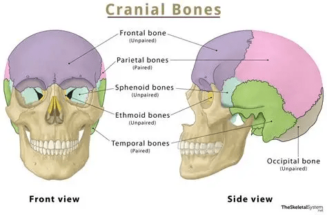
Head and neck GCT (2%) – The sphenoid, ethmoid, or temporal bones in the head.
The hyoid bone and the cartilaginous framework of the larynx (voice box) are only rarely affected.
A small set are malignant.
Ten per cent of cases are diagnosed within the second decade of life when multicentric tumours and spine tumours are more common.
Onset:
20 to 50 years of age.
Less than 3% of cases occur in children under 14.
13% over the age of 50.
Rarely in children and elderly.
Appearance:
The four E features: Epiphyseal, Eccentric, Expansile, and Eggshell crackling
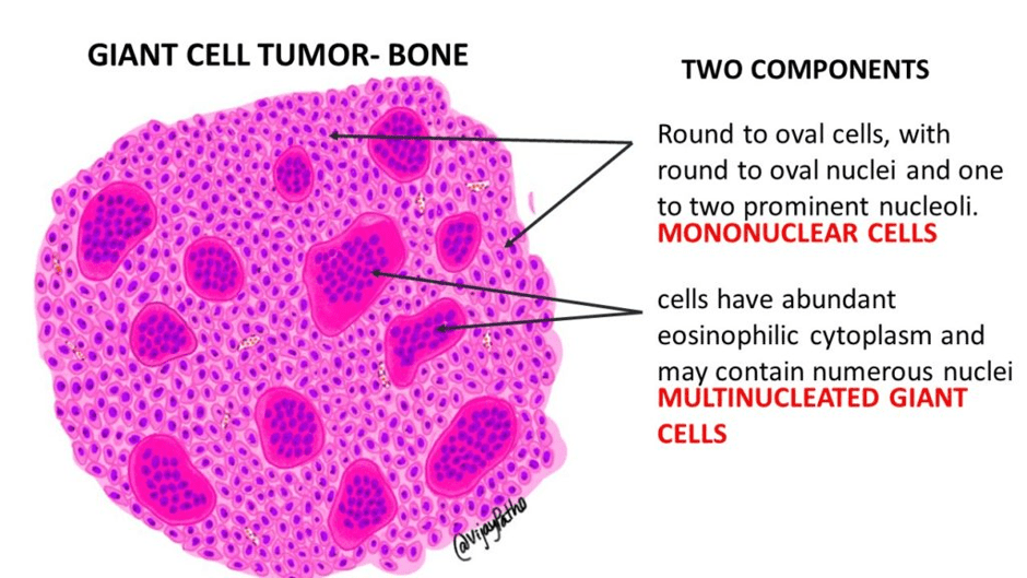
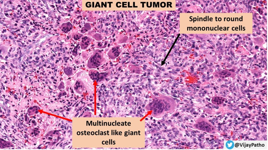
Researchers have indicated that the neoplastic cells are the osteoblast precursors that express receptor activator of nuclear factor kappa-B Ligand (RANKL). They recruit monocyte precursors and promote the formation of osteoclast-like giant cells.
Therefore, there is:
- Neoplastic mononuclear stromal cells with spindled fibroblast-like shapes.
- Macrophages
- Round or multinucleated giant cells with osteoclast-like giant cells.
- Bone also has irregular bone loss, cortex thinning, and perforation.
- Osteolytic lesions.
Risk Factors Of Giant Cell Tumour
A) Genetics:
Modifications of histones.
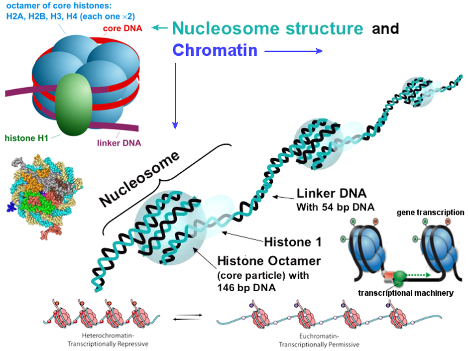
Histones are basic nuclear proteins that produce intricate packings around 146 bp DNA tightly called nucleosomes.
The nucleosomes are basic units of chromatin that make up thread-like structures called chromosomes.
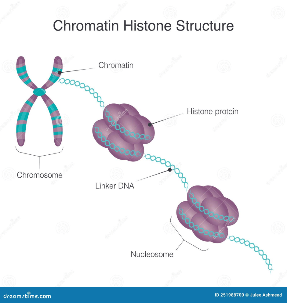
There are four core histones: H2A, H2B, H3 and H4.
Two of each of the core proteins form an octamer (8).
This means x2 (H2A, H2B, H3, H4).
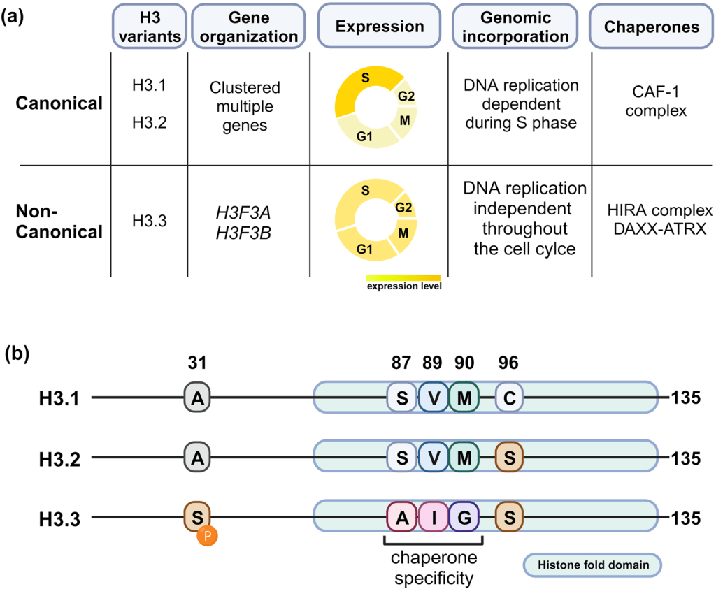
Moreover, H3 is the only histone protein that is subdivided into three variants: H3.1, H3.2 and H3.3.
H3.1 and H3.2 are primarily incorporated into nucleosomes during DNA synthesis (S phase of the cell cycle). It helps to maintain the nucleosome levels to ensure genomic integrity during cell division.
H3.3. Supports DNA replication independently and is present throughout the cell cycle, particularly during transcription and DNA damage repair.
H3.3 enters the DNA, where it replaces H3 in active genes and the ends of chromosomes called telomeres with a more accessible chromatin structure to help with gene transcription.
Histones regulate the chromatin structure through posttranslational modifications (PTMs), such as acetylation, methylation, phosphorylation, and ubiquitination, where there is an addition of an acetyl, methyl, phosphate, and ubiquitin, respectively. These chemical modifications determine how tightly the DNA is packed and whether the genes are on or off, which helps control gene expression, cell function, and development.
There is a linker histone called H1 that interacts with the linker DNA between the nucleosomes that compacts the protein.
Therefore, the histones are important for regulating transcription, repairing DNA, DNA replication, and providing stability to chromosomes.
What is the relevance of histones with Giant Cell Tumour?
One of the genes that encodes histone H3.3 is called H3F3A.
The gene mutation of H3F3A is found in conventional and malignant GCT and can facilitate differentiation from giant-cell enriched osteosarcoma.
p.Gly34Trp or p.Gly34Leu alterations.
Gly – Glycine amino acid
Trp – Tryptophan amino acid
Leu- Leucine amino acid
This H3F3A mutation is also found in chondroblastoma but has an alternative amino acid site.
p.Lys36Met.
Lys – Lysine amino acid
Met – Methionine amino acid
The location of the mutation determines tumour type.
The normal role of H3F3A differs from other histone genes.
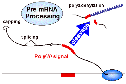
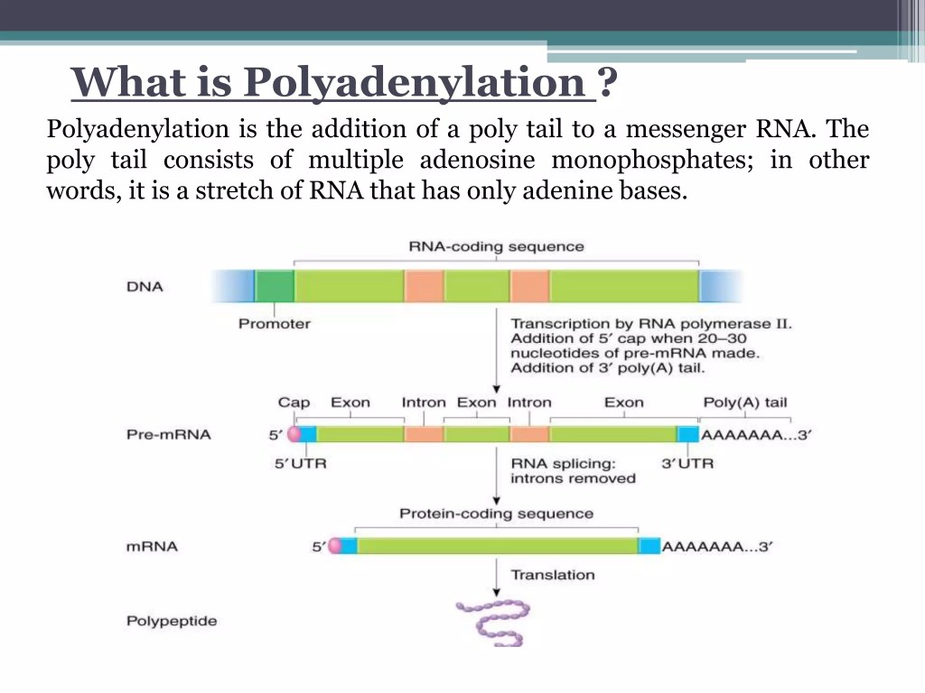
They have introns and produce a genetic material called messenger RNA (mRNA) that has undergone polyadenylation and has specific regulatory roles.
What is polyadenylation and how does it help with the role of H3.3?
Polyadenylation is a modification that occurs after transcription. This is where a poly A tail that consists of 50 to 250 adenine nucleotides is added to an RNA transcript. This helps to protect the mRNA from degradation and helps it to export from the nucleus and go to the ribosomes, where proteins are made through a process called translation, and enhances the efficiency of the process.
Other risk factors of Giant Cell Tumour
B) Gender
Equal distribution of sex.
More frequent in females.
C) Ethnicity:
Southern India and China, where GCT accounts for 20% of all primary bone tumours.
D) Other health conditions:
Paget’s disease – affects remodelling and the process of bone in the skull, facial bones, pelvis and spine.
Pathological fracture: 5 to 12%
Recurrence of GCT
Risk of local recurrence is up to 50%.
The main locations affected are the lungs that appear in 5% of the cases.
Appearance Under Microscope
Cell Morphology:
References
Alexiev, B. (2021) Bone & joints Osteoclastic giant cell rich tumors Giant cell tumor of bone, NOS, Available at: https://www.pathologyoutlines.com/topic/bonegiantcelltumor.html (Accessed: 4th April 2026)
Bădilă, A.E., Rădulescu, D.M., Niculescu, A.-G., Grumezescu, A.M., Rădulescu, M. and Rădulescu, A.R. (2021). Recent Advances in the Treatment of Bone Metastases and Primary Bone Tumors: An Up-to-Date Review. Cancers, [online] 13(16), p.4229. Doi: https://doi.org/10.3390/cancers13164229.
Biermann, J.S., Hirbe, A., Ahlawat, S., Bernthal, N.M., Binitie, O., Boles, S., Brigman, B., Callan, A.K., Cipriano, C., Cranmer, L.D., Davis, J., Donnelly, E., Ferguson, M., Graham, A., Groundland, J., Hess, M., Hiniker, S.M., Hoover-Regan, M.L., Hornick, J.L. and Jonard, B. (2025). Bone Cancer, Version 2.2025, NCCN Clinical Practice Guidelines in Oncology. Journal of the National Comprehensive Cancer Network, [online] 23(4). does: https://doi.org/10.6004/jnccn.2025.0017.
Biology Insights (2025) What Is the H3F3A Gene and Its Role in Human Disease? Available at: https://biologyinsights.com/what-is-the-h3f3a-gene-and-its-role-in-human-disease/ (Accessed: 4th May 2026)
Bone Cancer Research Trust (2026) Giant Cell Tumour of the Bone. Available at: https://www.bcrt.org.uk/information/information-by-type/giant-cell-tumor/ (Accessed: 4th April 2026)
Cancer Research UK (2025a) Types of bone cancer. Available at: https://www.cancerresearchuk.org/about-cancer/bone-cancer/types (Accessed 3rd April 2026)
Choi, J., Kim, T. and Cho, E.-J. (2024). HIRA vs. DAXX: the two axes shaping the histone H3.3 landscape. Experimental and Molecular Medicine/Experimental and molecular medicine, 56(2), pp.251–263. doi:10.1038/s12276-023-01145-3.
Cowan, P., Launico, M. and Kahai, P. (2024) Anatomy, Bones. Available at: https://www.ncbi.nlm.nih.gov/books/NBK537199/ (Accessed: 9th April 2026)
Genecards (2026) H3-3A Gene – H3.3 Histone A. Available at: https://www.genecards.org/cgi-bin/carddisp.pl?gene=H3-3A#summaries (Accessed: 9th April 2026)
Gerrand, C., Amary, F., Anwar, H.A., Brennan, B., Dileo, P., Kalkat, M.S., McCabe, M.G., McCullough, A.L., Parry, M.C., Patel, A., Seddon, B.M., Sherriff, J.M., Tirabosco, R. and Strauss, S.J. (2024). UK guidelines for the management of bone sarcomas. British Journal of Cancer. doi:https://doi.org/10.1038/s41416-024-02868-4.
Hosseini, H., Heydari, S., Kiavash Hushmandi, Salman Daneshi and Rasoul Raesi (2025). Bone tumors: a systematic review of prevalence, risk determinants, and survival patterns. BMC Cancer, 25(1). doi:https://doi.org/10.1186/s12885-025-13720-0.
Jha, Y. and Chaudhary, K. (2023). Giant Cell Tumour of Bone: A Comprehensive Review of Pathogenesis, Diagnosis, and Treatment. Cureus. doi:https://doi.org/10.7759/cureus.46945.
Pullan, J.E., and Lotfollahzadeh, S. (2024) Primary Bone Cancer. Available at: https://www.ncbi.nlm.nih.gov/books/NBK560830/ (Accessed: 9th April 2026)
Shankar, V. (2017) Giant Cell Tumor – Bone. Available at: https://ilovepathology.com/giant-cell-tumor-bone/ (Accessed: 24th July 2026)
WHO Editorial Board. WHO classification of bone tumours. In WHO Classification of Tumours Editorial Board. Soft tissue and bone tumours. Bovee J, Flanagan AM, Lazar AJ, Nielsen GP and Yoshida A (eds) pp 338. International Agency for Research on Cancer (2020)
Updated July 2026 Next Review July 2028


Leave a comment