Extensive efforts have been made recently to boost our understanding of the pathogenesis of bone cancer and develop therapeutic targets. This is an improvement since several decades ago; treatment protocols were medically challenged irrespective of their stage: bone sarcomas or metastasis of advanced cancers. There is a dire need for prompt treatment and critical attention. Conventional therapeutic interventions for primary and secondary bone cancer are namely: chemotherapy, radiotherapy, targeted therapy, and surgery. However, there are several complications: multidrug resistance, tumour recurrence, and side effects, which limit their application and efficacy. For instance, patients with osteosarcoma primarily have neoadjuvant chemotherapy (chemotherapy before surgery) followed by surgery and then another course of adjuvant chemotherapy (chemotherapy after surgery). There are different combinations of doxorubicin, cisplatin, ifosfamide and methotrexate to maximise efficacy and patient response to treatment (Hanaei et al. 2024). On the other hand, side effects may appear where toxicity damages the heart muscle (cardiomyopathy), the kidneys (nephrotoxicity), nerves (neurotoxicity), causes feeling sick (nausea), vomiting, and hypersensitivity reactions. Recent reports have also reported drug resistance. The side effects and chemotherapeutic resistance result in poor prognosis (Hanaei et al., 2024).
Another limitation is the development of large bone defects caused by trauma, tumour malignancy, tumour resection, and infection. Potential strategies have been examined, for instance, bone substitutes, regeneration of bone tissue, and carrier-mediated drug delivery instead of administering through systemic methods (Badila et al., 2021). The introduction of artificial intelligence (AI) has helped to speed up the identification of potential therapeutic compounds and the design of machine learning models to predict patient response to treatments. This has helped to make informed treatment decisions (Massive Bio, 2025). On the other hand, many potential therapies remain in the early stages of clinical trials to determine their safety, efficacy, and toxicity level before progressing onto human subjects. The development of 3D cellular models, genetic discoveries, targeted therapies to minimise side effects, biomarkers to improve prediction, and imaging methods facilitates revolutionising our understanding of the treatment of bone cancer and tailoring therapies to individual patient profiles.
New research into surgical treatment of bone cancer
For most bone cancers, surgery is the first line of treatment, and Cancer Research UK (2025) reported that a research team has developed a fluorescent dye to help surgeons remove all cancer cells and improve bone development. This helps to prevent tumour recurrences and improve functional stability.
The implantation of bone grafts is needed for bone reconstruction and repair for large bone defects, though bone is a type of tissue that is able to self-regenerate. The current methods employed are autograft, allograft, xenograft, and synthetic grafts (alloplasts). The difference in all four types is summarised in Table 1. Autograft is when the bone tissue is from the patient. Allograft is when the bone tissue is from a human donor. Xenograft is when the bone tissue is from another species. Alloplasts are from a synthetic material developed bone grafts that are fabricated in a laboratory and derived from different combinations of hydroxyapatite, β-TCP, polymers and/or bioactive glasses.
There are different chemical, physical, and mechanical properties that distinguish between all four types of grafting procedures and are summarised in Table 2. Osteoconductivity is the ability for a material to be a scaffold and support the growth of new bone tissue. It allows existing cells that form bones called osteoblasts to migrate, attach, and grow across a gap or defect. The scaffold can then help to guide how new bone from the edges of healthy bone enters into a defect and helps to regenerate bone. This highlights how the porous structure of a scaffold serves as a template for the attachment, differentiation, and proliferation of cells as well as tissue regeneration.
In contrast, osteoinduction is how a material can actively allow undifferentiated cells (stem cells), e.g., mesenchymal stem cells (MSCs), to grow and differentiate into specialised cells, e.g., osteoblasts, to form new bone tissue. This is commonly performed in bone surgery and trauma care. The gold-standard method involves using autografts, in which bone is harvested from the patient, to help regrow vertical or horizontal bone. It is mediated by growth factors in the bone microenvironment, e.g., Bone Morphogenetic Proteins (BMP), that bind to receptors on MSC and initiate the transition via signalling pathways from preosteoblasts and then to mature osteoblasts.
Several factors determine the successive rate of the impact of tissue reaction response to a bone substitute: granular size, granular shape, and pore size, which influence the fusion of regeneration and the expression of small proteins called cytokines produced by monocytes. Cytokines mediate immune response and facilitate how cells interact. Monocytes are a type of white blood cell that ingest pathogens (disease-causing microbes) (Badila et al., 2021). Osteoinductive materials can also help stimulate blood vessel formation and support extracellular matrix development. Osteogenesis is the process of forming new bone by living cells found in the graft material. Thus, autograft is suitable for osteoconduction, osteoinduction, and osteogenesis.
Similarly, allografts can recruit growth factors that partake in cellular functions, e.g., BMP2A and platelet-derived growth factor (PDGF), and are potentially osteoinductive. Other growth factors that act as bioscaffolds are fibroblast growth factors (FGF), vascular endothelial growth factor (VEGF), insulin-like growth factor (IGF), transforming growth factor-beta (TGF-β), interleukin-1 and 6 (IL-1, IL-6), and macrophage colony-stimulating factor. Nevertheless, autografts require extra surgical time, cost, risk of bacterial contamination, and have limited supply. This highlights the importance of growth factors that help promote osteogenesis and angiogenesis in scaffolds and acceptor repair.
On the other hand, xenografts are non-resorbable to prevent future resorption and have limited ability to induce bone formation. It is important to state that no single material can fulfil all requirements and requires two or more types of bone grafts to succeed. The ideal graft should be able to act as a scaffold, recruit stem cells for differentiation, and provide a scaffold for three-dimensional tissue growth.
Table 1: The classification of bone grafting materials (Miron, 2023)
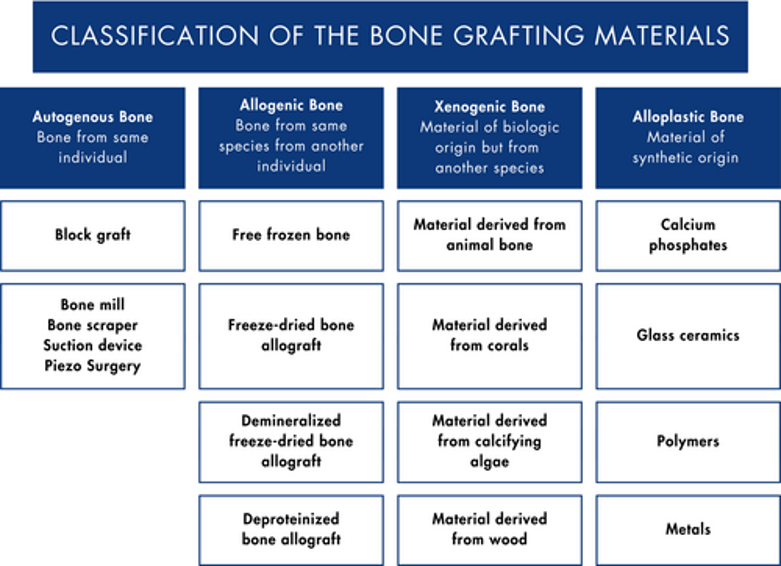
Table 2: The characteristics of the bone grafting materials (Miron, 2023)
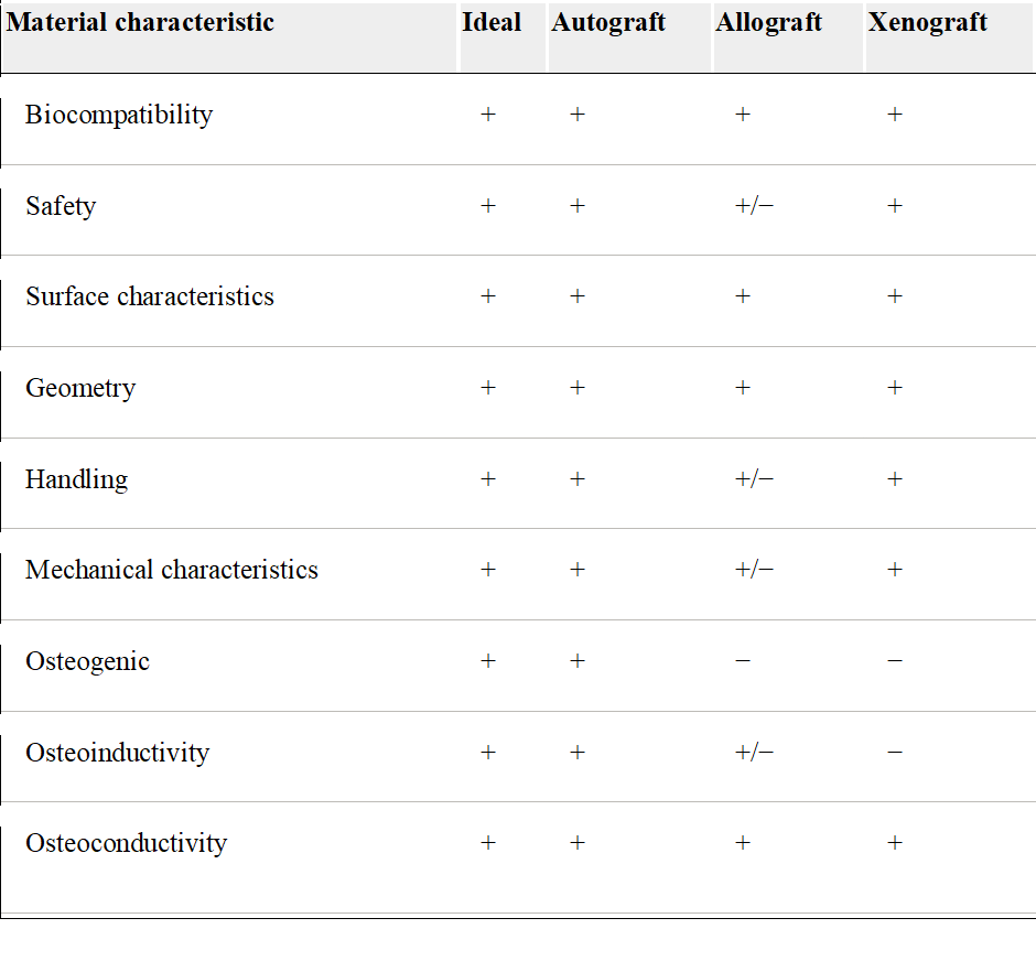
In the last decade, synthetic bone substitutes (alloplasts) have shown promise as an alternative to autografts, allografts, and xenografts, where patients are more willing to accept them during surgery. As mentioned in Table 2, synthetic bone grafts are biocompatible, biodegradable, and osteoconductive, but they do not have mechanical characteristics, osteogenic properties, and osteoinductivity (Miron, 2023). In contrast, Badila et al. (2021) contradict this view and suggest that artificial grafts can also support osteoinduction and osteointegration, in addition to osteoconduction. Osteointegration is the process of bone ingrowth into a metal implant. The differing findings in the literature may be attributed to variations in the types of synthetic graft materials used, differences in study design, or the presence of specific surface modifications and bioactive additives in some synthetic grafts, which can enhance osteoinductive or osteointegrative potential. Some studies may focus on more traditional inert synthetic materials, while others assess newer composite or hybrid materials designed to mimic natural bone properties more closely. Nevertheless, both researchers are in agreement regarding their biocompatibility, lack of toxicity, good tolerance, hydrophilic nature, affordability, ease of manipulation, sterility, and bioresorbability. Bioresorbable materials have a temporary function and need to be broken down by natural processes such as hydrolysis, corrosion, and enzymatic activity. They are then absorbed by the body over time without the need for removal surgery.
Furthermore, alloplasts can also assist with healing and structural support but depend on the type of material and body environment. Examples of types of material are polymers, metals, or ceramics. They differ from permanent implants in that they can be fully absorbed, safely metabolised, often as water or carbon dioxide. Researchers have also tried to combine alloplasts with antibiotics to prevent infections at the site of implantation. However, only a few are available because of fabrication. The main issue is exothermic polymerization, which is defined as the joining of molecules and giving off heat during material processes. This causes issues with antibiotics that are sensitive to heat.
On the other hand, bone substitutes made from ceramics ensure that there is good delivery of intraosseous tissue. Bioceramics is a type of tissue-engineered synthetic graft that can fabricate the bone scaffold and shares the same properties as the native extracellular matrix. This is achieved by their ability to adhere to cells, migrate and proliferate, and can also help to control porosity and physicochemical properties. Bioceramic scaffolds can also help to repair bone defects after surgery and destroy remaining tumour cells. Bioceramics can be classified based on their biological activity into bioinert ceramics (i.e., alumina and zirconia), bioactive ceramics (bioglass and hydroxyapatite), and biodegradable ceramics (β-tricalcium phosphate and calcium sulfate).
However, there are several drawbacks, including high temperature and antibiotic resistance. High temperature induced by scaffolds can cause breakdown of proteins, damage to the cell membrane, and delayed progression to apoptosis. This limitation can be overcome by photothermal or magnetothermal agents to further help improve bone formation in in vivo experimental studies. The aim is to maintain a high localized concentration in the target area while minimizing side effects.
There is a potential risk of antibiotic resistance in certain bacterial strains. Thus, there is antimicrobial activity without affecting osteointegration. This is prevented by combining ceramic with metallic compounds that have antibacterial properties to enhance scaffolding without needing antibiotics. Key examples are transition metals (copper, silver, iron, manganese, zinc, titanium) and alkali earth metals (strontium) added to bioceramic and hybrid scaffolds (Bădilă et al., 2021).
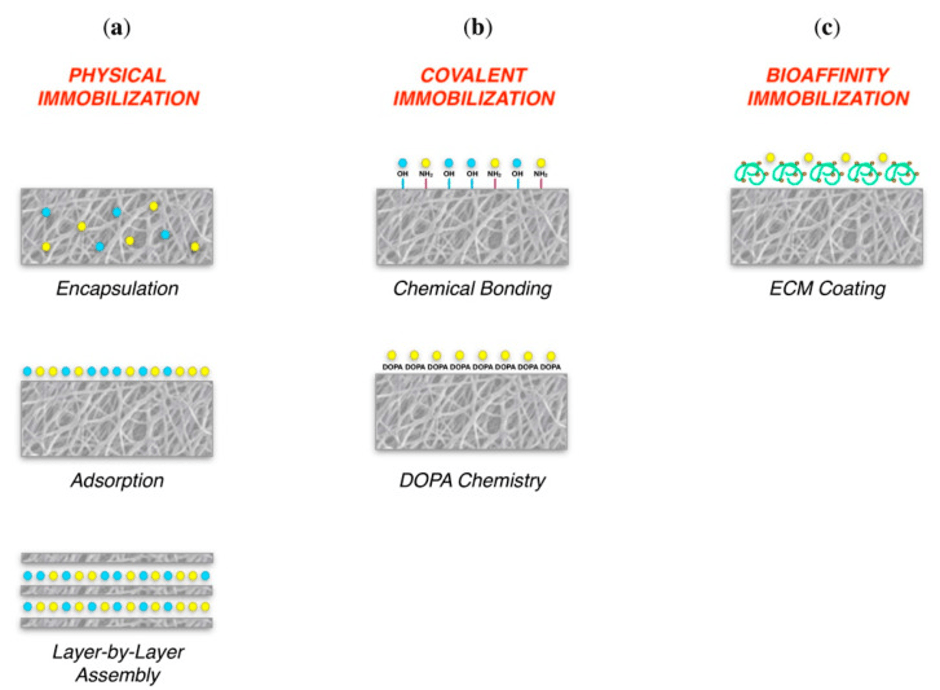
Several advancements to replace bone tissue. Key examples are alloys: titanium alloys, cobalt-chromium-molybdenum alloys, and stainless steel that can mimic their mechanical behaviour and give a large surface-to-volume ratio to help regenerate bone. They have different elastic and mechanical properties, which have high elastic properties between 100 and 115 GPa in contrast to cortical bone (3 to 30 GPa) and cancellous bone (0.02 to 2 GPa). Please see Figure 1. Immobilization is how molecules (antigen and antibody) can stick to a surface. This leads to loss of mobility and partial or complete protein activity because of random orientation and structural deformation (Creative Diagnostics, 2026). The choice of immobilization depends on the surface and protein. Physical immobilization has three steps: encapsulation, adsorption, and layer-by-layer. Covalent immobilisation associates biomolecules to a solid surface to further enhance stability and productability. The third type is bioaffinity, where specific interactions occur between biomolecules and surfaces, for instance, antibodies targeting antigens. ECM coating helps to enhance bioaffinity immobilization.
The Role Of Bioactive Glasses In Surgery
Aston University has developed a type of bioceramic called bioactive glasses that helps treat bone cancer. Bioactive glasses are a type of material that helps improve and strengthen bones and teeth. It is made by cooling high molten liquids (1450oC) to form glass. They are ground and sieved into tiny particles for treatment. This gives the properties of biocompatible, biodegradable, osteoconductive, osteoinductive, and osteogenic.
The antitumour properties of gallium were initially confirmed in 1971 by Hart and Adamon using calf thymus DNA. They discovered that gallium binds to DNA and disrupts its structure. Other molecular alterations include the condensation of chromatin and halting DNA replication. They also form complexes with surface transferrin receptors (Tfr) and block iron uptake, which causes cellular iron deprivation. This indicates the cytotoxic effects of gallium, where it can be absorbed by cancer cells and cause cell death (>99%).
Researchers have discovered that cancer cell death (apoptosis) is achieved via the mitochondrial pathway. Gallium can activate Bax, a protein that promotes apoptosis, which translocates to the mitochondria, an organelle responsible for the production of energy. The membrane that surrounds the mitochondria is pierced. This releases cytochrome C and activates the enzyme caspase-3. A cascade of enzymatic reactions with other caspases leads to apoptosis. Gallium ions decrease resorption (bone breakdown) by decreasing the activity of osteoclasts (Hanaei et al., 2024).
One of the gallium-based compounds, Ga(NO3)3 (gallium nitrate), has previously shown high efficacy to suppress tumours with low toxicity. It is commonly intravenously administered with a dosage up to 300 mg Kg−1 using protracted venous infusion. DelaFlor-Weiss and Muggia (1993) discovered that protracted venous infusion has the ability to indwell ports and central venous catheters and last longer using efficient pump technology. This causes less flushing during maintenance, supports surgical procedures, and can be used to administer analgesia for pain management and nutrition.
However, the gallium compound has several side effects; for instance, low levels of granulocytes grade 3 and 4, though there is a presence of growth factors. Other side effects include renal function alteration grade 3 or 4, hypocalcemia (low levels of calcium in the blood) grade 3 or 4, thrombocytopenia (low levels of platelets or thrombocytes), and temporary blindness have been reported in some of the subjects. Other gallium compounds have simple chemical forms, gallium chloride and gallium oxides, making it possible to be added to bioactive glasses.
Ga-doped bioactive glasses are a potential scaffold and alloplast for cancer therapy. Doping is introducing appropriate atoms/ions into the host lattice to create a hybrid material with new properties (Caie et al., n.d.). Hanaei et al. (2024) developed novel gallium-doped sodium calcium phosphor-silicate-based bioactive glasses to treat patients with high concentrations of gallium ions. Hanaei et al. (2024) conducted experimental studies on the effect of gallium on a non-cancerous mammalian cell line, normal human osteoblasts (NHOst), and a human-derived osteosarcoma cell (Saos-2), which were grown in media.
Hanaei et al. (2024) had six bioactive glasses with increasing concentrations of Ga2O3 (0, 1, 2, 3, 4 and 5 mol%). After 10 days, over 99% of Saos-2 cell death were treated with media containing 20 mg ml−1 of 5% Ga glasses. Saos-2 cells treated with 20 mg ml−1 of 4% Ga2O3 conditioned media showed less than 40% Saos-2 cell viability.
On the other hand, there was no significant difference in cell viability of the Saos-2 cell line when treated with 10 or 20 mg ml−1 of Ga-free (Bioglass 45S5/lack of gallium) conditioned media, even after 10 days, when compared to the negative control cells (NHOst). This illustrates that gallium ions are responsible for the cytotoxicity in Saos-2, where a decrease in cell viability compared to control in a dose-dependent manner. They also hold antibacterial and anti-inflammatory characteristics.
Moreover, another of Hanaei et al. (2024 )’s findings was the presence of an amorphous calcium phosphate/hydroxapaptite layer under physiological conditions on the bioactive glass particulates after incubation for seven days. This suggests that gallium can regenerate diseased bones and release calcium and phosphorus ions that form this layer. This illustrates that Ga-doped bioactive glasses have potential for bone grafting.
Conversely, researchers from Royal Orthopaedic Hospital (2024) have indicated that further studies are needed to determine the safety and effectiveness of biomaterials using bone metastatic cells from biopsies and developing a minimally invasive injectable paste for bone cancer (The Royal Orthopaedic Hospital, 2024). Moreover, there are minimal studies that tested bioactive glasses for bone cancer (Hanaei et al., 2024).
In addition, there is a difference in the efficacy of gallium ions and cytotoxic effects on different tissues. Bioavailability is low after oral administration. Bioavailability is defined as the extent and rate at which a drug or nutrient is absorbed by the systemic circulation (blood) and becomes available at the site of action. On the other hand, other studies claimed gallium has a dose-dependent repressing effect on the biosynthesis of haem, hepatic oxidative stress (liver), and the primary immune response.
The parenteral route, a route other than the digestive tract, e.g., through a needle, has bioavailability at a higher level. Renal toxicity is a limiting factor of its efficacy, e.g., gallium nitrate injection, such as a pump device for continuous infusion.
This indicates that bioactive-doped bioactive glasses are a safe method to deliver gallium ions that target cancer cells via TfR and degrade in aqueous media, releasing calcium, sodium, and phosphorus ions. Tumour cells have more of these receptors (three-fold higher) compared to non-cancerous cells, especially in highly proliferative cells. This also corresponds to higher iron intake in cancer progression. Other examples of cells include intestinal epithelium, basal epidermis, and activated immune cells. This causes TfR to be a potential target marker. High expression of TfR1 has been reported in 43.4% of patients with OS and is considered a prognostic factor for bone cancer patients.
Several genes are highly expressed in tumour cells, for example, FTH1, which is a cellular iron utilization gene, and FLCVCR1, an iron efflux gene, which has increased by more than five-fold. FLCVCR1 encodes a haem transporter protein needed for the production of erythropoiesis (production of red blood cells). It helps protect the development of erythrocytes (red blood cells), referred to as erythroid differentiation, from haem toxicity. It also helps transport haem from the cytoplasm to outside the cell; please see Figure 2. It is estimated that 360 billion erythrocytes produced daily require over 250 mg of haem to assemble into haemoglobin, the protein that binds to oxygen in the red blood cells.
FLCVCR1 is also a uniporter that mediates transport across the plasma membrane of choline and ethanolamine, which are precursors for phosphatidylcholine and phosphatidylcholine, respectively, into the cells to help make phospholipids (biosynthetic reactions – production of larger macromolecules).
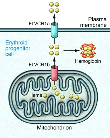
The export of Haem depends on the plasma protein haemopexin (HPX) to lower the risk of haem toxicity that may affect the liver, brain, and sensory neurons, and also to maintain an intracellular level of haem. During erythropoiesis, haem synthesis production is high. The export of intermediate products in the haem biosynthetic pathway, coproporphyrin and protoporphyrin IX, also takes place. The method is yet to be elucidated. The haem transporter encoded by FLCVCR1 transports haem from the mitochondria (the location where haem is produced, particularly in the mitochondrial membranes) to the cytoplasm. The protein is translated in the cytoplasm. The choline or ethanolamine gradient helps the haem to be transported across the plasma membrane, which is vital for iron absorption (Shoji et al., 2023).
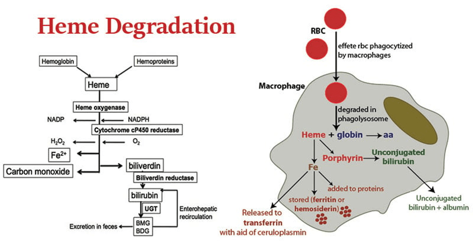
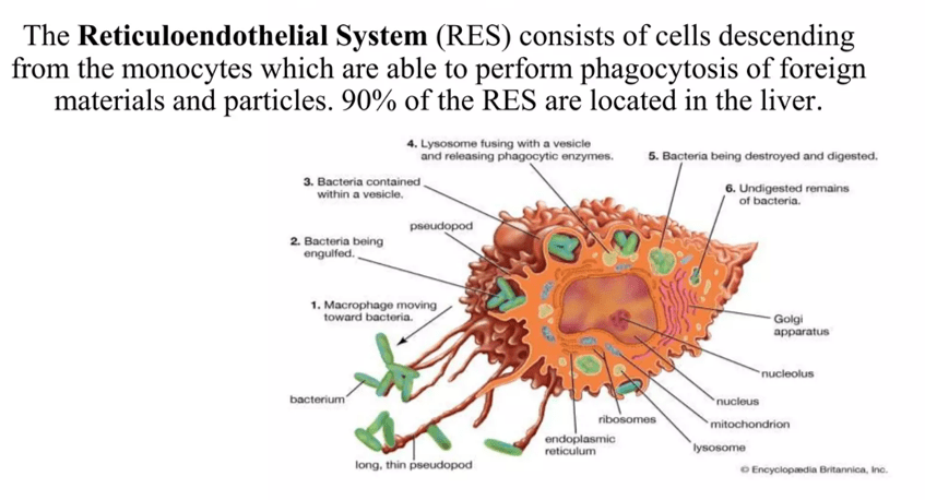
Red blood cells that no longer function are degraded by macrophages to form haem and globin. Iron recycling takes place in the reticuloendothelial system (RES) macrophage. The oxidation of haem to biliverdin, carbon monoxide and ferric iron is catalysed by haem oxygenase. The ferrous iron exporter, ferroportin (FPN1), mediates the iron recycled out of the macrophage. The process is regulated by the hormone hepcidin. The iron is then released to the plasma; a straw-coloured liquid forms part of the blood. The iron is bound to the glycoprotein transferrin/siderophilin. There are three outcomes for haem: (1) the production of red blood cells in the red bone marrow; (2) to be stored as ferritin or haemisiderin primarily in the liver; (3) Haem is needed for the production of haemoproteins that are involved in electron transfer and oxygen transport. They are reddish-brown conjugated proteins that have a haem group.
New Research In The Treatment Of Radiotherapy
Recent studies revealed that the three modalities—Photodynamic Therapy (PDT), Photothermal Therapy (PTT), and Photoimmunotherapy (PIT)—have anti-tumour effects against bone cancer. Photodynamic Therapy (PDT) generates reactive oxygen species (ROS) to elucidate its effects. In contrast, PTT, as the name suggests, uses heat in the localised area. PIT combines phototherapy and immunotherapy to boost tumour-specific immune responses and selectively kill cancer cells. However, these modalities also face challenges, for instance, difficulty in targeting tumours, inadequate ROS production, and physicochemical side effects.
PDT
The three main components involved in PDT are a specific wavelength of light, a photosensitizer, and molecular oxygen to kill cancer cells. It is a technique that involves injecting a light-sensitive agent into the blood, which remains in cancer cells for a prolonged period of time. The laser radiation produces an active form of oxygen to destroy the treated cancer cells. It can be used to treat cancers of the skin, bone in localised areas, eyes, and the lining of internal organs such as the bladder – Figure 4. This indicates that oxygen is a vital element during the anti-tumour process of PDT.
Nevertheless, some solid tumours have a hypoxic tumour microenvironment caused by an imbalance between oxygen supply and oxygen consumption within the tumour. Normally, oxygen levels in tissues (pO2) are between 4 and 7.5%; in contrast, solid tumours have lower values (0.3–4.2%). Hypoxia leads to increased cancer growth and irregular and non-functioning tumour vascular systems. The aberrant tumour vasculature is characterised by the attachment of cells called pericytes that surround the endothelial cells and elevated vascular permeability. As a result, proteins leak from the vessels, and interstitial fluid accumulates within the tumour matrix, causing abnormal tumour perfusion. Tumour perfusion is the ability of blood to flow within a tumour. This raises the tumour interstitial fluid pressure (TIFP) and hinders oxygen penetration. It further influences the delivery of oxygen and therapeutic agents (Cai et al., 2025).
The more the cancer progresses, the more restricted the rate of diffusion of oxygen gas. This is due to the increased distance to blood vessels. Though inadequate oxygenation within tumour cells can create carbon monoxide (CO) and chronic hypoxia. A hypoxic tumour microenvironment (TME) prevents the generation of ROS during PDT. This can be overcome through pharmacological and molecular interventions with the aim of producing oxygen, improving oxygen transport and distribution within the tumour, suppressing the hypoxia-inducible factor-1 (HIF-1) signalling pathway, and preventing cellular respiration (Cai et al., 2025).
Key examples of drugs that normalise vasculature are antiangiogenic drugs: multikinase inhibitor (Regorafenib), multi-targeted tyrosine kinase inhibitor (Lenvatinib), and EGFR inhibitor (Erlotinib). They help generate more ROS to remove the tumour. However, this poses a challenge because amongst the anti-tumour mechanisms of PDT is to induce vascular occlusion (closing or obstruction of the vessel) and thrombosis (when the blood changes from a liquid to a solid state, where it coagulates due to platelet formation). On the other hand, vascular occlusion can prevent the penetration of drugs and oxygen delivery within the tumour. In addition, antiangiogenic drugs like angiogenic II receptor blockers decrease the expression of VEGF, which directly lowers the tumour vascular supply, lowers the abnormal vessel density, raises the vessel wall thickness, and overcomes hypoxia. This can also lead to hypoxic TME. Further studies are needed to understand the mechanism of angiogenic drugs and their link with tumour vasculature (Cai et al., 2025).
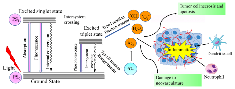
The light photons bind with chromophores, which are parts of molecules that absorb light in the visible spectrum or ultraviolet light in the electromagnetic spectrum. This causes the molecule to appear coloured. The rest of the molecule is reflected. A photon is a massless particle that performs waves as a speed of light. Absorption occurs when the energy of the photon equals the interaction between two electronic states (S0) and (S1) –please see Figure 4. The electron in the ground state (S0) interacts with the photon, up to the higher energy state (S1). The energy of the photon is transferred to the electron in S1 and returns to S0. The electrons in the excited state of S1 (high energy) undergo a nonradiative transition in the form of intersystem crossing. This forms a stable triplet state (T1) that returns to S0 through light energy release (phosphorescence – a type of radiative transition) or vibrational relaxation (a type of nonradiative transition). The T1 interacts with free radicals to generate reactive oxygen species (ROS). For instance, in the type 1 route, it depends on electron transfer that interacts with cellular substrates to generate superoxide anions (O2-), hydroxyl radicals (OH), and hydrogen peroxide (H202). Type 2 route relies on oxygen levels in the tissue, where it transfers triplet oxygen to produce singlet oxygen.
Moreover, there is a carbon-based nanoparticle, such as fullerene, that demonstrates it can form singlet oxygen via an energy transfer mechanism Type 2) and produce ROS (superoxide radicals, hydroxyl radicals, and others) via a Type 1 mechanism. This indicates its efficacy in cancers that are deprived of oxygen and presents its characteristic as photostability, and generates ROS efficiently (Cai et al. 2025).
Both routes contribute to cell death, destroy the microvascular system, and induce an inflammatory response. It also promotes the recruitment and maturation of antigen-presenting cells such as dendritic cells and macrophages to induce phagocytosis. Dendritic cells recognise the antigens found on the tumour cell surface and present them to T cells, which undergo a killing mechanism (CD8). Phagocytosis is the process in which a type of white blood cell called a macrophage, otherwise known as a phagocyte, engulfs microbes and other pathogens, like cancer cells, and breaks them down using the enzymes inside them. Cellular debris is then released.
The use of PDT in cases of osteosarcoma affecting deeper tissue, where surgical resection has a high risk of metastasis and recurrence. The 5-year overall survival rate is approximately 20 to 30% (Zhang et al., 2025). Circulating tumour cells seek advantage at infection sites where they recolonize, further increasing the risk of cancer recurrence (Zhang et al., 2025). One of the complications of utilising PDT is the restricted light penetration depth, where photons are unable to emit the energy in deeper lesions.
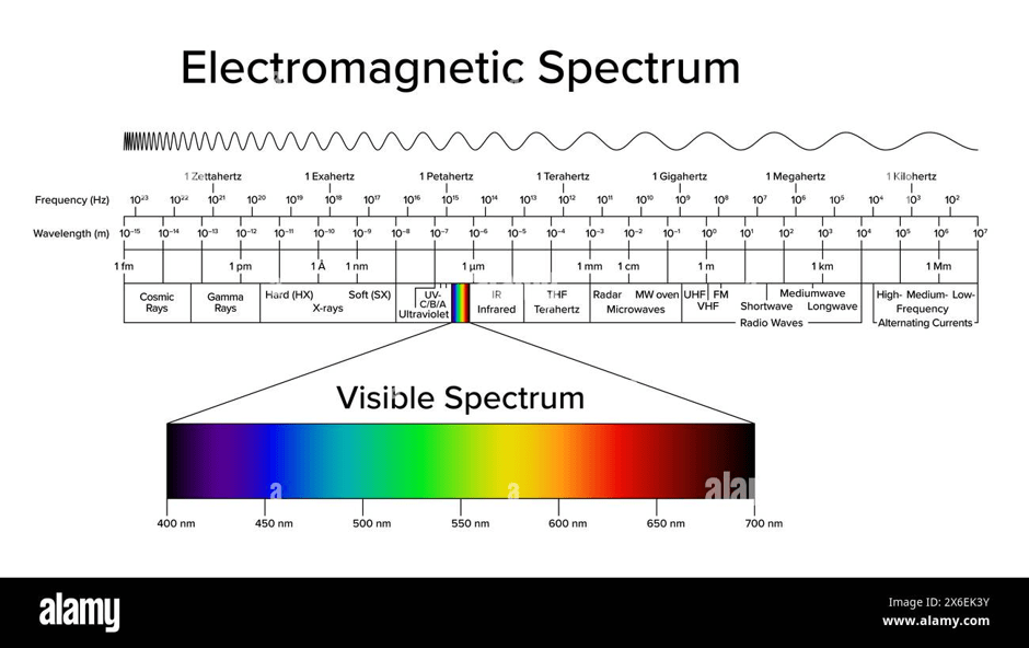
In some cases, a lack of light penetration depth limits reaching tumours outside the irradiated area. This is because different wavelengths of light have different tissue penetration depths. As the wavelength increases, the tissue penetration depth rises from UV to infrared radiation, as presented in Figure 5 in the electromagnetic spectrum. For instance, the UV light (150-380 nm), blue (390 – 470 nm), green (475-545 nm), yellow (545 to 600 nm), red (600 – 650 nm), and near-infrared (650 – 1300 nm). The tissue penetration depth ranges from less than 0.1 mm to 3mm. An efficient response from phototherapy is achieved with shorter wavelengths and high-energy photons.
This limitation can be overcome by using chemiluminescence and noble metal nanoparticles. A nanoparticle is a small particle whose size ranges between 1 and 100 nm and can be of organic and inorganic form. There are two types of chemiluminescence: direct and indirect, and in both cases utilise self-luminescent materials that produce light emission through chemical reactions like oxidation. The reactants or the intermediate product forms are unstable and are in an excited state (high energy). However, they differ in how energy is released. In direct chemiluminescence, the decomposition of the intermediate product releases energy by emitting photons. The excited state transfers to the ground state, producing chemiluminescence. Key examples of chemiluminescent molecules that emit light through direct chemiluminescence are luminol and acridine esters.
On the other hand, in indirect chemiluminescence, the energy is transferred through chemiluminescence resonance energy transfer (CRET). CRET is a type of non-radiative transfer of energy from a chemiluminescence reagent (donor) to a suitable energy acceptor with appropriate absorption within 10 nm in a chemiluminescence reaction. In this case, a fluorescence dye is a suitable energy acceptor (Yan et al., 2020). The emission of the substrate (CL reagent) will excite or be quenched by the acceptor. Thus, indirect chemiluminescence does not require excitation from an external energy source, nor does the intermediate emit photons. The fluorescent molecules become excited and cause them to emit light (Cai et al., 2025).
Another way to overcome the impediments of conventional PDT is by the manufacturing of a minimally invasive and implantable biphotonic wireless system that provides phototherapy using an optoelectronic device to generate ROS in deep localised zones without the need for external light, nor do you have to worry about the safety and durability of wired connections and battery-operated power sources. The flexible circuit board in the multi-layered patch-type biophotonic device primarily consists of a polyimide (PI) substrate, copper coils, and polydimethylsiloxane (PDMS) in the following manner: PI, Cu, PI, Cu, PI, and PDMS. The PI is the substrate that allows flexibility, thinness, and light weight. The biophotonic device is lightweight (16 mg), thin (160 μm), and has adjustable spatial dimensions to control the optical energy delivery to the target area. The two layers of copper coils offer malleability, less reactivity, and electroconductivity. The PDMS is an encapsulant material that is transparent, flexible, and chemically resistant. Through in vivo experimental studies, >90% of osteosarcomal cell growth has been eradicated. Other advantages include antibacterial activity (>99%), negligible damage to surrounding tissue, enhanced spatial utilization, transmission speed at a faster weight, resistance to interference, minimal thermal damage, and a lower risk of oedemas and burns from long-term direct irradiation of external light sources; no immune reactions were observed in major tissues after implantation (Zhang et al., 2025).
Moreover, the biophotonic device operates by emitting green fluorescence with an output power of >5 W and a distance of 2cm from the excitation light source (Zhang et al., 2025).
Furthermore, one of the free radicals that produces ROS in a type 1 route is hydrogen peroxide, where overexpression is linked to tumourigenesis, DNA damage, and tumour metastasis. Decomposition of hydrogen peroxide by the oxidoreductase enzyme catalase to form oxygen and water. Researchers hypothesize that tumours with high levels of hydrogen peroxide can be treated with catalase to generate more oxygen in situ. This increases ROS production via PDT.
Several microorganisms can increase the rate of water splitting, for instance, cyanobacteria, chlorella, and spirulina. Chlorella was studied by Wang et al. (2021b) to control the light-controlled PDT and increase oxygen production by chlorella upon irradiation. They were collected and enriched with perfluorocarbons (PFC) and delivered. Oxygen has a higher solubility in PFC than haemoglobin. Haemoglobin can only bind to four oxygen molecules. PFC, however, does not form a chemical bond with oxygen, they have weak intermolecular interactions to dissolve. Oxygen dissolved in PFC is utilised by 90% of tissues.
PTT
In PTT, after excitation by light at specific wavelengths, the photothermal agent (PTA) undergoes oscillatory relaxation of electron-excited energy. This is where the electron in the S1 state relaxes non-radiatively back to the S0 state. This is known as internal conversion. A series of collisions may occur between the chromophore and the surrounding environment. These releases decay as thermal energy (a type of non-radiative transition). The increase in temperature in the tumour region causes damage. The light and heat can be absorbed by proteins, lipids, and nucleic acids (DNA and RNA).
However, the location of the PTA varies with thermal conductivity. Outside the cell, the light energy is directly absorbed and converted into heat energy, maximising the effect, but the rapid dispersion of heat can prevent the aggregation or accumulation needed to produce effective cytotoxic action. On the other hand, in the cell membrane, there is low heat, which prevents heat from being dispersed easily and more directly and readily induces cell death.
At low to moderate temperatures (41 to 43 degrees Celsius), the heating is low enough to kill tumour cells directly. Conversely, it can aggregate and cause damage to proteins. This increases vascular permeability in the tumour area and impairs the transport of drugs and oxygen. Heat shock proteins (HSPs) may be activated to protect cells from thermal damage and increase the efficacy of chemotherapy or radiotherapy (Cai et al., 2025).
At 43 to 45 degrees Celsius, the heat causes cellular stress and further increases protein denaturation. This affects the cell membrane and releases contents from inside the cells. It also affects the regulation of gene expression. This elevates heat-mediated apoptosis and affects collagen fibers in the stroma, eliciting structural malfunctions (Cai et al., 2025).
At even higher temperatures (55 to 60 degrees Celsius), coagulative necrosis of tumours may appear and cause damage to the kidneys and the heart due to necrosis. This can lead to overstimulation of heat shock proteins (HSP 70/90) that suppress the anticancer activity of the immune system (Cai et al., 2025).
In cases of bone cancer, PTT is recommended because it is minimally invasive, focused on the damaged area, allowing space and time (specific spatiotemporal selectivity). Recently, iron nanoparticles have had a photothermal effect when releasing iron. This helps to decompose hydrogen peroxide inside the tumour, facilitating the production of reactive oxygen species that cause further damage to DNA, proteins, and lipids (Badila et al., 2021). It also elicits an immune response through damage-associated molecular proteins (DAMPs) and cytokines.
Cai et al (2025) discovered that several noble metals, gold (Au) and silver (Ag), can absorb laser light and excite electrons from the ground state to an excited state through the release of energy via non-radiative decay. Radiative decay can also be induced to generate ROS. For instance, this is due to their size, shape, and atomic number, and can be adjusted. Thus, the noble metal nonmaterial can facilitate the application of PTT and PDT, respectively.
Cai et al. (2025) further revealed that the energy from localised surface plasmon resonance (LSPR) is transferred to molecular oxygen (O2) to form singlet oxygen (1O2). The LSPR refers to the process where light interacts with metallic nanoparticles, forming the light-matter coupling. Electric fields are greatly intensified at the nanoparticle’s surface. The oscillating electric field of light causes free-moving conduction electrons on the nanoparticle surface to shift and oscillate, too.
The noble metal nanomaterial can make singlet ions by adsorbing molecular oxygen on its low-energy surface states of the nanoparticles and can efficiently transfer energy to molecular oxygen. On the other hand, higher-energy surface states of the nanoparticles have less efficiency in transferring the energy of LSPR to molecular oxygen. The LSPC can maximise light absorption and scattering at different frequencies. Thus, energy is released through electron-phonon relaxation processes, thereby exhibiting high photothermal conversion efficiencies. Metallic nanoparticles, when illuminated, indicate the impact made. Thus, nanomaterials’ high specific surface area allows drug delivery. Hence, phototherapy and chemotherapy enhance anti-cancer efficiency compared to phototherapy alone.
In addition, PTT can cause damage to non-cancerous tissues. This can be overcome using sonodynamic therapy (SDT) that operates via ultrasound (sound waves) rather than photons (light) to penetrate the tissue. Please see Figure 6. It has a higher tissue penetration capability (> 10 cm), and it can also control time, intensity, and radiation frequency, thus targeting deeper tumour tissues and lowering damage to surrounding areas. The sonosensitizer is taken up by cancer cells more than normal cells. The ultrasound energy activates the sonosensitizer, which produces singlet oxygen and free radical oxygen in cancer cells. This leads to cancer cell death.
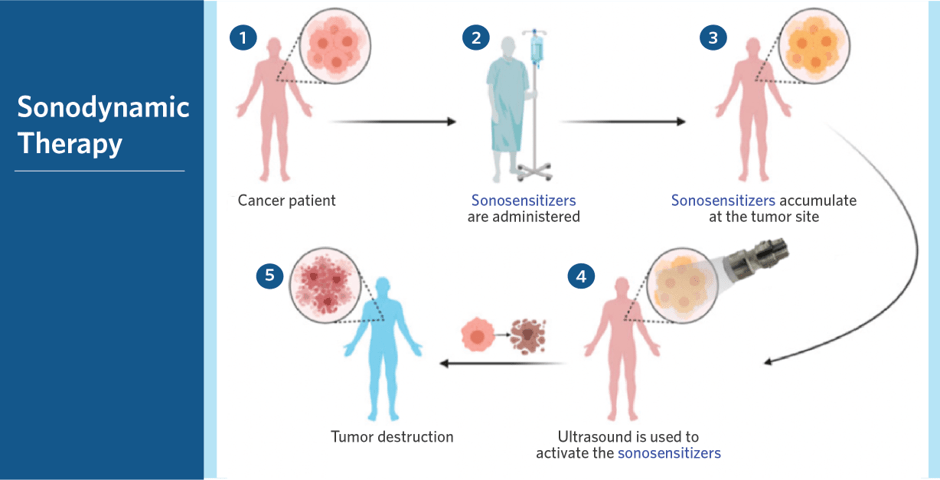
Sonodynamic Therapy Explained: How Ultrasound Targets Unhealthy Cells by IV elements.
Sonodynamic therapy – A noninvasive therapy to destroy tumors. By AI and Healthcare
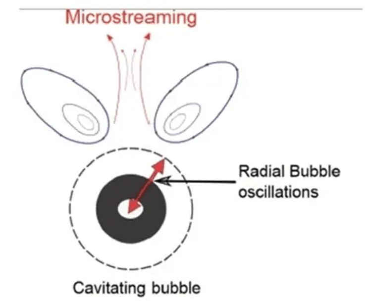
There are four key steps involved in SDT: cavitation, sonoporation, sonoluminescence, and ultrasonic microstreaming. Cavitation is the formation of microbubbles in the tissue fluid due to alterations in the mechanical pressure. Please see Figure 7 of a cavitation bubble. In non-inertial cavitation, low-intensity ultrasound creates the microbubbles in the tissue fluid that remain stable. There are small radial oscillations that affect the surrounding cells and molecules. This increases the permeability of the cells and also helps drug influx into tumour cells at a higher concentration.
On the other hand, inertial cavitation occurs when the high intensity of ultrasound causes the microbubbles to absorb more energy. There are also faster oscillations, expansion, collapse, and high temperature and pressure – all these processes cause a release of lots of energy.
Sonoluminescence is the process by which sonosensitizers become excited via energy transfers. The energy is released from the ultrasound and can activate the sonosensitizers from the ground state to the excited state (high energy). This leads to the formation of electrons and ROS production. For instance, electron transfer interacts with cellular substrates to generate superoxide anions via the type I route of PDT. The energy transfer is via the type II route.
An inorganic sonosensitizer is Ce6, or Chlorin e6, a second-generation photosensitizer derived from chlorophyll in plants. Chlorophyll makes leaves green and absorbs light and can generate high levels of ROS when activated through light and ultrasound.
The steady flow of a liquid medium or fluid causes the propagation of sound. The energy from the rupture of the microbubble can directly cleave surrounding water molecules.
Ultrasonic microstreaming, otherwise known as acoustic microstreaming, is produced by the ultrasonic cavitation microbubbles. It is the interaction between a minimum of two modes of bubble oscillations (Wu et al. 2025). This produces ROS that kill tumour cells.
PIT
The phototherapeutic agents in combination with immunotherapy induce tumour cell death and immunogenic cell death (ICD). It affects the endoplasmic reticulum, causing stress. The endoplasmic reticulum is found inside the cell and is involved in the folding and metabolism of proteins. It also has roles in lipid (fat) metabolism and cell-to-cell communication.
The cells respond by starting an unfolded protein response (UPR) to maintain homeostasis and promote tumour survival. The UPR helps to maintain the folding of proteins. Protein folding is the process by which amino acids, the building blocks of proteins, are held together by various bonds and forces to make a stable 3D protein structure. The ICD releases high levels of DAMPs and cytokines and recruits antigen-presenting cells (APCs) and T cells to mediate destruction. Heat shock proteins offer a protective measure, whereas CALR, found on the cell surface, associates with the C91 receptor. The secretion of adenosine triphosphate (ATP) as an energy source overlaps with CRT. It involves the enzyme caspase 8 and drives apoptosis through cleavage and activation of downstream caspases 3, 5, and 7 (Cai et al. 2025).
Nanocarrier delivery systems
To overcome resistance to chemotherapy, nanoparticles as a carrier delivery system have been developed to ensure the correct dose is being administered to the target tissue. This prevents rapid clearance and adverse effects, such as being non-immunogenic, inert, and biocompatible. Key chemotherapeutic targets are cisplatin, doxorubicin, and paclitaxel, where encapsulating them helps to deliver the anticancer drug safely to the bone and maximise its toxicity and intracellular retention.
A good example of a nanocarrier system is chitosan, a natural polymer derived from chitin, a type of polysaccharide (sugar molecule). The nanoparticle of chitosan can bring the hydrophobic drug closer to the target point due to the mucoadhesive property. Hydrophobic molecules are ‘water-disliking’. Mucoadhesive refers to the tendency for a substance to remain adhesive to the inner lining of the cell, called the mucosa. The role of the mucosa is to lubricate and protect, and it consists of water, mucin, proteins, lipids, sugar, and electrolytes (ions) (Badila et al., 2021).
Another characteristic of chitosan nanoparticles is their ability to be modifiable. It can be combined with sulphate groups to obtain a heparin-like polysaccharide structure that can associate with basic amino acids of BMP-2. Heparin is a protein produced in liver cells and some white blood cells and functions as an anticoagulant by inhibiting the enzyme thrombin. Coagulation is the formation of blood clots.
There are 20 amino acids, and three of them belong to the Basic amino acids group. Basic amino acids are positively charged with an amine (NH2) side chain. Key examples of basic amino acids are lysine, histidine, and arginine. Other examples of delivery systems include collagen and fibrin (Badila et al. 2021). The inactive precursor fibrinogen is converted into fibrin via the enzyme thrombin as part of the coagulation process.
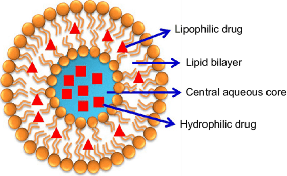
An example of a hydrophobic chemotherapeutic drug is paclitaxel, and lipid-based nanoparticles like liposomes can entrap these drugs in the lipid layer. Please see Figure 8. A liposome is an aqueous core surrounded by a phospholipid bilayer. Hydrophilic drugs are ‘water-loving,’ and they are encapsulated in an aqueous region and are protected from degradation by enzymes and premature clearance. Other substances are peptides and nucleic acids that are sensitive to enzymatic degradation. Premature clearance may arise. On the other hand, the presence of hydrophobic drugs helps improve solubility, stability, and lower systemic metabolism. The ability to carry both hydrophobic and hydrophilic drugs indicates liposome is a highly versatile delivery system for small molecules, peptides, proteins, and nucleic acids (BOC Sciences, 2026). PEGylation helps to overcome rapid clearance via the reticuloendothelial system. Please see Figure 9.
’
Drug delivery using liposomes involves several sequential steps, each of which contributes to improving delivery efficiency and targeting specificity. These steps include antibody or ligand modification, PEGylation, drug encapsulation, circulation in the bloodstream, targeted binding to receptors, intracellular drug release, and endocytosis or membrane fusion (see Figure 10). Antibody or ligand modification enables the surface of liposomes to specifically recognize and bind to target cells. This increases the precision of drug delivery and minimizes off-target effects. PEGylation coats the liposome in a protective polyethylene glycol layer, which lowers immune system recognition and protein adsorption. This maximizes the circulation half-life of the liposome and provides more time for the carrier to reach the target tissue.
Drug encapsulation ensures that the therapeutic compound is efficiently packed inside the liposome, protecting it from degradation and allowing for controlled release. Circulation in the bloodstream transports the drug-loaded liposome throughout the body, and an extended circulation time boosts the likelihood of accumulation at the desired site.
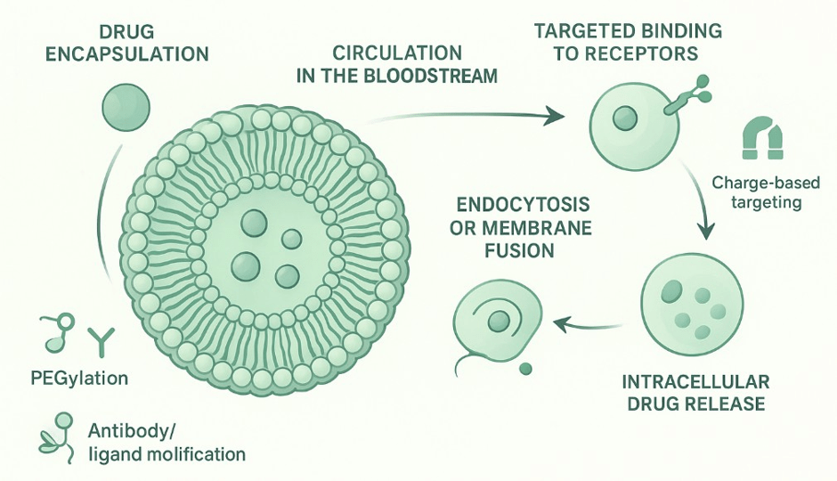
After circulating in the bloodstream, active targeted binding between the ligand and receptors takes place. The surface of the liposomes has ligands that recognise receptors or antigens on target cells. This helps to improve cellular uptake and facilitate local drug concentration. It also helps to minimise side effects, especially with highly potent chemotherapeutic drugs. Key examples are antibodies that have high specificity for overexpressed tumours. Peptides that increase receptor specificity. Small molecules called aptamers enable selective binding and are commonly used in gene therapy (BOC Sciences, 2026).
Intracellular drug release is controlled by factors like pH, temperature, or enzymes in the target environment, allowing for on-demand release and improved therapeutic outcomes. The optimisation of the therapeutic agent effects spatial (space) and temporal (time) delivery (BOC Science, 2026). It minimises uptake by the liver and spleen by macrophages. Neutral or slightly negative liposomes lower off-target interactions by decreasing non-specific binding. On the other hand, positive charges may increase uptake by cells, but the risk of clearance is high.
Other stimuli that contribute to the release of the chemotherapeutic drug from the liposome are external triggers, e.g., light, ultrasound, and hyperthermia (high temperature), which can activate the liposome at the target site and minimise drug exposure. Small liposomes (70 to 120 nm) penetrate tissues more effectively, whereas large vesicles are sequestered by the spleen and liver.
Lastly, internalization via endocytosis or direct membrane fusion ensures that the drug reaches the cytoplasm of target cells, further optimizing delivery and minimizing unintended toxicity. PEGylation coating lowers the ability of the liposome to be recognised by the immune system and also protein adsorption, and maximises half-life, allowing time for the liposome to reach the target goal. This helps to increase local drug concentration (BOC Sciences, 2026).
The main method of internalization of liposomes is through endocytosis, that depend on particle size, composition of lipids, type of target cell, and surface chemistry. Clathrin-mediated endocytosis is employed by small liposomes (50 to 150 nm), and receptor formulation is suitable to deliver small molecules to specific receptor-expressing cells. Please see Figure 11. Caveolae-mediated uptake evades degradation by the lysosome and enables cytosolic delivery. Caveolae are used by endothelial and epithelial cells. Larger liposomes and aggregated particles undergo micropinocytosis. Pinocytosis is a type of endocytosis where there is the formation of vesicles from the plasma membrane. The uptake of extracellular fluid and solutes by a cell. This takes place in white blood cells, the liver, and other types of cells. It is active in tumour cells and phagocytes and enables high-volume internalization. Post-internalization, liposomes remain trapped in the endosome and limit the functional ability of drugs. The endosome is a structure found inside the cells that functions as a vesicle and is bound to the membranes. It helps in the transport of proteins and lipids across the cell.
This can be overcome using pH-sensitive lipids and the proton sponge effect (BOC Sciences, 2026). The pH-sensitive lipids break down the liposomal bilayer in acidic conditions, releasing contents. The proton response effect is when positive ions (cations) lead to swelling via the process of osmosis in endosomes. This leads to rupture and release of contents inside the cytoplasm.
Membrane fusion is another way in how target is released. It is more common when liposomes bind to the plasma membrane and release contents into the cytosol faster style e.g., nucleic acid therapy, e.g., siRNA (silencing of RNA), mRNA, proteins, as sensitive to endosomal degradation. An example is fusogenic lipids like dioleoylphosphatidylethanolamine (DOPE), which destabilize the bilayer to promote fusion and efficient cytosolic delivery.
Liposomes increase the accumulation of drugs into target cells, enhancing drug stability and lowering side effects. Its surface can be modified by the addition of glutamic hexapeptide and folic acid, and has shown great efficacy against metastatic bone cancer (Badila et al., 2021; Wang et al., 2020). This helps to overcome its adverse side effects, like poor water solubility and a lack of specificity to target the bone surface. These delivery approaches helped to suppress bone tumours.
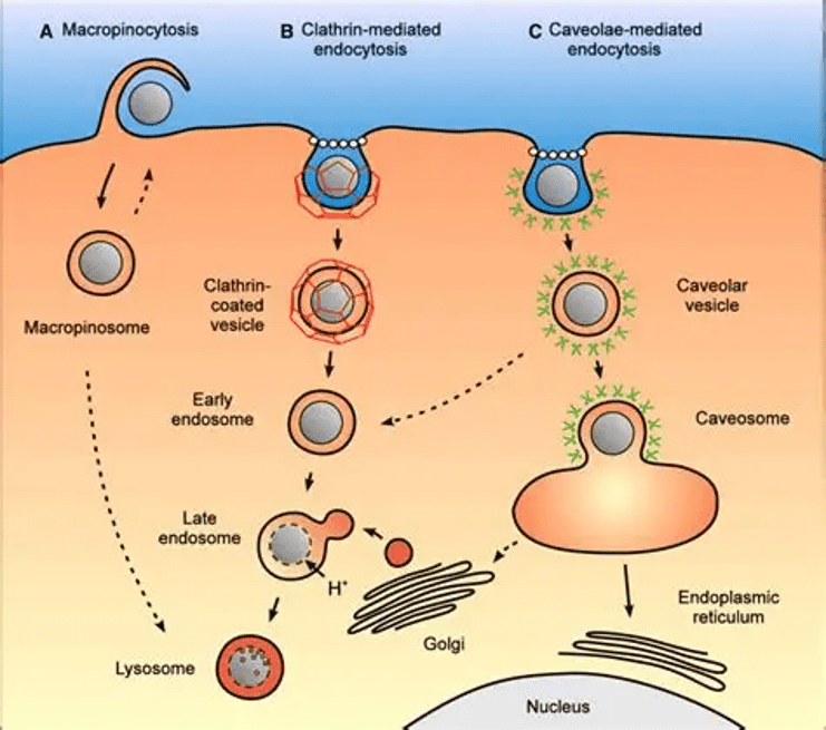
Therefore, PEGylation and ligand modification enhance liposome circulation and targeting. Stimuli-responsive release and membrane fusion improve intracellular drug delivery. Surface modifications can increase efficacy against specific cancers, such as metastatic bone cancer.
Targeted Therapy
Cancer Research UK (2025) recently indicated that researchers are searching for ways to improve the diagnosis and treatment of primary bone cancers. This reduces the likelihood of relapsed cancers (cancers coming back) and refractory cancers (continuing to grow). Targeted therapy aims to find genes, proteins, and substances that enable the immune system to respond better in bone cancers, stop cancer cells from proliferating, evading angiogenesis (blood vessels that provide nutrients for growth), and strengthen the efficacy of chemotherapy. Current research is reviewing sunitinib, nivolumab, and Lenvatinib (Cancer Research UK, 2025a).
Lenvatinib and sunitinib are tyrosine kinase inhibitors. Tyrosine kinases are proteins that facilitate cellular growth. However, tyrosine kinase inhibitors are commonly prescribed for distinctive bone cancer types. Sunitinib for chordoma if imatinib is not working as the first regimen, whereas Lenvatinib is prescribed for malignant primary bone cancer called osteosarcoma. Nivolumab is a programmed death-1 (PD-1) inhibitor that boosts the immune response (Cancer Research UK, 2025a, b).
Another example of a PD-1 inhibitor is Pembrolizumab. Nivolumab and Pembrolizumab facilitate the treatment of bone cancer, particularly osteosarcoma and chordoma. Nivolumab is a fully human IgG4 monoclonal antibody, while pembrolizumab is a humanized IgG4 variant. A monoclonal antibody is an antibody produced in a laboratory that mimics the effects of one type of antibody produced by the parental lymphocyte, boosting the immune response.
PD-1 inhibitors can recognise and attack cancer cells. Activated T cells, a type of immune cell, express PD-1 on their surface. The ligand of PD-1 is PD-L1 and is expressed in macrophages and tumour cells (Mira et al., 2019). PD-1 functions as a checkpoint receptor that limits T-cell activity when it binds to PD-L1 or PD-L2. PD-1 causes the immune system to be overactive, which may occur in autoimmune reactions. Therefore, blocking PD-1 enables T cells to attack cancer cells more effectively.
Both checkpoint inhibitors, Nivolumab and Pembrolizumab, have shown improvements in overall survival and progression-free survival compared to standard treatments (Thisvs.That. n.d). There is also multiple evidence from observational data, retrospective and meta-analytical studies that support their efficacy when combined rather than alone. These are studies conducted by researchers on large patient groups to understand how well the drugs work, their mode of action, and side effects, but not head-to-head, as they have similar clinical effects.
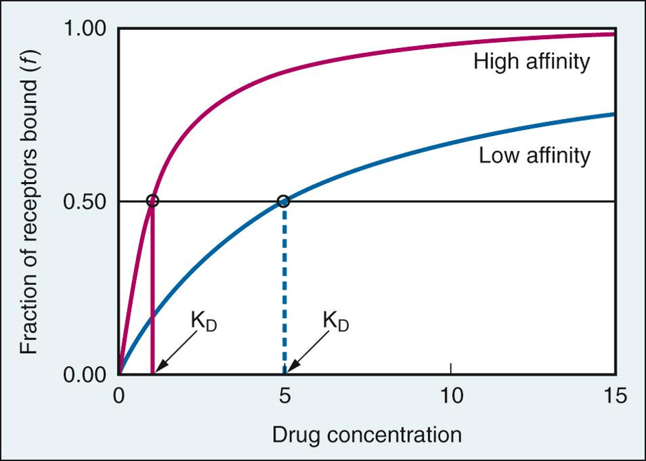
However, one would ask why doctors are focusing on the checkpoint inhibitor Nivolumab as a novel bone cancer treatment research, rather than Pembrolizumab, which has the same function and has been approved for use in a wider range of cancers than Nivolumab. One possible reason is Nivolumab’s higher affinity to bind to the PD-1 receptor compared to Pembrolizumab. Drug affinity is one of the key factors that determines how efficient a drug is and its safety profile. It is defined as the strength of the drug’s binding to its target and is measured by the dissociation constant (Kd). The Kd value is an indicator of the concentration of the drug needed to occupy half of the binding sites present on the target. Please see Figure 12. The lower the Kd value, the higher the affinity because the drug binds more tightly to its receptor, which influences the potency of the drug and the dosage needed to induce a therapeutic response.
Another possible reason is the efficacy of Nivolumab in combination with bone-targeting agents (BTA) in patients with bone metastases, secondary to primary cancers like renal cell carcinoma (RCC – kidney cancer). Poor prognosis has been established in bone metastatic RCC upon treatment with antiangiogenic agents.
On the other hand, Velev et al. (2023) discovered that Nivolumab induces shorter progression-free survival and lower objective response rate in RCC patients with bone metastasis. For instance, in patients with RCC and bone metastasis, the median progression-free survival was 2.8 months, and the objective response rate (ORR) was 14.8%. Progression-Free Survival (PFS) refers to the time from the start of treatment or being randomised until the disease has progressed or death occurs from any cause. ORR is defined as the proportion of patients in each cohort with the best overall response. This is shorter in comparison to patients without bone metastasis. The progression-free survival was 4.6 months, and the ORR was 23.3%. This indicates that BTA with targeted immunotherapy may help increase survival and decrease the incidence of skeletal-related events.
Another difference between Nivolumab and Pembrolizumab is the administration schedule by intravenous (IV) infusion. They can also be injected subcutaneously (under the skin) (Anderson, 2026). Nivolumab is administered every 2 to 4 weeks, whereas Pembrolizumab is administered every 3 weeks. This is dependent on the type of cancer, the patient’s medical history, the clinical characteristics of cancer, and the patient’s overall health status. For example, Pembrolizumab is given at a fixed dose, whereas the weight of a patient determines the dose given for Nivolumab (This vs. That, n.d).
On the other hand, though both PD-1 inhibitors are well-tolerated, they can cause immune-related adverse effects, most commonly fatigue, rash, fever, dizziness, nausea, joint/muscle aches, wheezing, and diarrhoea. Conversely, Nivolumab has a higher incidence of causing inflammatory conditions like pneumonitis and colitis that affect the lungs and intestines, respectively, in comparison to Pembrolizumab. They can also cause issues with the thyroid gland and elevated levels of liver enzymes.
Therefore, the choice between them depends on the type of cancer, the drugs used in combination, previous treatments, cost considerations, and other facts given in the guidelines provided by the health authority.
Pembrolizumab would work more efficiently in sarcomas with elevated expression of PD-1. High levels of PD-1 have been found on CD4+ T helper cells in patients with metastatic cancers than in non-metastatic cancers (Mira et al., 2019). High levels were also found in cytotoxic CD8+ T-killing cells.
Previous in vivo studies, like Lussier et al. (2015), revealed that anti-PD-L1 antibodies combined with anti-cytotoxic T lymphocyte antigen-4 (anti-CTLA-4) antibody improved the overall survival of osteosarcoma more than anti-CTLA-4 alone. Overall Survival (OS) is the time from diagnosis or the start of treatment until death from any cause. Expression of PD-L1 also correlated with tumour-infiltrating lymphocytes (TIL). This indicates that PD-1, PD-L1, and CTLA-4 immune checkpoints partake in the pathogenesis and progression of osteosarcoma (Mira et al., 2019).
Tawbi et al. (2017) conducted a multicenter phase 2 trial on Pembrolizumab on 84 subjects. There was an equal number of patients with bone sarcoma (n=42) and soft tissue sarcoma (n=42). They discovered that 18% of 40 subjects with bone and soft sarcoma had an ORR, and the response varied by histological subtype in relation to complete response (CR) or partial response (PR). The duration of the overall response was objectively recorded and measured from the time criteria were met for CR or PR until the first date that recurrent or progressive disease occurred. 40% ORR was for the soft tissue sarcoma: undifferentiated pleomorphic sarcoma (1 CR+3PR/10), 2 PR/10 in dedifferentiated liposarcoma (DDLPS), 1PR/10 in synovial sarcoma (SS), and 0/10 in leiomyosarcoma (LMS) (Tawbi et al., 2017).
For bone sarcoma, the median follow-up was 17.8 months, ORR 5%, with 1PR/22 in osteosarcoma (OGS), 1PR/5 in chondrosarcoma, and 0/13 in Ewing sarcoma (Tawbi et al., 2017).
All subjects in Tawbi et al. (2017) were treated with Pembrolizumab at 200 mg intravenously every 3 weeks until disease progression or unacceptable toxicity. This was followed by imaging as computed tomography (CT) or magnetic resonance imaging (MRI) (Tawbi et al., 2017).
Results indicated that objective responses in 18% in soft tissue sarcomas, while the response rate was only 5% in bone cancers (Tawbi et al., 2017).
In additional the 12-week progression-free survival rate in soft tissue sarcoma was 55% than 40% 12-weeks PFR required for an active regimen (p=0.001). This emphasises that though the response rate in soft tissue sarcoma was short and below the required 8 responses out of 40, there was clinical efficacy in the treatment group (Tawbi et al., 2017).
Moreover, Tawbi et al. (2017) revealed that there was a partial response in most histological subtypes, particularly UPS and LPS. For instance, there was an improvement in the 12-week progression-free survival rate in UPS (70%). The median duration of responses for UPS was 30 weeks; the median overall survival was not reached. These results also support the efficacy of Pembrolizumab in blocking the immune checkpoint activity.
On the other hand, the expression of the ligand of PD-1, PD-L1, was found in two subjects with UPS and responded well to Pembrolizumab. Similarly, though there were two responses in bone sarcoma (osteosarcoma n=1; Chondrosarcoma n=1). There was a good clinical response because there was >50% reduction in size, and it was durable for a minimum of 6 months.
On the contrary, there was no clinical response in patients with Ewing Sarcoma (n=13). A possible explanation for this is the highly suppressive immune microenvironment. Nevertheless, with any treatment, a small proportion of patients is likely to experience adverse effects. Several patients had grade 3 to 4 complications, namely fatigue (n=11), anaemia (n=9), lymphopenia (n=8). Some were advanced and immune-related, such as adrenal insufficiency (n=2), pneumonitis (n=2), and one case of interstitial nephritis (Tawbi et al., 2017). Lymphopenia is the low white blood cells (lymphocytes) count. Anaemia is caused by lack og haemoglobin that carries oxygen around the body. Pneumonitis is an inflammatory conitions caused by a bacteria called Streptoccocus Pneumonia. It affects the lungs and causes them to become solid. Interstitial nephritis is the inflammation of the kidneys especially the outer line called interstitium.
Moreover, one aspect that is pivotally established from this data is the role of immunotherapy to eradicate bone cancer cells, where enhanced efficacy can be found in Immunotherapy in combination with chemotherapy or radiotherapy than alone (Massive bio, 2026). It also further indicates how the clinical response to sarcomas is histologically distinctive and is linked to the molecular and immunological characteristics.
Other immunotherapeutic options for osteosarcoma are dendritic cell immunotherapy (Cowan, Launico, and Kahai, 2024). Dendritic cells form part of the innate immune response, where they recognise and present antigens found on the cell surface of pathogens (disease-causing microbes) and cancer cells. This allows B lymphocytes, CD4-positive T helper lymphocytes, and CD8-positive cytotoxic T lymphocytes to elicit an immune response (Miwa et al., 2019).
Amongst the cytokines released by other immune cells, such as macrophages and mast cells, is interferon-alpha (IFN-α). In vivo studies revealed that it prevents many of the hallmarks of cancer that enable cancer cells to progress. For instance, it prevents angiogenesis, promotes apoptosis, and decreases the rate of proliferation.
The pegylated version of IFN-α (IFN-α-2b) was investigated with and without the combined chemotherapeutic regimen: methotrexate, doxorubicin, and cisplatin (MAP). Results revealed that there was a slight increase in the 5-year overall survival rate with the pegylated IFN-α-2b (84%) than without. This shows promise of prolonging the survival (Miwa et al., 2019).
Moreover, another member of the small protein family of Interferons is Interferon-gamma (IFN-γ), which is produced by T cells and Natural Killer cells. IFN-γ is associated with its receptor IFNGR and stimulates macrophages and the differentiation of other immune cells. Mira et al. (2019) revealed that treatment of dendritic cells with tumour lysate increased the levels of cytotoxic T cells and IFN-γ and lowered the risk of forming metastases in the lungs and CD8-positive (CD8+) T lymphocytes in areas of metastasis. This increases systemic immune response without severe toxicity.
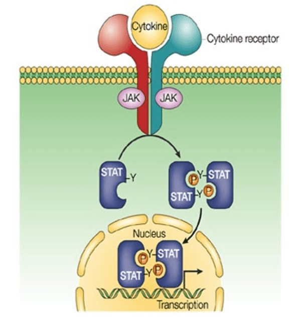
The interferon mode of action involves the stimulation of the Janus kinase/signal transducer and activator of transcription (JAK-STAT) from the cell membrane to the nucleus. Once the interferon is activated with its receptor on the plasma membrane, a group of enzymes called Janus kinases (JAKs) phosphorylates the interferon receptor complex. Phosphorylation is the addition of phosphate groups. This creates docking sites for the transcription factors called STAT. The JAKs phosphorylate the STATs, where they detach from the interferon receptor complex and bind with the interferon regulatory factor 9 (IRF9) protein. The STAT-IRF9 complex moves into the nucleus of the cell, where they associate with DNA response elements, which are DNA sequences, and induces transcription of target genes called Interferon-Stimulated Genes (ISGs).
Some of the target genes encode for proteins involved in growth, proliferation, repair, and decausde viral and cellular DNA (Biology Insights, 2025). This indicates the role of targeted therapy that can analyse patients at a molecular level to identify the expression of genes, proteins, and signalling pathways that cause elevated induced growth. This allows patients to have access to new treatments of bone cancer that analyse individual analysis and the importance of precision medicine (Massive Bio, 2026).
Another monoclonal antibody that aids with bone tumour management is Denosumab. It primarily treats giant cell tumour (Badila et al. , 2021; Cancer Research UK, 2025b; Massive bio 2026). It targets the protein RANKL1 that regulates the activity of osteoclasts. Osteoclasts are bone cells that break down old bone (demineralization). Denosumab halts RANKL1 from doing so, delaying the occurrence of fractures and strengthening bone activity. This also lowers the risk of skeletal-related events when combined with Zoledronic acid, a type of bisphosphonate (Badila et al., 2021). Bisphosphonates are small-molecular drugs that inhibit bone demineralisation at high doses to penetrate the target area. It also has high overall prolonged survival (Badila et al., 2021). Bisphosphonates elicit a uniform distribution of bone cells.
Moreover, small bivalent molecules, EB-TCIP, have been designed by Bond et al. (2025) to drive the transcriptional expression of genes that promote apoptosis in paediatric bone cancers like Ewing sarcoma. The bivalent compound targets the transcriptional factor EWSR1::FLI1 and recruits it to DNA sites of BCL6, increasing its expression and remodelling chromatin.
Evading apoptosis is one of the hallmarks of cancer. Apoptosis also occurs in normal cells, where there is a fine balance between growth, differentiation, and apoptosis to ensure there is the correct number of cells. Aberrant genetic expression and the ability to evade apoptosis can give rise to tumours.
The family of Bcl-2 proteins is divided into two groups: one group promotes apoptosis, whereas the other group evades apoptosis. The BCL2 protein associates and suppresses the pro-apoptotic proteins: Bim, Bax, and Bak. Normally, the Bax/Bak form pores in the mitochondrial membrane and release cytochrome C. This leads to a series of cascade events by protein kinases called caspases that induce cell death.
On the other hand, BCL6 is a transcriptional repressor that is pivotal in the development of follicular helper T cells (CD4) and conventional dendritic cells (cDCs) in the spleen. BCL6 is upregulated in pre-CDC and its proliferation. However, levels decrease during cDC maturation. BCL6 is an oncogene that promotes the progression of human B-cell lymphomas, a type of blood cancer where there is excessive production of lymphocytes in primary and secondary lymphoid organs, e.g., bone marrow, spleen, and thymus, respectively (Basso and Dalla-Favera, 2010). In addition, they also differ in pattern under the microscope: BCL2 is cytoplasmic, whereas BCL6 is nuclear.
Furthermore, oncolytic viruses have shown effectiveness in preclinical studies against osteosarcoma. They are genetically engineered viruses that aim to replicate inside cancer cells and effectively kill them. It has shown potential effects against osteosarcoma. Their usefulness can be enhanced by using nanocarriers to the tumour site and facilitating targeting of tumour cells. Further studies are needed to establish the role of oncolytic viruses and the tumour microenvironment and immune system, and the cause of limited extravasation of oncolytic viruses. A release of oncolytic viruses enables the release of antigens that elicit an immune response (Badila et al., 2021).
Oncolytic Viruses By Life Science Animation
Overall, there is ongoing progress in modern oncology. Precision medicine is pivotal for bone cancer patients to develop individualised care plan that is dependent on the type, stage, grade, and clinical characteristics of the cancer and the patients’ clinical history. Novel and improved techniques have been notable for conventional treatment such as phototherapy, surgery, and targeted therapy, which, in combination with chemotherapy, has better overall performance. The application of nanocarrier systems has made the target area more focused and less damage to non-cancerous sites through electron and energy transfer, which are common routes to sensitise the tumour sites. Ongoing research is made to improve the quality of life and enhance patient clinical outcomes. The downside of bone cancer treatment is the adverse side effects, where corticosteroids are commonly prescribed to alleviate the impact.
References
Anderson, L.A. (2026) Pembrolizumab vs. nivolumab: How do they compare? Available at: https://www.drugs.com/medical-answers/pembrolizumab-nivolumab-compare-3554067/ (Accessed: 25th June 2026)
Bădilă, A.E., Rădulescu, D.M., Niculescu, A.-G., Grumezescu, A.M., Rădulescu, M. and Rădulescu, A.R. (2021). Recent Advances in the Treatment of Bone Metastases and Primary Bone Tumors: An Up-to-Date Review. Cancers, [online] 13(16), p.4229. doi:https://doi.org/10.3390/cancers13164229.
Basso, K. and Dalla-Favera, R. (2010). BCL6. Advances in Immunology, 105; pp.193–210. doi:https://doi.org/10.1016/s0065-2776(10)05007-8.
Biology Insights (2025) The Three Types of Interferons and How They Work. Available at: https://scienceinsights.org/the-three-types-of-interferons-and-how-they-work/ (Accessed: 25th June 2026)
BOC sciences (2026) How liposomal drug delivery works: Mechanisms and Targeting strategies Available at: https://liposomes.bocsci.com/resources/how-liposomal-drug-delivery-works-mechanisms-targeting-strategies.html (Accessed: 25th June 2026)
Bond, M.J., Golden, R.P., DiGiovanni, G., Howard, B., Sarott, R.C., Karim, B.A., Gourisankar, S., Alexe, G., Ross, K., Jones, H.M., Dwyer, B.G., Gray, N.S. and Stegmaier, K. (2025). Rewiring the Fusion Oncoprotein EWSR1::FLI1 in Ewing Sarcoma with Bivalent Small Molecules. Journal of the American Chemical Society, 147(49), pp.44739–44758. doi:https://doi.org/10.1021/jacs.5c05634.
Cai, Y., Chai, T., Nguyen, W., Liu, J., Xiao, E., Ran, X., Ran, Y., Du, D., Chen, W. and Chen, X. (2025). Phototherapy in cancer treatment: strategies and challenges. Signal Transduction and Targeted Therapy, [online] 10(1), pp.1–46. doi:https://doi.org/10.1038/s41392-025-02140-y.
Cancer Research UK (2025a) Research into primary bone cancer Available at: https://www.cancerresearchuk.org/about-cancer/bone-cancer/research-clinical-trials/research-bone (Accessed: 30th March 2026)
Cancer Research UK (2025b) Targeted cancer drugs for bone cancer Available at: https://www.cancerresearchuk.org/about-cancer/bone-cancer/treatment/Targeted-cancer-drugs (Accessed: 25th June 2026)
Cowan, P., Launico, M. and Kahai, P. (2024) Anatomy, Bones. Available at: https://www.ncbi.nlm.nih.gov/books/NBK537199/ (Accessed: 9th April 2026)
Creative Diagnostics (2026) ELISA Development Guide – Developing an ELISA. Available at; https://www.creative-diagnostics.com/ELISA-Development-guide.htm (Accessed 25th June 2026)
DelaFlor-Weiss, E., Uziely, B. and Muggia, F.M. (1993). Protracted drug infusions in cancer treatment: An appraisal of 5-fluorouracil, doxorubicin, and platinums. Annals of Oncology, 4(9), pp.723–733. doi:https://doi.org/10.1093/oxfordjournals.annonc.a058656.
Fleming, M.D. and Hamza, I. (2012). Mitochondrial heme: an exit strategy at last. Journal of Clinical Investigation, 122(12), pp.4328–4330. doi:https://doi.org/10.1172/jci66607.
Hanaei, S.B., Murugesan, R.C., Souza, L., Juan Ignacio Cadiz-Miranda, Jeys, L., Wall, I.B. and Martin, R.A. (2024). Multifunctional gallium doped bioactive glasses: a targeted delivery for antineoplastic agents and tissue repair against osteosarcoma. Biomedical Materials. doi:https://doi.org/10.1088/1748-605x/ad76f1.
Lussier, D.M., Johnson, J.L., Hingorani, P. and Blattman, J.N. (2015). Combination immunotherapy with α-CTLA-4 and α-PD-L1 antibody blockade prevents immune escape and leads to complete control of metastatic osteosarcoma. Journal for ImmunoTherapy of Cancer, 3(1). doi:https://doi.org/10.1186/s40425-015-0067-z.
Massive Bio (2026) Latest Research and Clinical Trials on Bone Cancer https://massivebio.com/latest-research-and-clinical-trials-on-bone-cancer-bio/ (Accessed: 11th June 2026)
Miron, R.J. (2023). Optimized bone grafting. Periodontology 2000. doi:https://doi.org/10.1111/prd.12517.
Miwa, S., Shirai, T., Yamamoto, N., Hayashi, K., Takeuchi, A., Igarashi, K. and Tsuchiya, H. (2019). Current and Emerging Targets in Immunotherapy for Osteosarcoma. Journal of Oncology, 2019, pp.1–8. doi:https://doi.org/10.1155/2019/7035045.
Shoji, O., Yuichiro Aiba, Shinya Ariyasu and Onoda, H. (2023). Heme-containing proteins: Structures, functions, and engineering. Elsevier eBooks, pp.194–214. doi:https://doi.org/10.1016/b978-0-12-823144-9.00167-9.
Tawbi, H A-H, Burgess, M.A., Bolejack, V., Van, B.A., Schuetze, S.M., Hu, J.C., D’Angelo, S.P., Attia, S., Riedel, R.F., Priebat, D.A., Sujana Movva, Davis, L.E., Okuno, S.H., Reed, D.R., Crowley, J., Butterfield, L.H., Salazar, R., Rodriguez-Canales, J., Lazar, A.J. and Wistuba, I.I. (2017). Pembrolizumab in advanced soft-tissue sarcoma and bone sarcoma (SARC028): a multicentre, two-cohort, single-arm, open-label, phase 2 trial. Lancet Oncology, 18(11), pp.1493–1501. doi:https://doi.org/10.1016/s1470-2045(17)30624-1.
Thisvs.That (n.d) Nivolumab vs. Pembrolizumab What’s the Difference? Available at: https://thisvsthat.io/nivolumab-vs-pembrolizumab#google_vignette (Accessed: 25th June 2026)
Thr Royal Orthopaedic Hispital (2024) Lab tests show novel bone cancer therapy has 99% success rate. Available at; https://roh.nhs.uk/news-events/lab-tests-show-novel-bone-cancer-therapy-has-99-success-rate (4th April 2026)
UniProt (2026) Q9Y5Y0 · fC1_HUMAN. Available at: https://www.uniprot.org/uniprotkb/Q9Y5Y0/entry (5th April 2026)
Velev, M., Dalban, C., Chevreau, C., Gravis, G., Negrier, S., Laguerre, B., Gross-Goupil, M., Ladoire, S., Borchiellini, D., Geoffrois, L., Joly, F., Priou, F., Barthelemy, P., Laramas, M., Narciso, B., Thiery-Vuillemin, A., Berdah, J.-F., Ferrari, V., Dominique Thomas, Q. and Mione, C. (2023). Efficacy and safety of nivolumab in bone metastases from renal cell carcinoma: Results of the GETUG-AFU26-NIVOREN multicentre phase II study. European Journal of Cancer, 182, pp.66–76. doi:https://doi.org/10.1016/j.ejca.2022.12.028.
Wang, H., Guo, Y., Wang, C., Jiang, X., Liu, H., Yuan, A., Yan, J., Hu, Y. and Wu, J. (2021b. Light-controlled oxygen production and collection for sustainable photodynamic therapy in tumor hypoxia. Biomaterials, [online] 269, p.120621. doi:https://doi.org/10.1016/j.biomaterials.2020.120621.
Wang, S.-Y., Hu, H.-Z., Qing, X.-C., Zhang, Z.-C. and Shao, Z.-W. (2020). Recent advances of drug delivery nanocarriers in osteosarcoma treatment. Journal of Cancer, 11(1), pp.69–82. doi:https://doi.org/10.7150/jca.36588.
Wang, X., Luo, D. and Basilion, J.P. (2021a). Photodynamic Therapy: Targeting Cancer Biomarkers for the Treatment of Cancers. Cancers, 13(12), p.2992. doi:https://doi.org/10.3390/cancers13122992.
Wu, Y. and Wang, C. (2025). Microstreaming from acoustic cavitation bubble: modeling and simulation. Modeling and Simulation of Sono-Processes, pp.139–163. doi:https://doi.org/10.1016/b978-0-443-23651-8.00009-7.
Yan, Y., Wang, X., Hai, X., Song, W., Ding, C., Cao, J. and Bi, S. (2020). Chemiluminescence resonance energy transfer: From mechanisms to analytical applications. TrAC Trends in Analytical Chemistry, 123, p.115755. doi:https://doi.org/10.1016/j.trac.2019.115755.
Zhang, J., Nie, R., Jia, Q., Qin, G., Li, Y., Zhang, Y. and Li, P. (2025). Implantable, flexible biophotonic device for wireless photodynamic therapy of postoperative infection and tumor recurrence. Device, [online] 3(7), p.100754. doi:https://doi.org/10.1016/j.device.2025.100754.
Updated July 2026 Next Review July 2028


Leave a Reply