There have been several recent discoveries that have come to our attention, which significantly impact our understanding of bone cancer and open new avenues for detecting cancer, especially in cases where bone cancer is less likely to be linked to modifiable risk factors. Current risk factors for bone cancers arise from genetic conditions, chemical or radiation exposure, or conditions like Paget’s disease that disrupt bone homeostasis—specifically, the balance between bone synthesis via osteoblasts and bone resorption via osteoclasts. Genetic testing has made a substantial impact on modern bone cancer diagnosis and treatment, enabling greater accuracy in identifying cancer subtypes that have unique characteristics, improving our ability to predict disease progression, and allowing healthcare providers to offer more targeted therapies as part of precision medicine to improve clinical outcomes (Cancer Research UK, 2025; Massive Bio, 2026). For example, patients with specific genetic mutations may now receive therapies uniquely suited to the genetic profile of their tumour, which can increase treatment effectiveness and reduce unnecessary side effects compared to traditional methods.
Similarly, the advent of artificial intelligence (AI) in recent decades has transformed clinical practice, from enhancing the diagnostic accuracy of medical imaging to optimizing treatment planning; AI algorithms now rapidly analyze imaging data to highlight subtle signs of bone tumours, assisting clinicians in making earlier and more accurate diagnoses. This integration of genetic and AI advances into routine care has reduced diagnostic delays, allowed for earlier intervention, and supported continuous monitoring of treatment responses, leading to improved clinical outcomes for patients with bone cancer. The aim of this article is to provide an insight into the research study by Anderson et al. (2026), who used the zebrafish as an experimental model that incorporates the histological and molecular hallmarks of Ewing sarcoma (ES) to study the Ewing sarcoma breakpoint region 1 (EWSR1) gene and the Friend leukemia virus integration 1 (FLI1) fusion gene that encodes an aberrant transcription factor that harbours 90% of ES cases.
An insight into Ewing Sarcoma
Ewing sarcoma is the second most common primary bone malignancy in children and adolescents and is the leading cause of paediatric cancer death. The most common site is the paravertebral region, followed by the leg (40%) and extraosseous tissue (15-20%). Under the microscope, the histological features commonly observed with haematoxylin and eosin (H and E) staining in ES are undifferentiated cells with little cytoplasm. Please see Figure 1. There is a small, round, deep blue cell tumour (Anderson et al., 2026). The cytoplasm has a high content of the storage of glucose (sugar) called glycogen. Glycogen is stained positive for periodic acid-Schiff (PAS). Though it is not a specific characteristic, it can be stained positive for the cell-surface glycoprotein biomarker CD99 (Anderson et al., 2026).
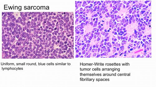
Figure 1: The histological features of Ewing Sarcoma
The translocation of the EWSR1-FLI1 fusion
Several types of genetic mutations and chromosomal translocations have been identified as key drivers of the three main primary bone cancers: Ewing sarcoma, osteosarcoma, and chondrosarcoma. Of particular relevance to Ewing sarcoma, chromosomal translocations occur when sections of thread-like structures that contain genetic information in the form of genes called chromosomes are rearranged. This can lead to the formation of abnormal fusion genes that promote cancer growth. There are two main types of chromosomal translocations: simple and reciprocal translocation. Please see Figure 2. A simple translocation is where one piece of a chromosome is attached to another chromosome. On the other hand, reciprocal translocation is when two different types of chromosomes exchange pieces; therefore, the two chromosomes are carrying translocations.
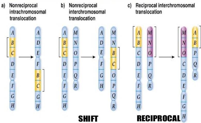
Figure 2: An illustration on the difference between simple and reciprocal chromosome translocation
Genetic mutations, or changes to the DNA sequence, can similarly alter how proteins function in cells. By definition, a gene is a short section of the deoxyribonucleic acid (DNA) that contains the genetic information to encode a protein. A change in the gene can influence the structure and function of a protein, rendering it nonfunctional. Some examples of genetic mutations include insertion, deletion, translocation, and duplication; these changes can also occur in chromosomes. In bone cancers, such alterations are especially important when they disrupt genes that regulate cell growth and differentiation.
The chromosomal translocation, t(11;22)(q24;q12), generates the EWSR1-FLI1 fusion gene. EWSR1 is a member of the ten-eleven translocation (TET) family of RNA-binding proteins. It contributes to other chromosomal translocations where its N-terminal transactivation domain associates with the DNA-binding domain of FLI1, a member of the E26 transformation-specific (ETS) family of transcription factors. The structures of both components of the fusion gene are shown in Figure 3. Transcription factors are proteins that regulate gene expression by associating with specific DNA sequences and controlling whether genes are turned on or off. Thus, this fusion gene functions as an aberrant transcriptional regulator that dysregulates the genetic expression of target genes and drives oncogenic transformation of Ewing sarcoma and other soft tissue sarcomas.
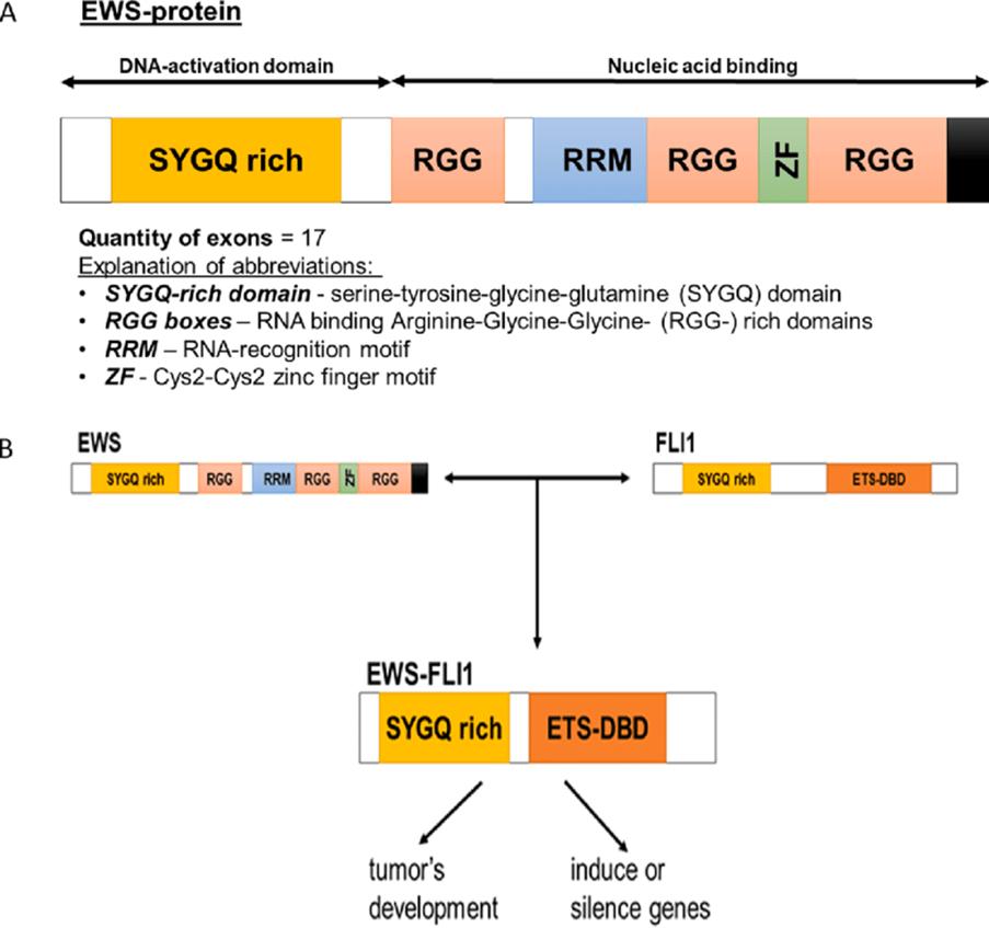
Figure 3: The formation of the EWS-FLI1 fusion protein SYCQ -serine-Tyrosine-Glycine-Glutamine-rich domain (synonym -activation domain); RGG boxes -RNA binding Arginine-Glycine-Glycine-rich domain; RRM -RNA-recognition motif; ZN -Zinc finger motif; ETS-DBD -E26 transformation-specific gen’ DNA binding domain. (Karlina et al., 2022)
The zebrafish as an experimental model
Minimal progress has been made despite the identification of the oncogene more than three decades ago because of the lack of reliable and robust animal models that incorporate the histological and molecular hallmarks of Ewing sarcoma and the inherent toxicity of the fusion protein. Key examples of previous sources were genetically engineered mice and mammalian cell lines. However, Anderson et al. (2026) findings suggest that zebrafish show promise in exhibiting EWSR1:FLI1 expression, particularly the t(11;22)(q24;q12) chromosomal translocation. Please see Figure 4.
Anderson et al. (2026) discovered that the exon 2 in the human type II collagen gene (col2a1a) promoter is what drives the expression of the EWSR1-FLI1 fusion oncoprotein in zebrafish. Thus, the Col2a1a promoter is also a strong marker for early and proliferating chondrocytes in the growth plate. Chondrocytes are cells that produce cartilage. Their expression is inversely proportional to hypertrophic differentiation; the more hypertrophic differentiation, the less cellular expression of Col2a1a. There was minimal expression of Col2a1a in the late-stage chondrocytes. The Col2a1a promoter is also active in other tissues, namely embryonic skeletal, neural crest, and notochord tissues.
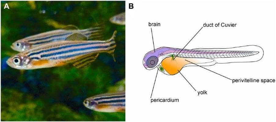
Figure 4: The zebrafish (Vittori, Motaln and Turnsek, 2015)
Hypertrophic differentiation using the example of osteoarthritis
Hypertrophy is the increase in size of cells or tissue rather than number, and differentiation is the process where cells become more specialised and have a specific function. To understand the importance of hypertrophic differentiation in disease states, an insight into the analogous signalling events involved in this process and in normal chondrocyte proliferation is reviewed. Chondrocytes resist proliferation and terminal differentiation in healthy articular cartilage. On the other hand, in disease conditions, like osteoarthritis, the chondrocytes continue to proliferate and develop hypertrophy (Dreier, 2010). This results in loss of joint mobility and chronic pain. Moreover, there is a change in the normal expression of molecules that reside in the extracellular matrix (ECM) and proteases that influence cartilage homeostasis. This leads to degenerative changes (Dreier, 2010). Other characteristic features of osteoarthritis are bone formation at the joint margins (osteophytes), limited inflammation (synovitis), and changes in subchondral bone structure (sclerosis).
The major fibrillar components of undifferentiated mesenchymal progenitor cells are collagen (Type I, III and V). Other types of collagenous proteins (I, II, III,V, IX and XI) and proteoglycan (aggrecan) are expressed and produced post-differentiation into chondrocytes. IX and XI are specific for cartilage, but the others are for connective tissues (Goldring, n.d.). The collagen network of cartilage facilitates tensile strength, whereas aggrecan provides resistance to compression (Goldring, n.d).
The resting chondrocytes during differentiation have low proliferation rates and are small and uniform. On the contrary, the ECM takes more space than these cells. Additional expression of collagen (VI, II, IX and XI) and matrilin is found in the proliferative stage, where chondrocytes undergo cell division and support flat cells arranged in a long column (Anderson et al., 2026).
The rate and progression of chondrocyte differentiation in growth cartilage consists of interacting with signalling molecules from cells in the surrounding tissue, such as perichondrium or subchondral blood vessels, to mediate positive and negative feedback mechanisms to maintain homeostasis. Cell surface receptors also partake in cellular events in their surrounding matrix: proliferation, maturation, and survival of cartilage cells. Hydrolytic enzymes such as proteinases also intervene in regulatory function by removing Erp57 (endoplasmic reticulum protein 57) and activating cytokines like transforming growth factor beta (TGFB) and releasing vascular endothelial growth factor (VEGF) from ECM stores.
A molecule called Indian hedgehog (Ihh) is expressed during prehypertrophy. The differentiation from prehypertrophic to hypertrophic chondrocytes solely induces collagen X. Low levels or termination of other subtypes of collagen II, IX, and XI are produced by hypertrophic chondrocytes. The enzymes involved are matrix metalloproteinase (MMP-13) and alkaline phosphatases. Other molecules also contribute to the process, for instance, vascular endothelial growth factor (VEGF), transcription factor Runx2, and osteopontin (Anderson et al., 2026).
Moreover, there are some similarities with chondrocyte differentiation that appear in cartilage in other bone tissues and articular cartilage, but it is at a lower metabolic degree of activity because the production of articular cartilage is eternal. The normal articular cartilage is characterised by a milky, shelled-almond (hyaline) appearance (Goldring, n.d.). For example, there is interaction between molecules in their surrounding ECM that consist of collagens (II, VI, IX and XI) and multiple types of proteoglycans (aggrecan, decorin, biglycan and fibromodulin) and other types of matrix proteins. Please see Figure 5 Proteoglycans possess large amounts of water bound to hydrophilic glycosaminoglycans; long chains of sugar molecules. This cartilaginous ECM bound with water exhibits resistance to deformation by compressive forces (Goldring, n.d).
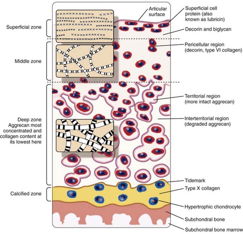
Figure 5: The structural network of articular cartilage (Goldring, n.d.)
There is minimal proliferation during the resting phase of articular chondrocytes in normal conditions, but some articular chondrocytes in diseased states have the potential to proliferate and undergo hypertrophic differentiation leading to cell death and mineralization. This is marked by high levels of alkaline phosphatase, collagen X, and MMP-13. Several components are involved in chondrocyte differentiation to osteoarthritic-like cells in cartilage with contrasting effects. Bone morphogenic protein (BMP) is stimulated by MMP-13, causing cartilage loss and influencing signalling, whereas Fibroblastic growth factor-2 (FGF2) supports cartilage homeostasis. This is due to the overexpression of extracellular heparan sulfatases Sulf-1 and Sulf-2, which are overexpressed in osteoarthritic cartilage. This enhances BMP signalling but inhibits FGF signalling in osteoarthritic cartilage by phosphorylation of ERK1/2 enzymes. This indicates how they support the repair of cartilage but also maintain homeostasis (Dreier, 2010).
TGF-Beta signalling and Wnt signalling also result in osteoarthritic-like changes with terminal differentiation of chondrocytes. The discoidin domain receptor (DDR-2) is activated by interaction of collagen type 2 with chondrocytes, which increases expression of itself and MMP-13, which can occur in normal and osteoarthritis. This illustrates that the upregulation of degradative enzymes like MMP-13 or aggrecanases along with a reduction in cartilage collagen production are mediators for the pathogenesis and progression of disease states such as osteoarthritis. Furthermore, it highlights the dual role of growth factors, where they are involved in the proliferation and differentiation of chondrocytes but also partake in the pathogenesis of osteoarthritis, where hypertrophic differentiation occurs.
Growth hormone also stimulates longitudinal bone growth and proliferation of the resting zone and can induce chondrocyte hypertrophy. This is mediated by insulin-like growth factors. Other contributors to longitudinal bone growth and control in chondrocyte proliferation and differentiation include glucocorticoid, thyroid, androgens, and oestrogen hormones.
In addition, the transcription factors Runx2 and 3 positively regulate the transformation of proliferating chondrocytes to hypertrophic chondrocytes in the skeleton and are also strongly proportional to VEGF expression during bone development and the production of blood vessels (angiogenesis) that supply oxygen and nutrients. Similarly, the transcription factor CCAAT/enhancer binding protein beta (C/EBPβ) can facilitate the hypertrophic differentiation of chondrocytes by directly activating p57Kip2 and influencing collagen type X expression during bone development (Drierir, 2010).
Thus, in relevance to Ewing sarcoma, low expression of the Col2a1a promoter causes more hypertrophic differentiation that can exhibit the above cellular and molecular events that influence the growth and proliferation of chondrocytes from early to maturation.
The role of Col2a1a promoter in Ewing sarcoma
Anderson et al. (2026) revealed that the Col2a1a promoter was first detected in the notochord at 10 hours post fertilization. The notochord is a flexible, elastic, rod-like structure found in chordates that provides early structural support during embryonic development. The notochord in zebrafish is situated dorsal to the gut and ventral to the neural tube. Please see Figure 6. The neural tube will later form the brain and spinal cord during maturation. The notochord is derived from the axial mesoderm and consists of vacuolated cells surrounded by collagen and elastin that gives its characteristics of rigidity and flexibility. These cellular features are similar to human development. However, the notochord, which is also found in vertebrates, is temporary, and it matures and forms the nucleus pulposus. The nucleus pulposus is found in the intervertebral discs of the spine during development. Please see Figure 7. This indicates why zebrafish are suitable as a model for understanding human bone development in normal and disease states.
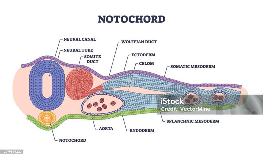
Figure 6: The anatomical structure of the zebrafish
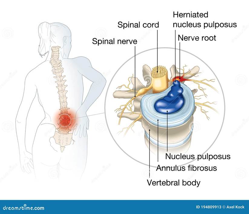
Figure 7: The structure of the vertebral disc in the spine.
At the one-cell stage, the research team injected the Tg(col2a1a: EWSR1::FLI1:pA) and Tg(ubi: EWSR1::FLI1:pA), which had a green lens fluorescence marker, into wild-type zebrafish. The former construct has the Col2a1a promoter, whereas the latter construct is promoter-less for comparative studies. The induction of these constructs into zebrafish is called transgenic fish. At 2 days post-fertilization (dpf), the fluorescence marker was detectable. By 3 dpf, there were contrasting results with each construct. 97% of the injected embryos with Tg(ubi:EWSR1::FLI1:pA) were congenitally malformed or dead. This indicates the level of lethality of the fusion oncoprotein.
By contrast, Tg(col2a1a: EWSR1::FLI1:pA) injected embryos were alive at 3 dpf and showed no gross congenital malformations. However, over time, morphological phenotypes were observed. At first, some of the transgenic fish had enlarged ears but were not classified as grossly abnormal, though it affected them unilaterally and bilaterally. By 7 dpf, malformed features began to present cranial defects in the brain. This illustrates that the expression of col2a1a in zebrafish affects the resting and proliferating chondrocytes in the endochondral growth plates. This leads to defects in the craniofacial skeleton, otic vesicle of the ear, and notochordal defects. This indicates how the embryonic state of the bone environment facilitates the expression of the EWSR1:FLI1 oncoprotein.
Additional changes appeared where 60% who did not develop a swim bladder by 7 dpf passed away and were smaller in size compared to non-transgenic fish. However, these features were not linked to either the ear or craniofacial defects. Alternatively, the transgenic zebrafish that maintained their swim bladder (40%) had no significant difference in size or function compared to non-transgenic fish. Other notable features that appeared later are the narrowing of the angles of the ceratohyal and Meckel’s cartilage, which is illustrated in Figure 8. The ethmoid plate was shorter. These structures normally appear during embryonic development. Some of these structures also appear in humans, such as the ethmoid bones in the face and Meckel’s cartilage in the mouth (mandible). This illustrates the significant differences between the normal zebrafish and transgenic fish. It also presents some of the similarities in the anatomical features of the embryonic development in both zebrafish and humans, which further implies how zebrafish is a good model for experimental studies.
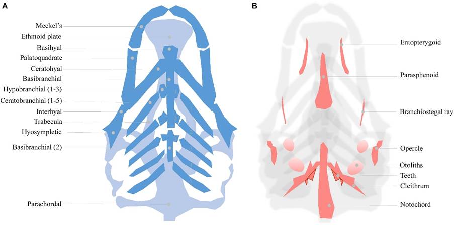
Figure 8: The ventral angle of the normal structural locations in the neurocranium and viscerocranium of the zebrafish (Raterman et al., 2020). The human skeletal features of Meckel’s cartilage for the lower jaw (mandible), the ethmoid plate for the hard palate, and the palatoquadrate for the middle ear are also illustrated. (B) The dorsal angle of the Intramembranous bones of the 5 dpf zebrafish head.
Moreover, one of the key features of Ewing sarcoma is the presence of flesh-gray-white lobulated tumours. This feature was also found in the zebrafish with Tg(col2a1a: EWSR1::FLI1:pA) near the bony tissues of the fins after 7 dpf. Anderson et al. (2026) revealed that 70% of notochord tumours occurred within the first 72 to 96 hours and were driven by the col2a1a promoter. Five percent of the surviving fish exhibited features of Ewing sarcoma, where they developed round blue cell tumours at 9 months post-fertilization upon Haematoxylin and eosin staining, positive PAS staining, and positive CD99 staining. This suggests that expression of the fusion oncoprotein within Col2a1a-expressing cells allows for the development of Ewing sarcoma.
Furthermore, several genes involved in the production of chondrocytes (chondrogenesis) were expressed. Sox9 regulates proliferation and differentiation of non-hypertrophic chondrocytes. They also regulate the expression of collagen proteins (II and XI) and proteoglycan (aggrecan). It also serves as a negative regulator of chondrocyte hypertrophy, cartilage vascularization, and bone marrow formation (Anderson et al., 2026)
Overall, the col2a1a promoter is a strongly expressed indicator to understand the pathogenesis and genetic disposition of Ewing sarcoma using a stable tissue-specific transgenic model of a zebrafish. The EWSR1-FLI1 fusion oncoprotein acts as an aberrant transcription factor and is driven by the col2a1a promoter, leading to dysregulated gene expression and oncogenic transformation. There are multiple regulators involved in the proliferation and differentiation of chondrocytes, and it has been largely associated with the stimulation of matrix metalloproteinases (MMP13), that deduced collagen production and increases degradation. This has also been associated with the pathogenesis of degenerative conditions like osteoarthritis. Moreover, genetic testing and AI are advancing bone cancer diagnosis and treatment.
References
Anderson, R.A., Chen, X., Oyarbide, U., Alvarez, N.J., Sievers, A., Schwartz, G.K. and Corey, S.J. (2026). Tissue-Specific Expression of the EWSR1::FLI1 Fusion Protein Identifies col2a1a-Positive Cells as a Source of Ewing Sarcoma-like Tumors in Zebrafish. International Journal of Molecular Sciences, 27(7), p.3131. doi:https://doi.org/10.3390/ijms27073131.
Cancer Research UK (2025) Research into primary bone cancer Available at: https://www.cancerresearchuk.org/about-cancer/bone-cancer/research-clinical-trials/research-bone (Accessed: 30th March 2026)
Dreier, R. (2010). Hypertrophic differentiation of chondrocytes in osteoarthritis: the developmental aspect of degenerative joint disorders. Arthritis Research & Therapy, [online] 12(5), p.216. doi:https://doi.org/10.1186/ar3117.
Goldring, M. (n.d.) Cartilage and Chondrocytes. Available at:
https://musculoskeletalkey.com/cartilage-and-chondrocytes/ (Accessed: 12th June 2026)
Karlina, I.A., Schroeder, B., K.I. Kirgizov, Romantsova, O.O., A.L. Istranov, Andrey Anatolievich Nedorubov, Timashev, P.S. and Ulasov, I.V. (2022). Latest developments in the pathobiology of Ewing sarcoma. [online] Journal of Bone oncology 35, pp.100440–100440. doi:https://doi.org/10.1016/j.jbo.2022.100440.
Massive Bio (2026) Latest Research and Clinical Trials on Bone Cancer https://massivebio.com/latest-research-and-clinical-trials-on-bone-cancer-bio/ (Accessed: 11th June 2026)
Raterman, S.T., Metz, J.R., Wagener, F.A.D.T.G. and Von den Hoff, J.W. (2020). Zebrafish Models of Craniofacial Malformations: Interactions of Environmental Factors. Frontiers in Cell and Developmental Biology, [online] 8, p.600926. doi:https://doi.org/10.3389/fcell.2020.600926.
Vittori, M., Motaln, H. and Turnšek, T.L. (2015). The Study of Glioma by Xenotransplantation in Zebrafish Early Life Stages. Journal of Histochemistry & Cytochemistry, 63(10), pp.749–761. doi:https://doi.org/10.1369/0022155415595670.
Updated July 2026 Next Review July 2028


Leave a Reply