Tests and Procedures To Diagnose Bone Cancer PART 1: Physical Examination to Radiology
There are a range of tests and procedures to help detect bone cancer.
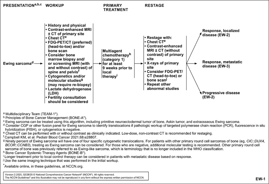
EW-1. NCCN Clinical Practice Guidelines in Oncology (NCCN Guidelines) for Bone Cancer, Version 2.2025.
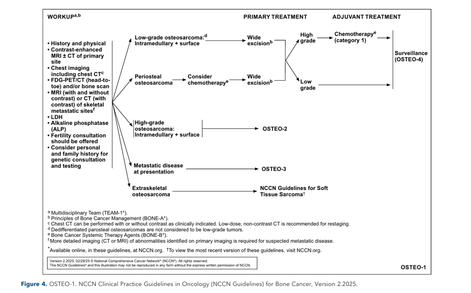
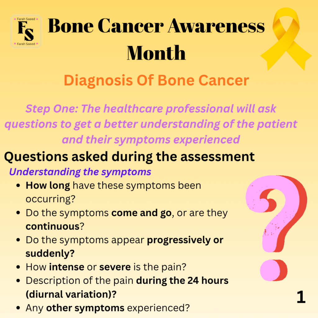
Step One and Two
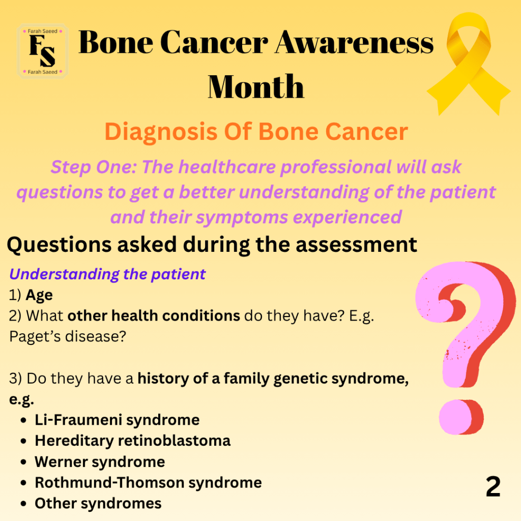
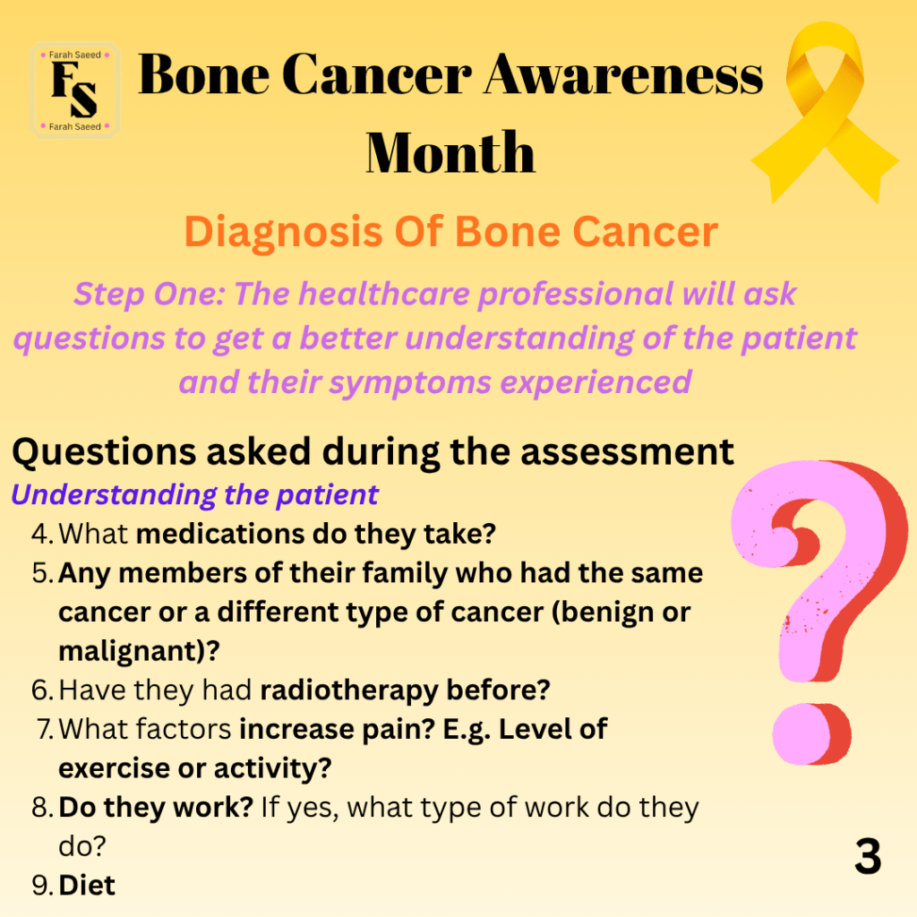
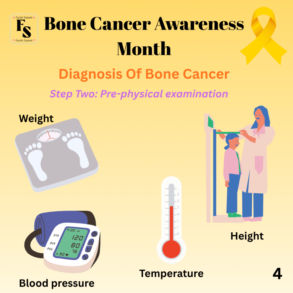
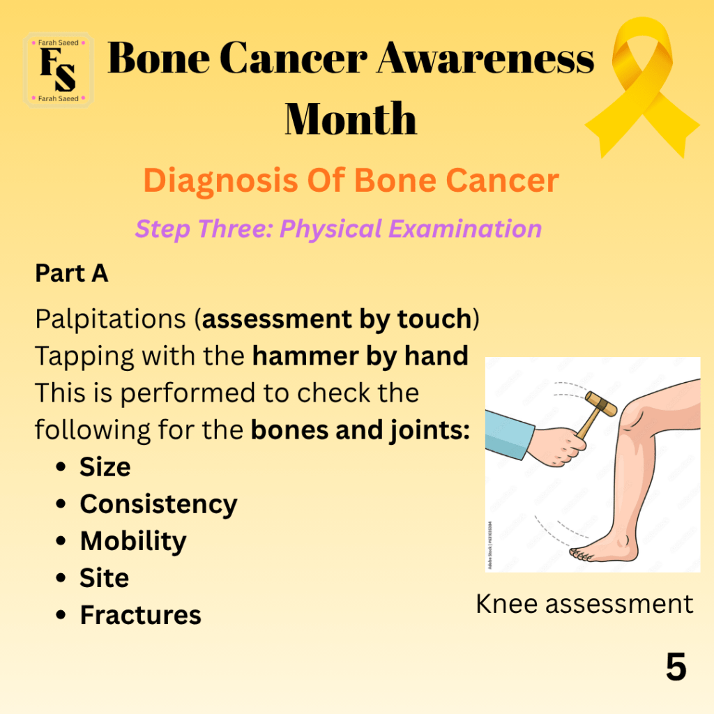
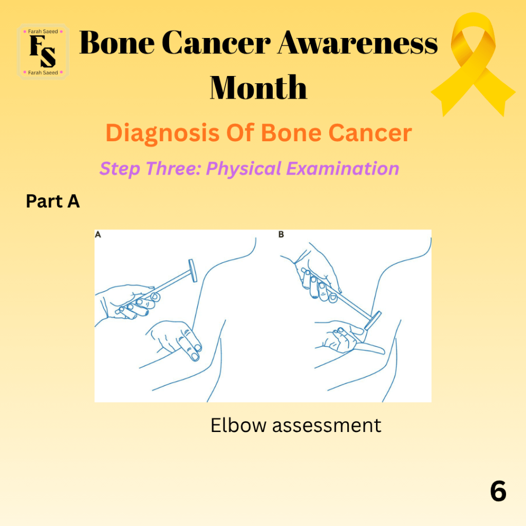
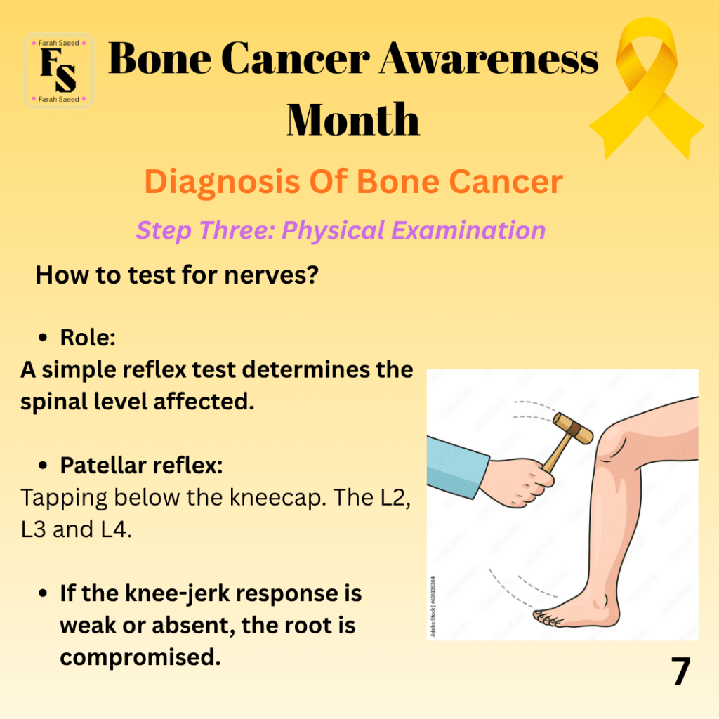
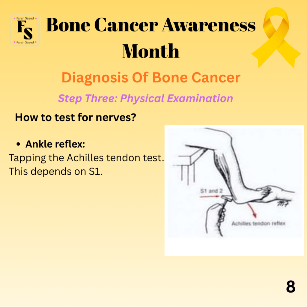
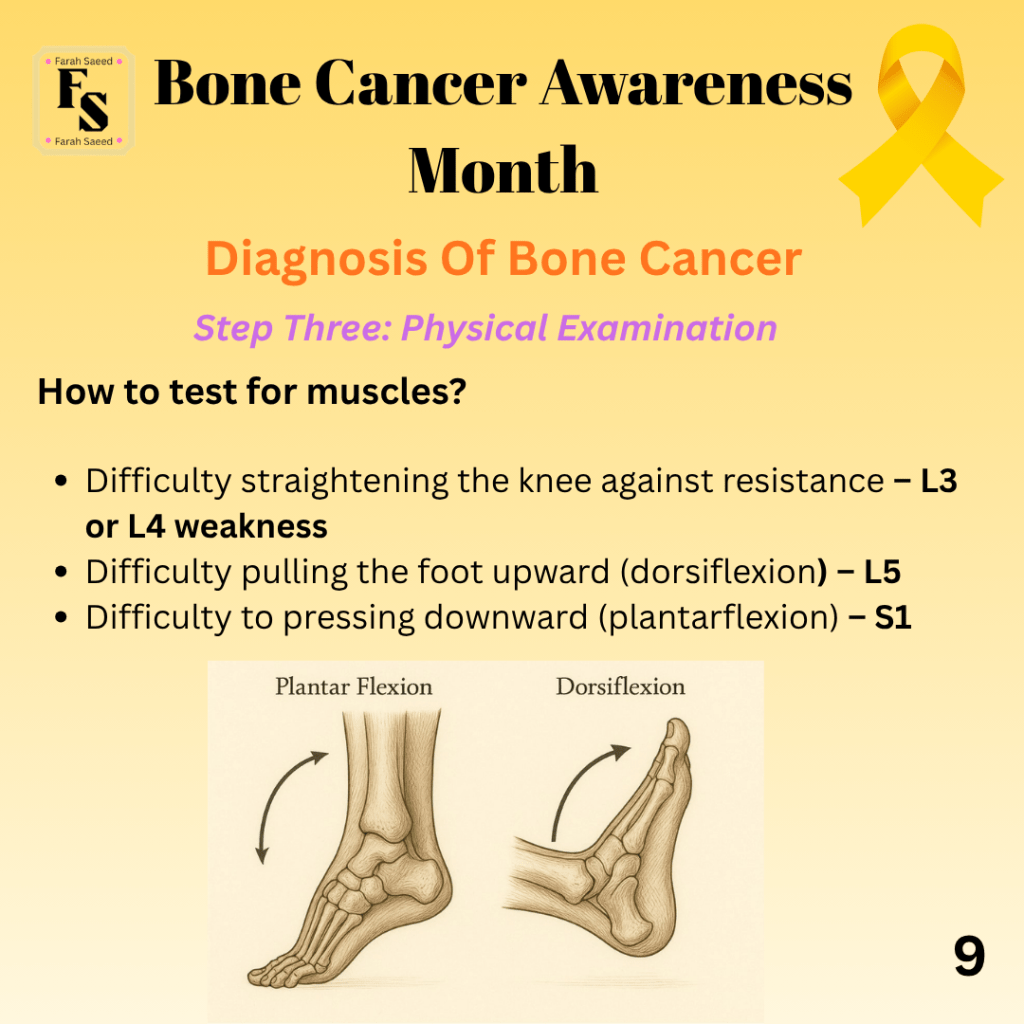
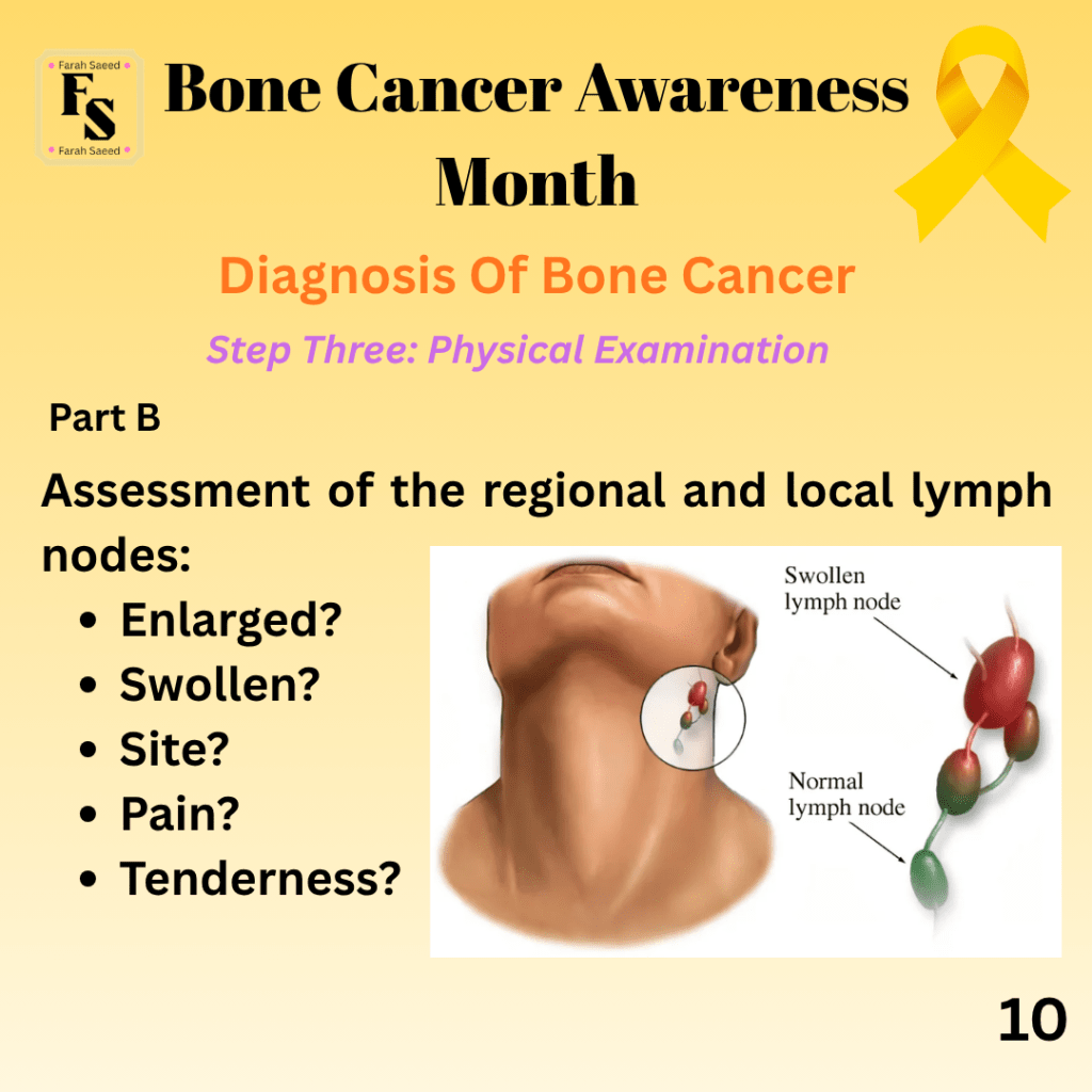
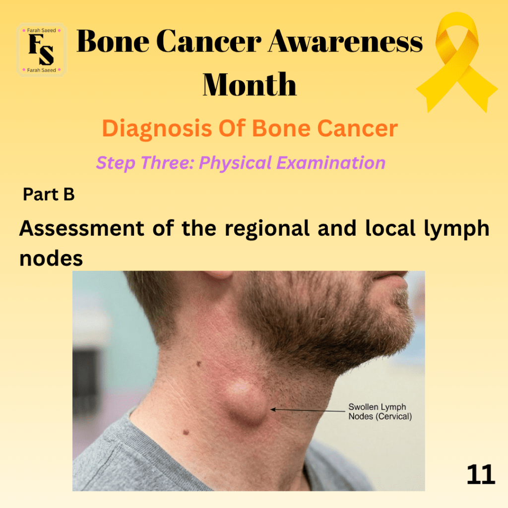
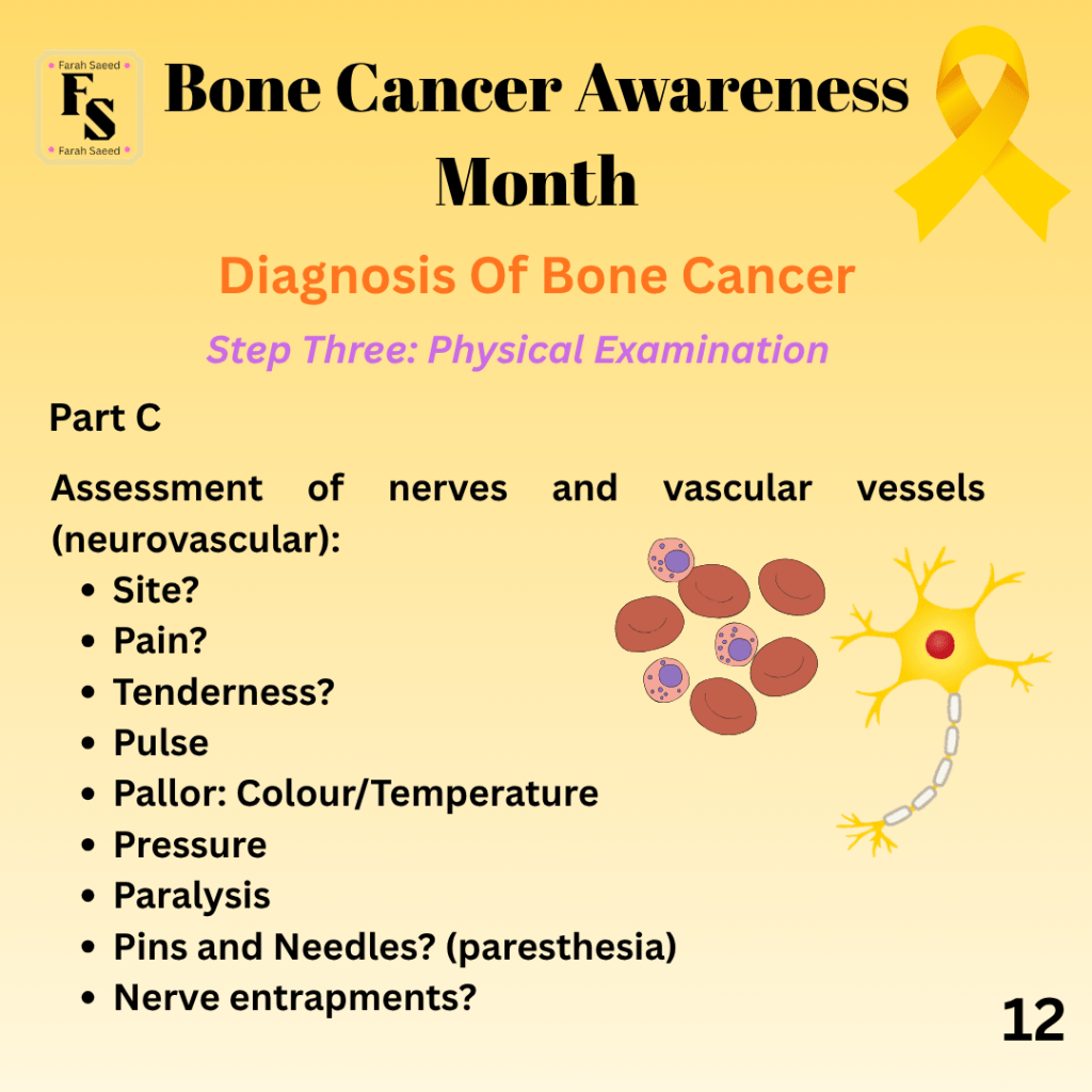
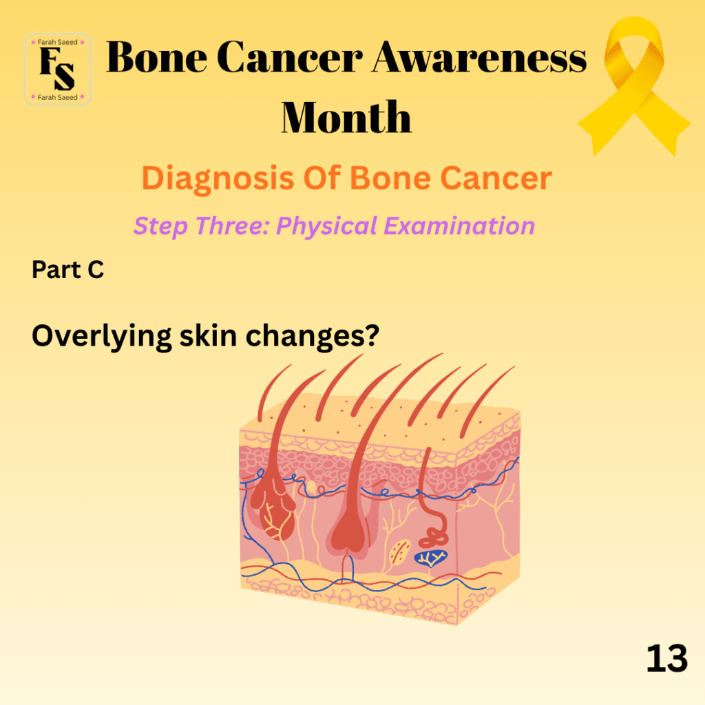
Supporting images for Step 1 and 2
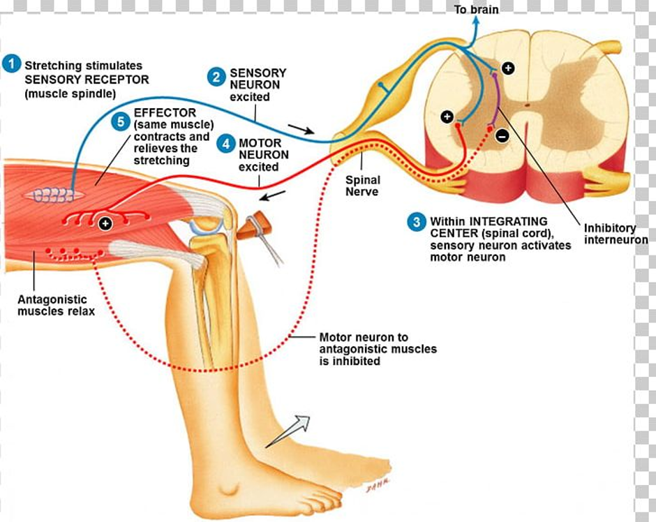
The 6 P’s of Neurovascular Assessment By Dr Picmonic
Further guidance on referrals for adult bone cancers (NHS Scotland)
Further guidance on referrals for paediatric bone cancers (NHS Scotland)
Step Three Blood Test
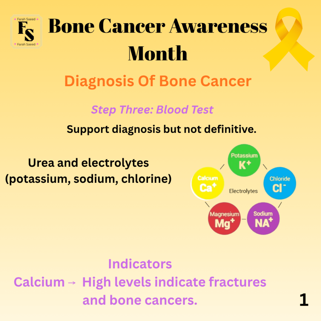
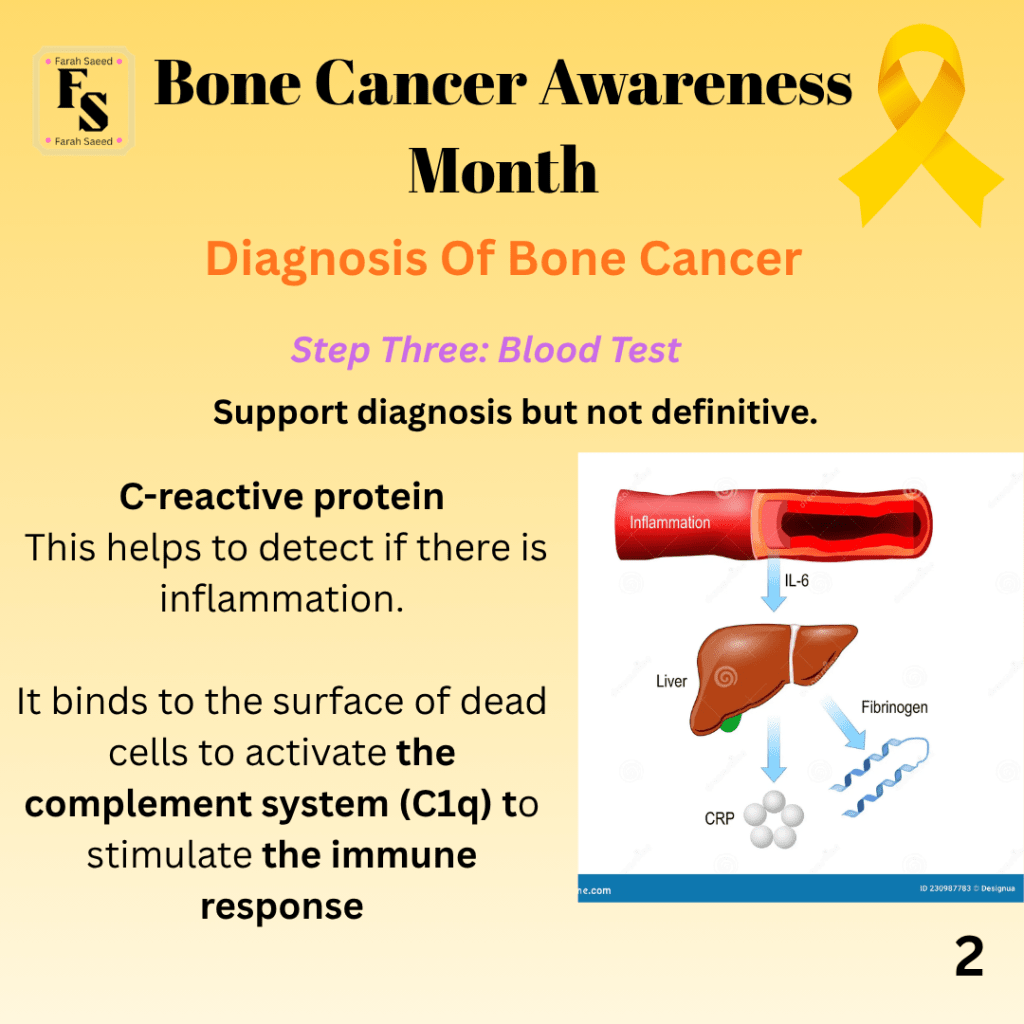
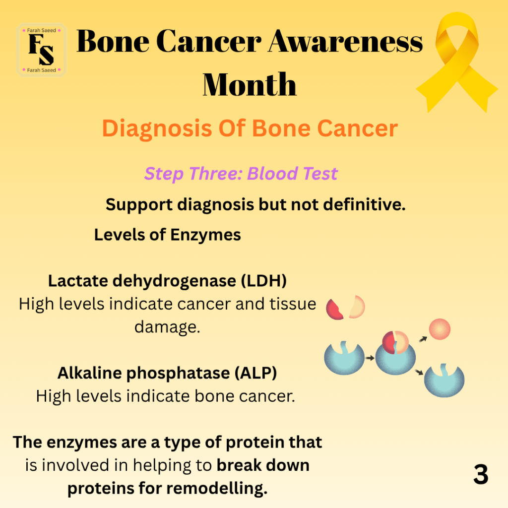
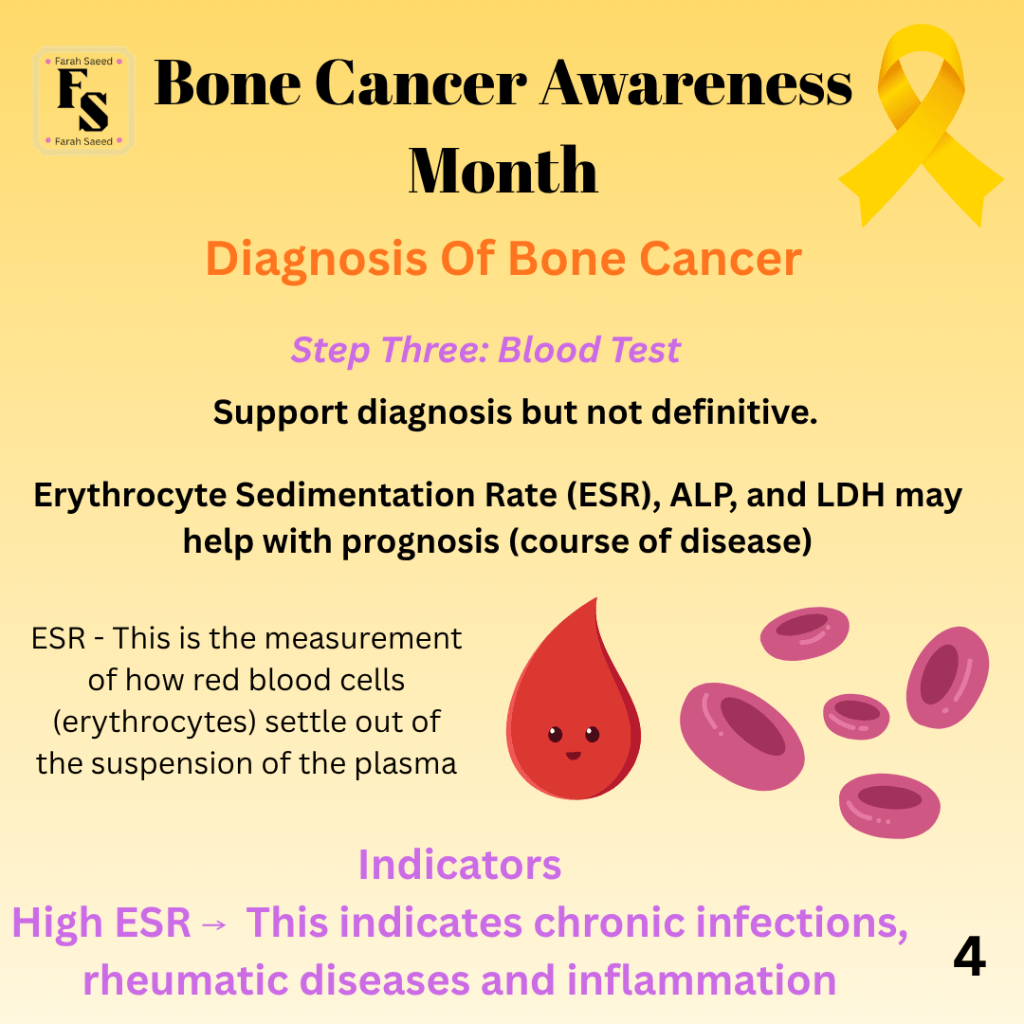

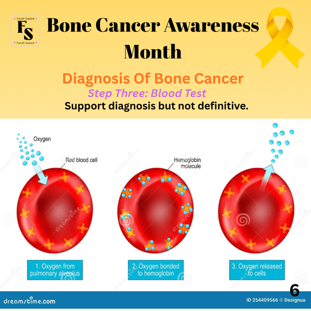
Supporting Images For Step Three
Blood Markers By Bone Cancer Relief Guide
Step Four: Urine Tests

Supporting Images For Step Four
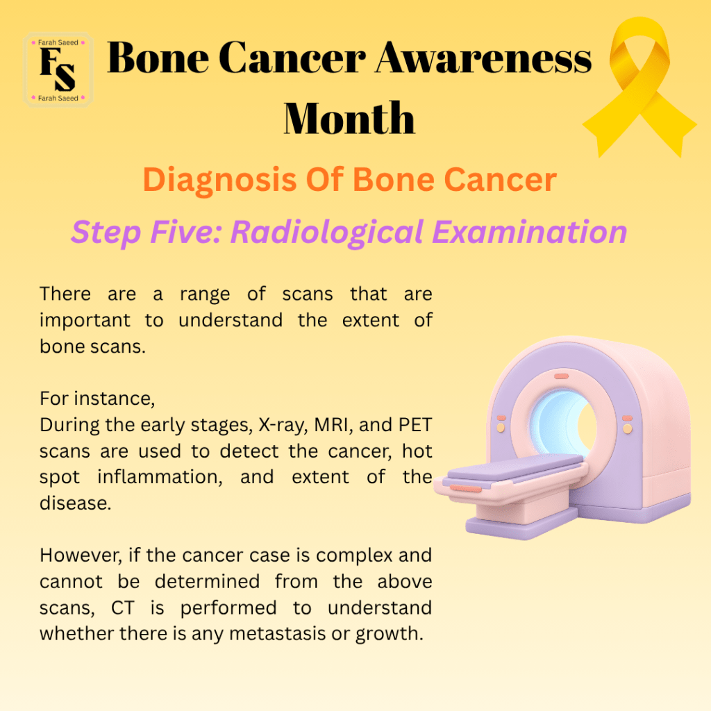
Step Five: Radiological Examination
How Does a Bone Cancer Scan Take Place? By Cancer Research UK
NICE Guidelines 2025: Primary care investigations for bone and soft tissue sarcoma.
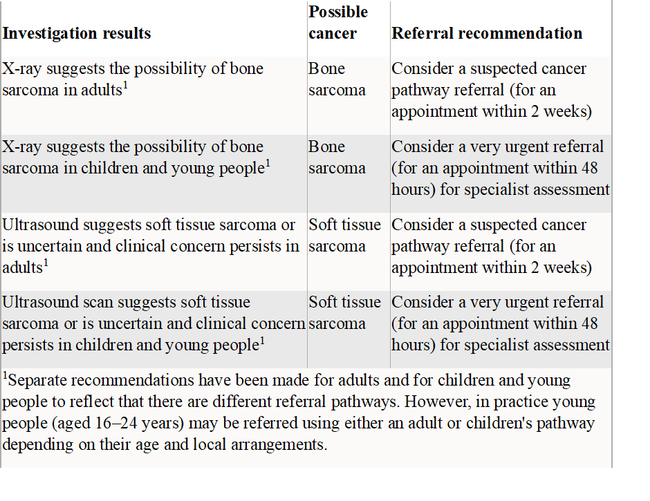
Further guidance on referrals for adult bone cancers (NHS Scotland)
Further guidance on referrals for paediatric bone cancers (NHS Scotland)
How To Assess Imaging
Three key factors to assess:
The extent and presence of hotspots of inflammation
The presence of fractures
The extent of bone disease. Are there any growths?
Guidance on how to read X-rays of bone tumours
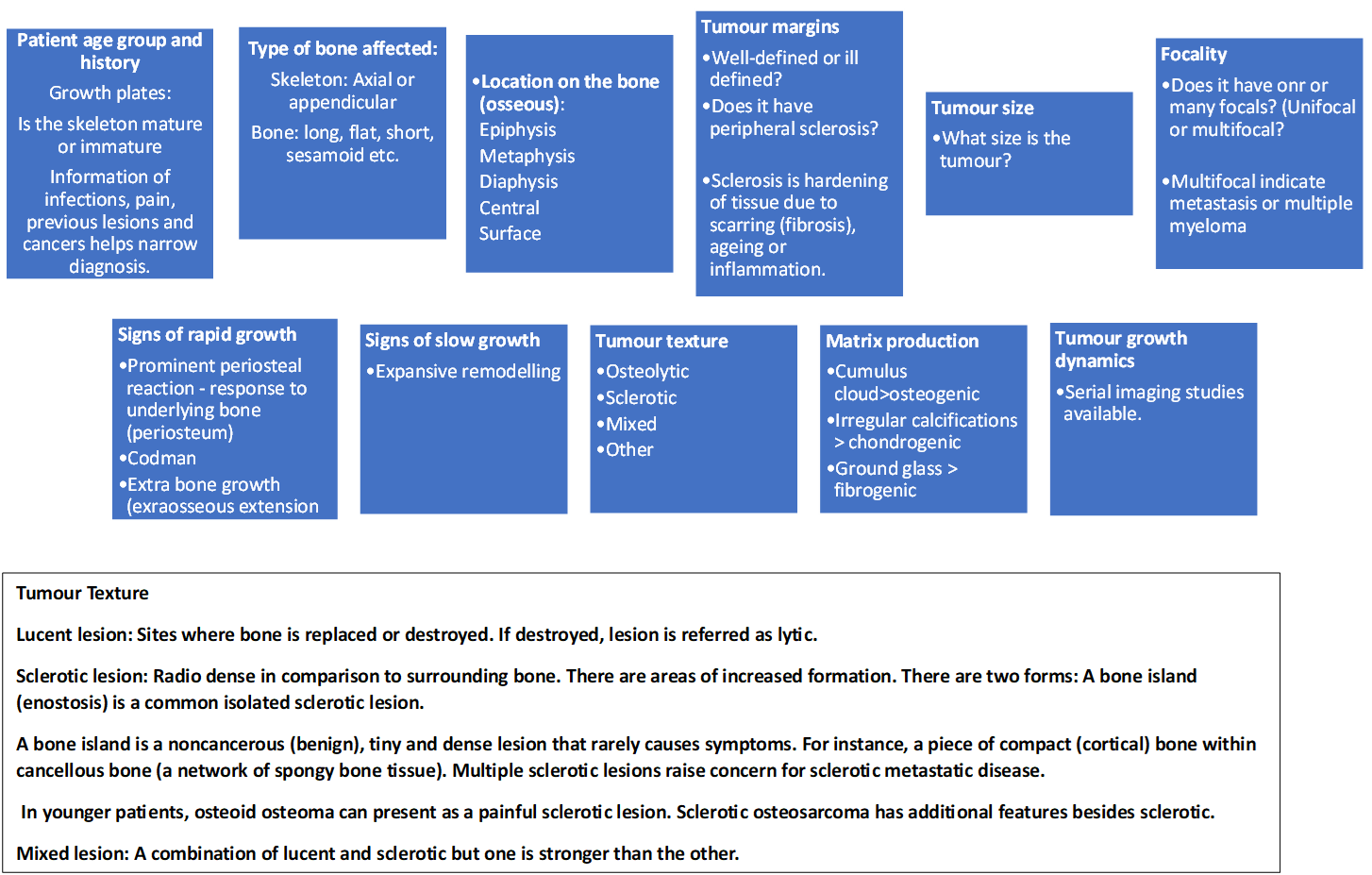
Supporting Image
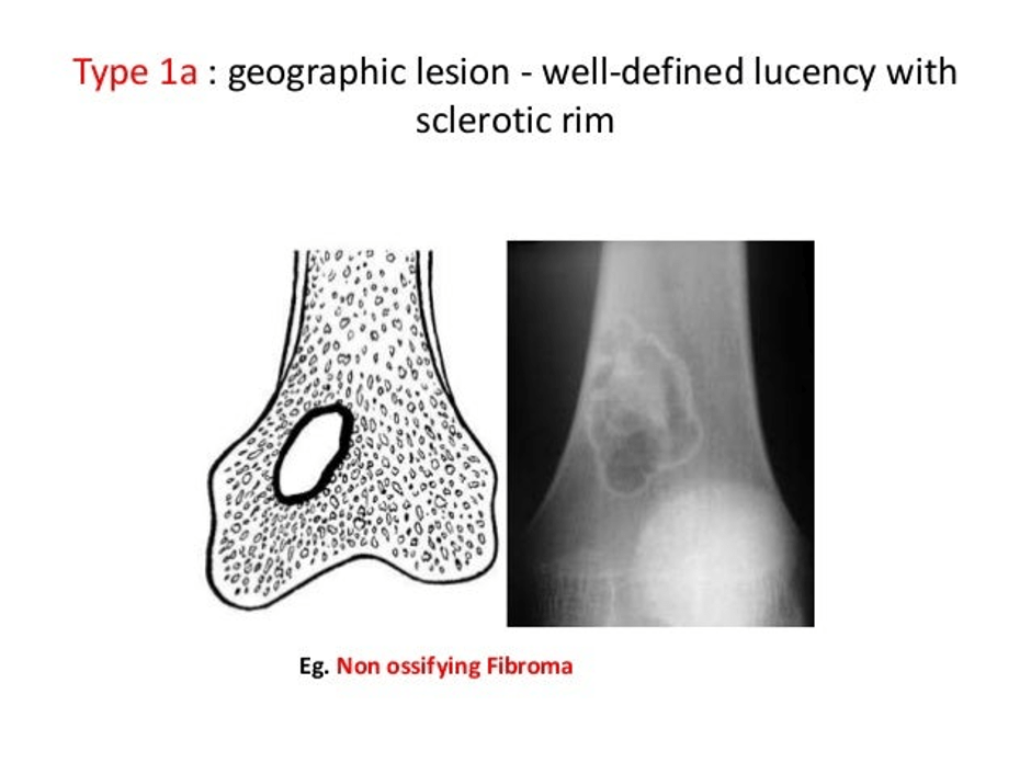
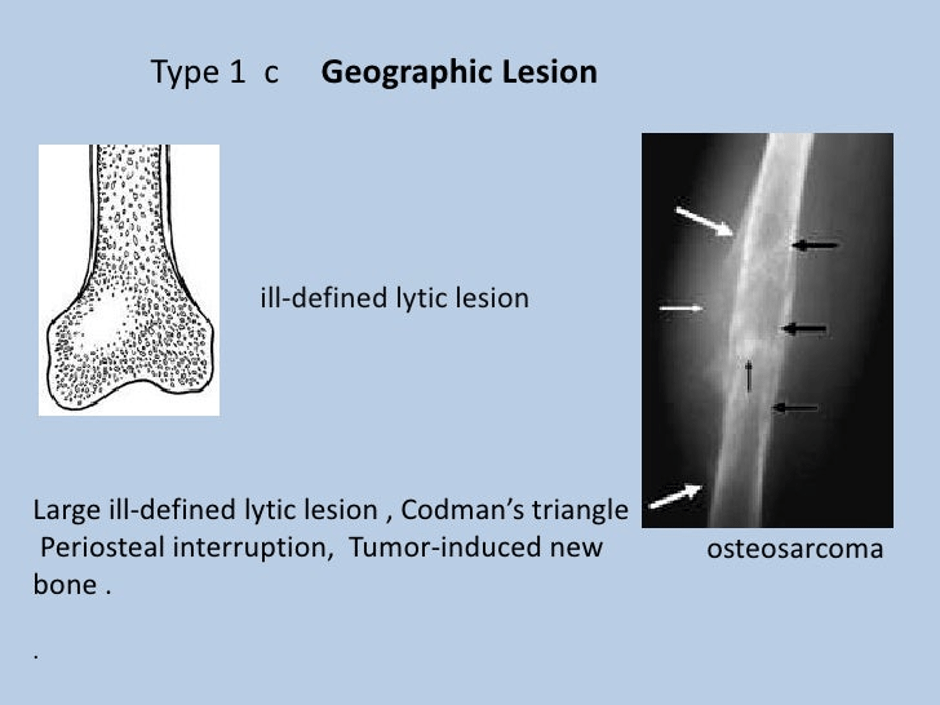
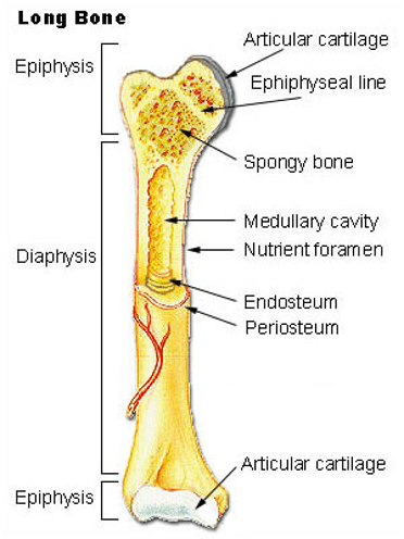
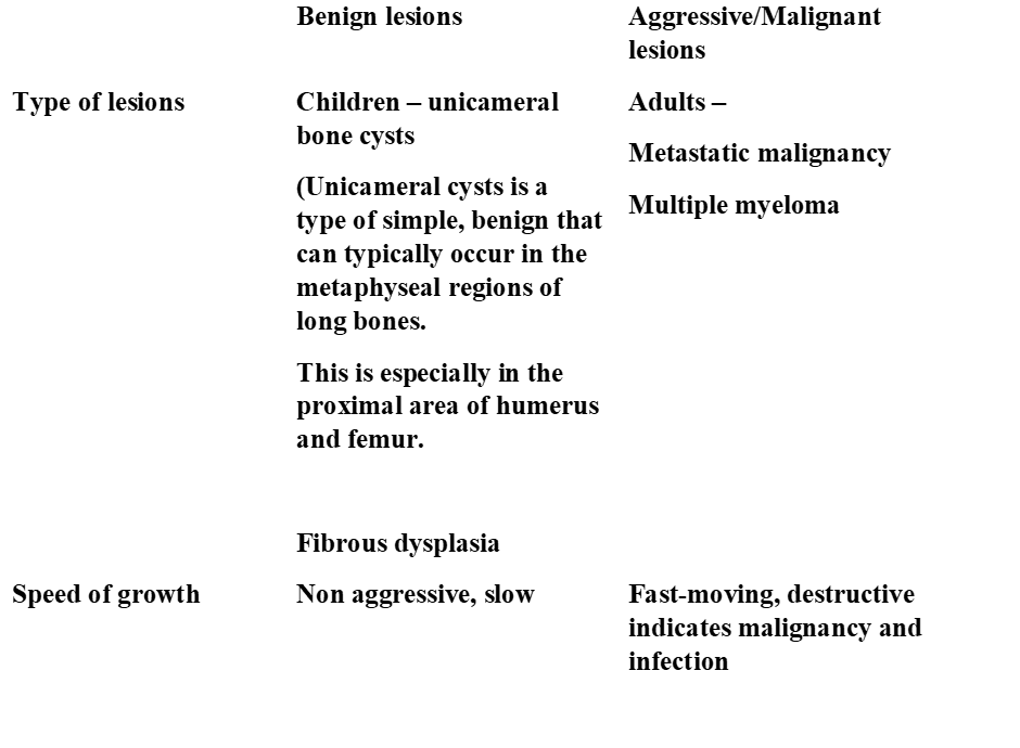
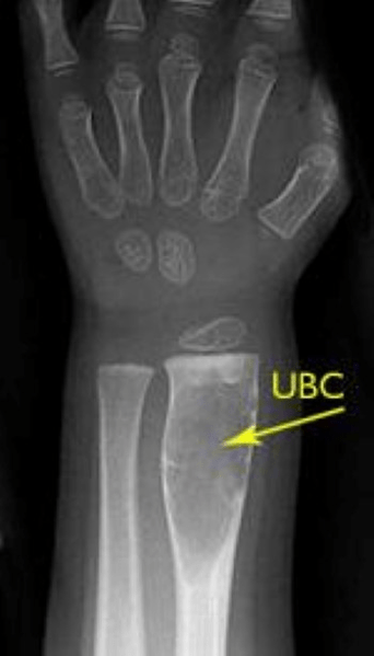
Patients under 40 with suspected primary malignant bone tumours (PMBT).
X-ray
- This is performed as an urgent X-ray of the affected area to determine the following:
- Lytic lesions: joint pain, trauma.
- Benign: non-aggressive lesions, no further intervention
- Aggressive: fast-moving, destructive à malignancy and infection.
- Bone malfunctions: thinning of the bone, loss of the line on the bone, destruction
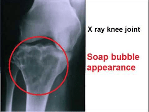
- Moth-eaten appearance: This indicates joint destruction. This is commonly seen in Ewing sarcoma and telangiectatic osteosarcoma.
- New bone formation: Osteoblastic, osteolytic, or a combination of both. This is accompanied by calcification.
- Swelling of the soft tissue
Periosteum features
- Periosteum is the covering of the bone; the outer layer contains lots of blood vessels, and the inner layer contains osteoblasts and fewer blood vessels.
- Sunburst/spiculated: Inflammation of the periosteum (periostitis) and vertical osteoid calcification.
- Onion skinning/lamellated: Periosteal reactions – tumour is lifting partially formed periosteum in Ewing sarcoma.
- Codman triangle: Periosteum lifted off bone and osteoid laid down.
Permeative appearance: Tumour progression through bone. There is an ill-defined zone between tumour and health bony, e.g. Ewing sarcoma.
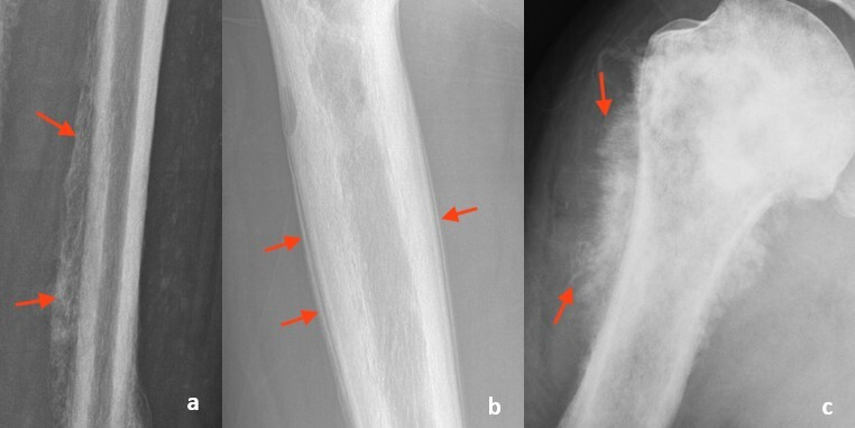
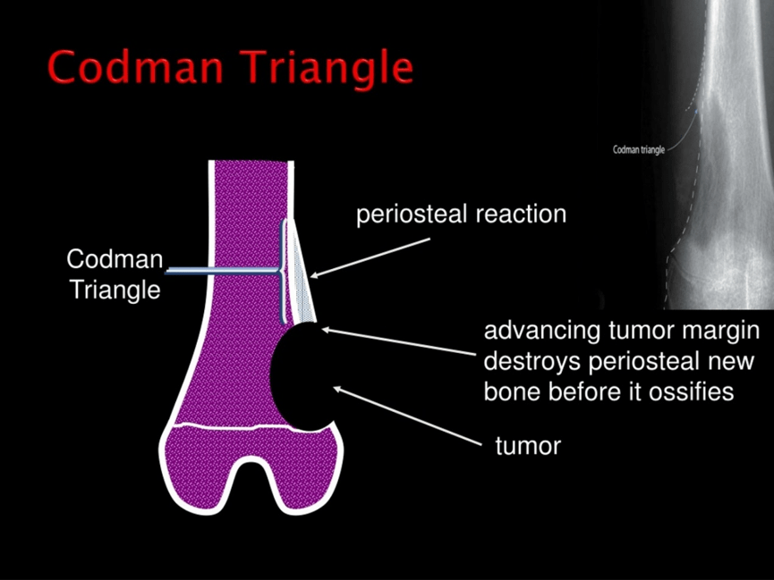
A normal X-ray does not exclude primary malignant bone tumours (PMBT).
An urgent magnetic resonance imaging (MRI) or referral is needed if there is ongoing pain or mass in the area.
Patients above 40 years with suspected primary malignant bone tumours (PMBT).
Metastatic carcinoma in the bone.
Computed Tomography of the chest, abdomen (belly) and pelvis.
Bone scan or MRI to detect the whole skeleton in the body.
If solitary bone lesions are present, they are referred to bone sarcoma to exclude PMBT on urgent cancer pathway.
Other Scans
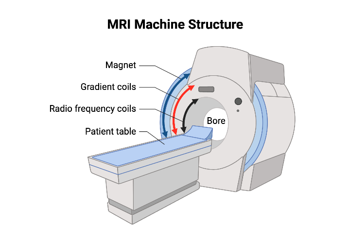
Magnetic Resonance Imaging
This is the gold standard method to assess the extent of local tumour, e.g. bones and adjacent joints affected.
For instance: benign cystic and fibrous lesions from aggressive lesions.
The extent to which it has affected blood vessels, nerves and soft tissue.
MRI with intravenous contrast can provide images of bone lesions and around them, characterise and assess extent.
Dynamic contrast-enhanced MRI – this helps to identify any cancerous areas that are high grade. This is commonly guided with biopsy. An example of cancer is chondrosarcoma.
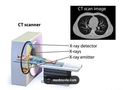
Computed Tomography
This is commonly used if the results are unclear or MRI is contraindicated.
Microcalcification
Periosteal bone formation
Cortical destruction.
Other Scans
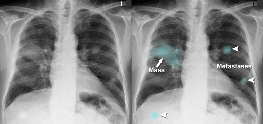
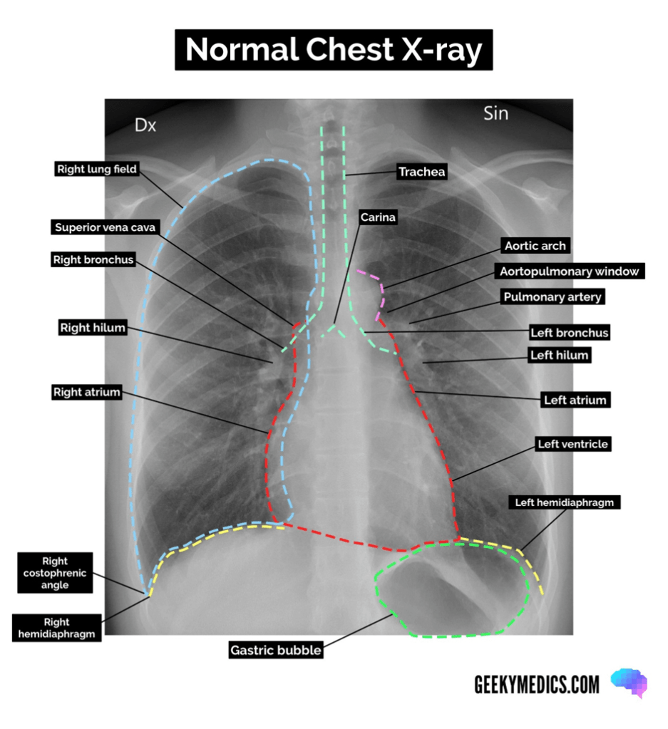
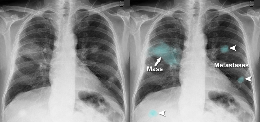
Chest X-ray
It helps detect metastasis in the lungs.
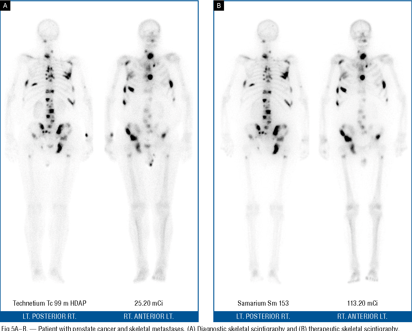
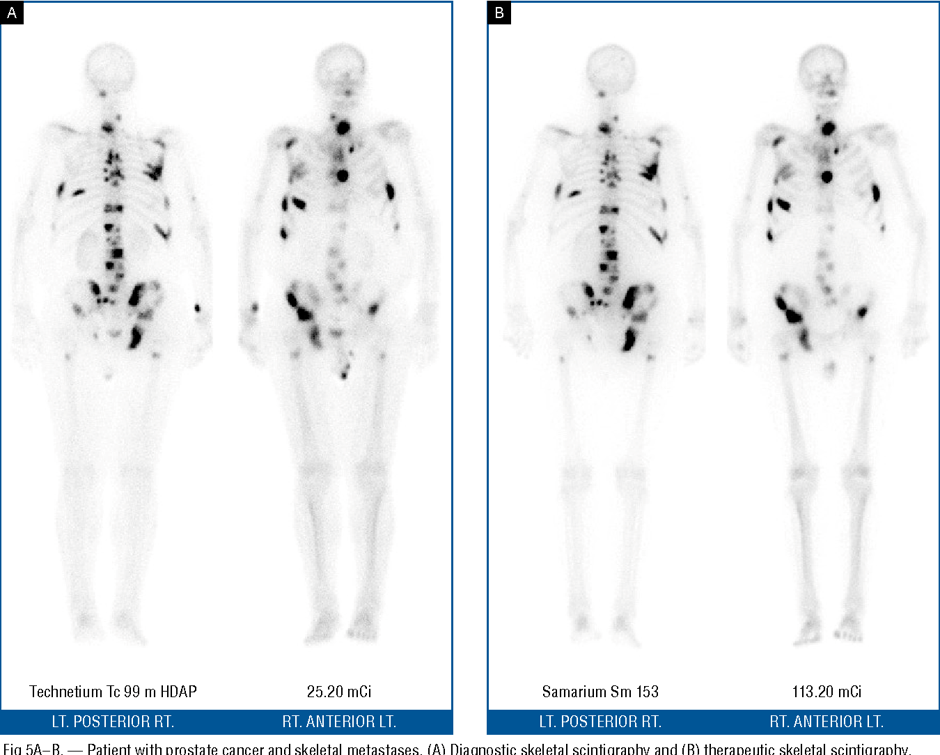
Bone scintigraphy – spine
Radiologically active agent: technetium-99m
To detect osteoblastic activity
To determine if there is malignancy and/or metastasis.
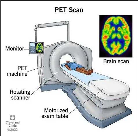
Positron Emission Tomography
It measures the metabolic rate of tumour cells by measuring how much radiolabeled F-18 fluoro-deoxy-glucose is taken up.
It is used for the initial staging of primary bone tumours.
It is also used with a CT scan in the follow-up.
Supporting Images For Step Five: Radiology
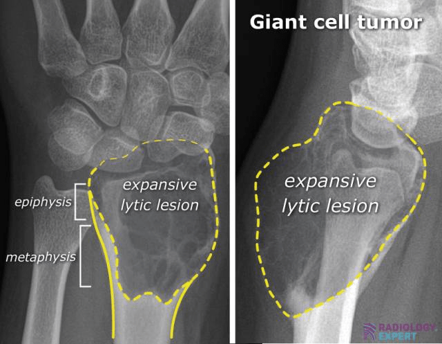
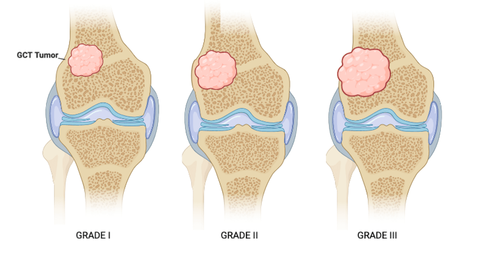
Example One: Giant Cell Tumour
- X-ray and CT = assess bone component in tumour
- MRI scan – evaluate intramedullary and soft tissue expansion.
- Traits: Donut sign, strong radio-nuclear uptake in the periphery due to hyperaemia and photopenia in the centre in response to necrosis.
- Hyperaemia – excessive blood volume; there are two forms:
- Active hyperaemia – the small blood vessels (arterioles) are relaxed
- Passive hyperaemia – there is an obstruction in the blood vessel.
- Photopenia – low or absence of radioactive tracers
Recommended Reading
References
Alexiev, B. (2021) Bone & joints Osteoclastic giant cell rich tumors Giant cell tumor of bone, NOS, Available at: https://www.pathologyoutlines.com/topic/bonegiantcelltumor.html (4th April 2026)
Biermann, J.S., Hirbe, A., Ahlawat, S., Bernthal, N.M., Binitie, O., Boles, S., Brigman, B., Callan, A.K., Cipriano, C., Cranmer, L.D., Davis, J., Donnelly, E., Ferguson, M., Graham, A., Groundland, J., Hess, M., Hiniker, S.M., Hoover-Regan, M.L., Hornick, J.L. and Jonard, B. (2025). Bone Cancer, Version 2.2025, NCCN Clinical Practice Guidelines in Oncology. Journal of the National Comprehensive Cancer Network, [online] 23(4). DOI: HTTPs://doi.org/10.6004/jnccn.2025.0017.
Cleveland Clinic (2022) Bone Island (Enostosis). Available at: https://my.clevelandclinic.org/health/diseases/22360-bone-island-enostosis (Accessed: 2nd June 2026)
Cleveland Clinic (2025) Chondrosarcoma. Available at: https://my.clevelandclinic.org/health/diseases/22112-chondrosarcoma (Accessed: 2nd June 2026)
Gerrand, C., Amary, F., Anwar, H.A., Brennan, B., Dileo, P., Kalkat, M.S., McCabe, M.G., McCullough, A.L., Parry, M.C., Patel, A., Seddon, B.M., Sherriff, J.M., Tirabosco, R. and Strauss, S.J. (2024). UK guidelines for the management of bone sarcomas. British Journal of Cancer. DOI: HTTPs://doi.org/10.1038/s41416-024-02868-4.
MacKay, B.J., McCormack, R.A., Blank, A.T., Bettiol, P., Cox, C., Brindley, G. and Rapp, T.B. (2021). Diagnosis and management of primary malignant tumors in the upper extremity. Orthopedic Reviews, 12(4). doi: https://doi.org/10.4081/or.2020.8345.
Montilla-Soler, J.L. and Makanji, R. (2017). Skeletal Scintigraphy. Cancer Control: Journal of the Moffitt Cancer Center, [online] 24(2), pp.137–146. doi:10.1177/107327481702400206.
National Institute for Health Care and Excellence (2025a). What symptoms are suggestive of bone or soft tissue sarcoma? Available at: https://cks.nice.org.uk/topics/bone-soft-tissue-sarcoma-recognition-referral/diagnosis/ (Accessed: 30th March 2026)
National Institute for Health and Care Excellence (2025b). What symptoms are suggestive of bone or soft tissue sarcoma? Available at: https://cks.nice.org.uk/topics/bone-soft-tissue-sarcoma-recognition-referral/diagnosis/symptoms-investigations/ (Accessed: 30th March 2026)
Oliveira, I., Chavda, A., Rajakulasingam, R. and Saifuddin, A. (2020). Chondral tumours: discrepancy rate between needle biopsy and surgical histology. Skeletal Radiology, 49(7), pp.1115–1125. doi:https://doi.org/10.1007/s00256-020-03406-y.
Pullan, J. and Lotfollahzadeh, S. (2024). Primary Bone Cancer. Available at: https://www.ncbi.nlm.nih.gov/books/NBK560830/ (Accessed: 9th April 2026)
Taylor, H.H, Huffman, H.S., Parker, K. and Morris, R. (2024) Top 10 Facts to Know about Bone Lesions Identified on Radiographs. Available at: https://jmsma.scholasticahq.com/article/124334-top-10-facts-to-know-about-bone-lesions-identified-on-radiographs (Accessed: 2nd June 2026)
 The Farah Saeed Project