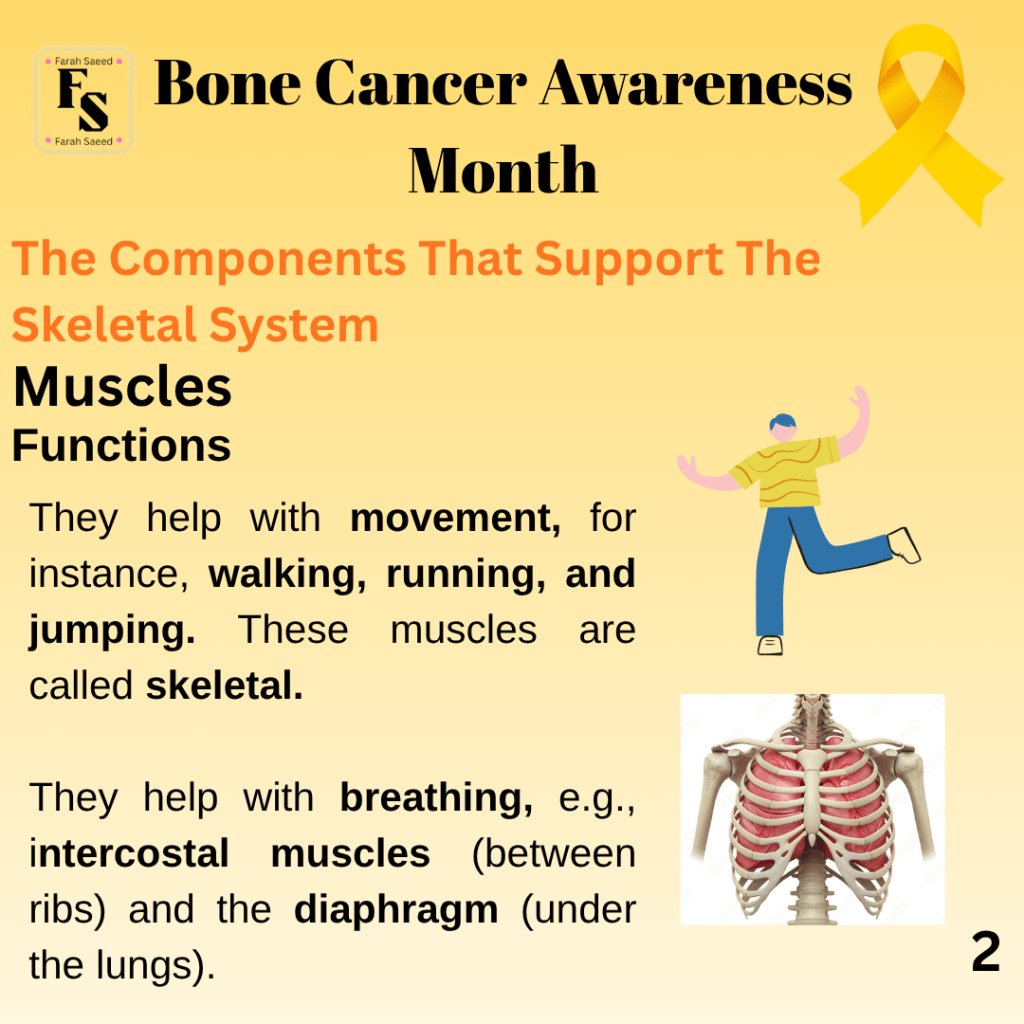
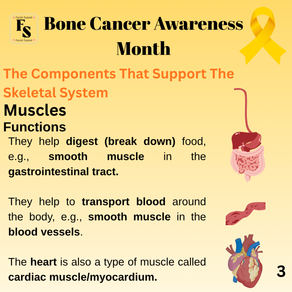
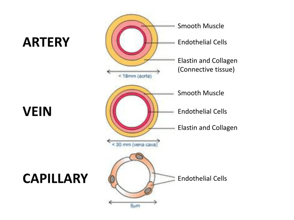
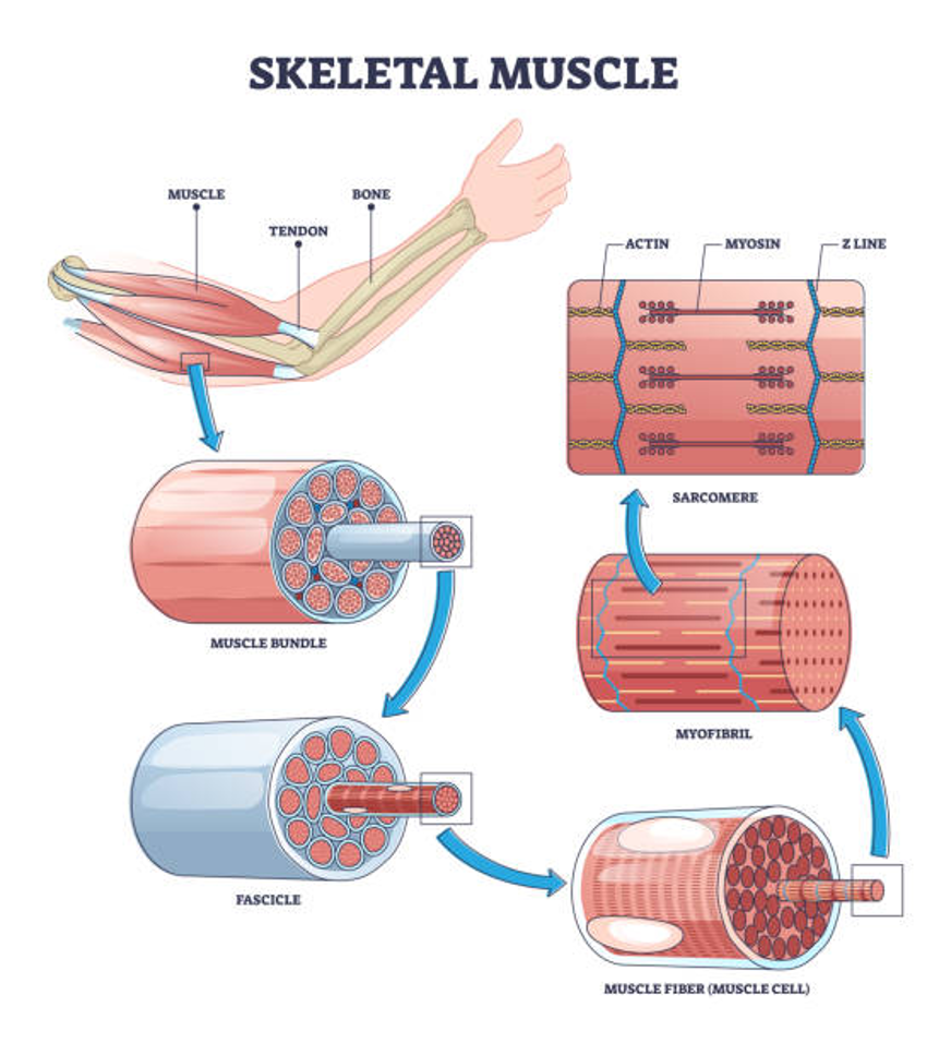
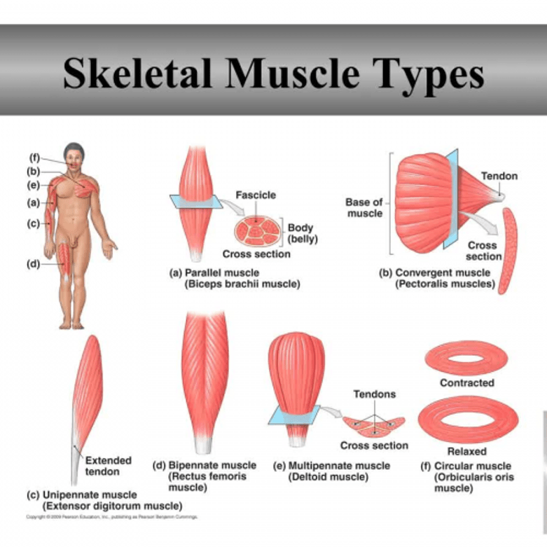
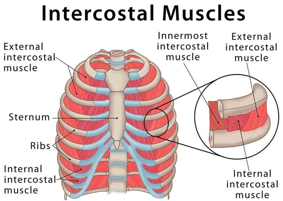
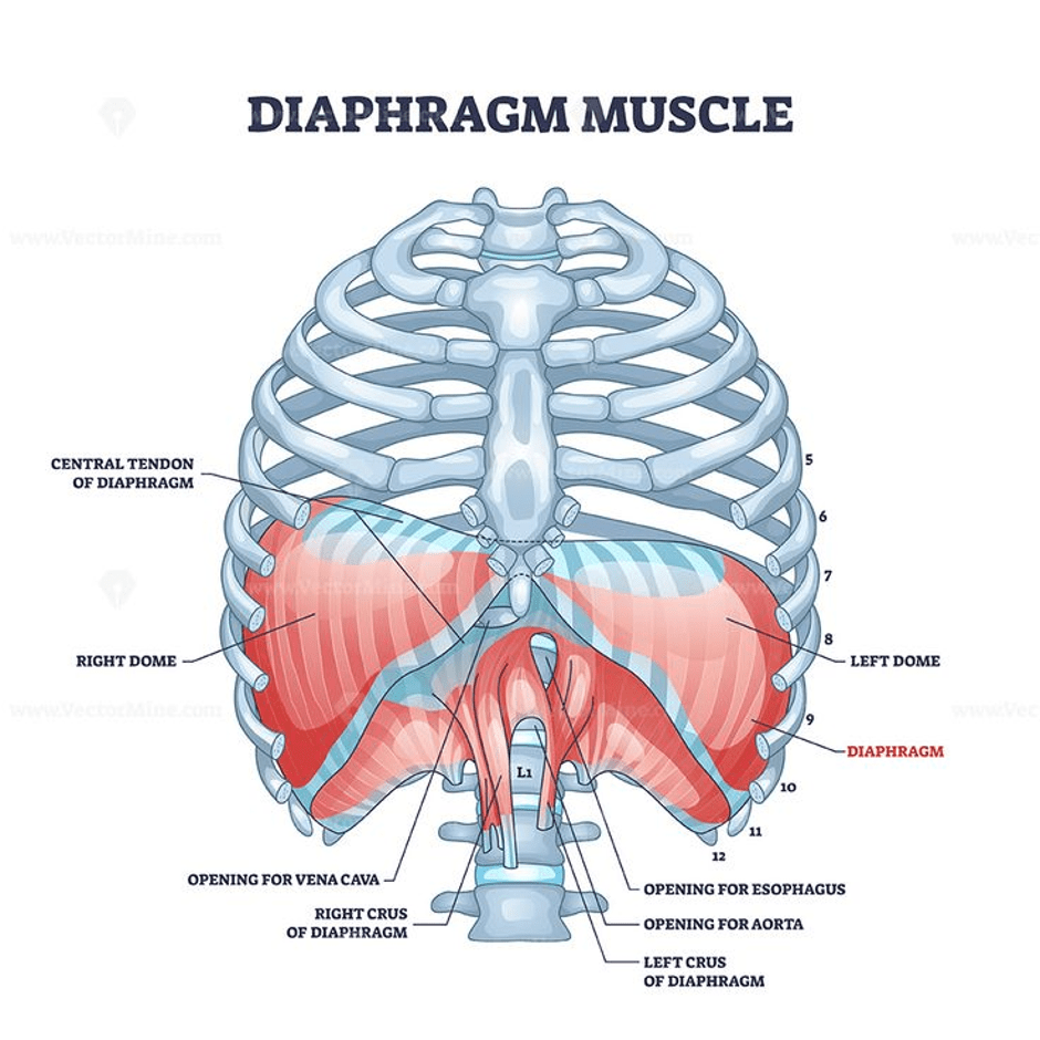
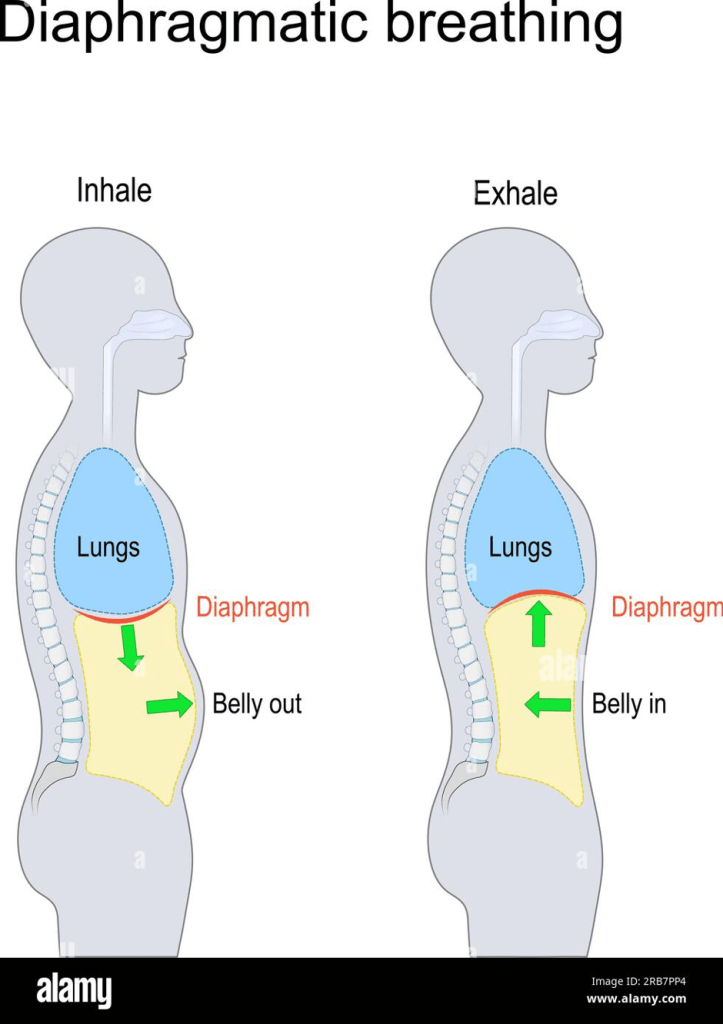
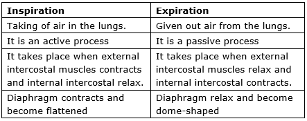
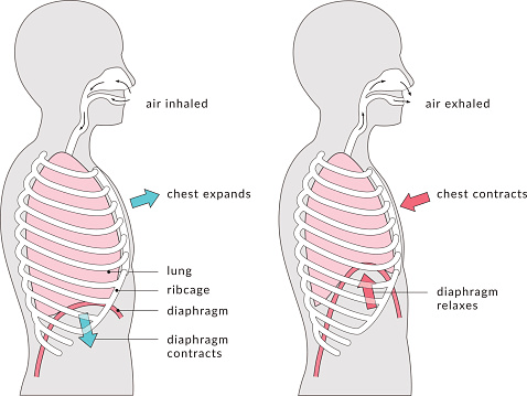
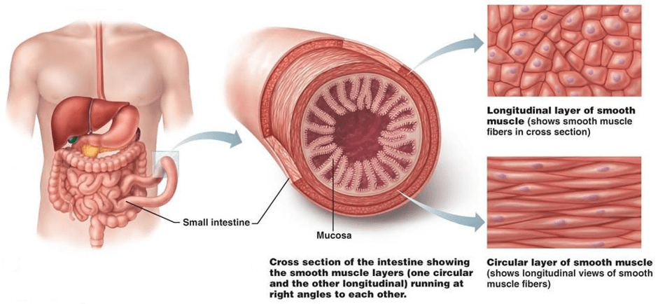
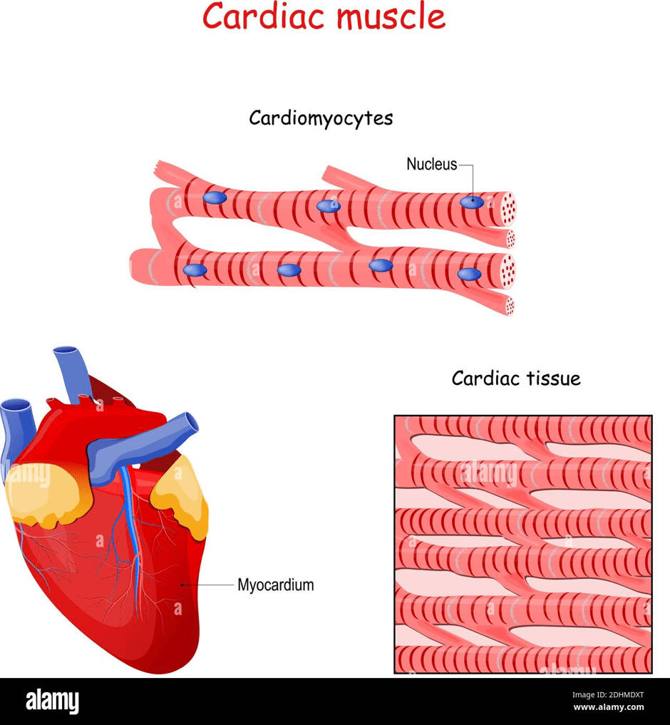
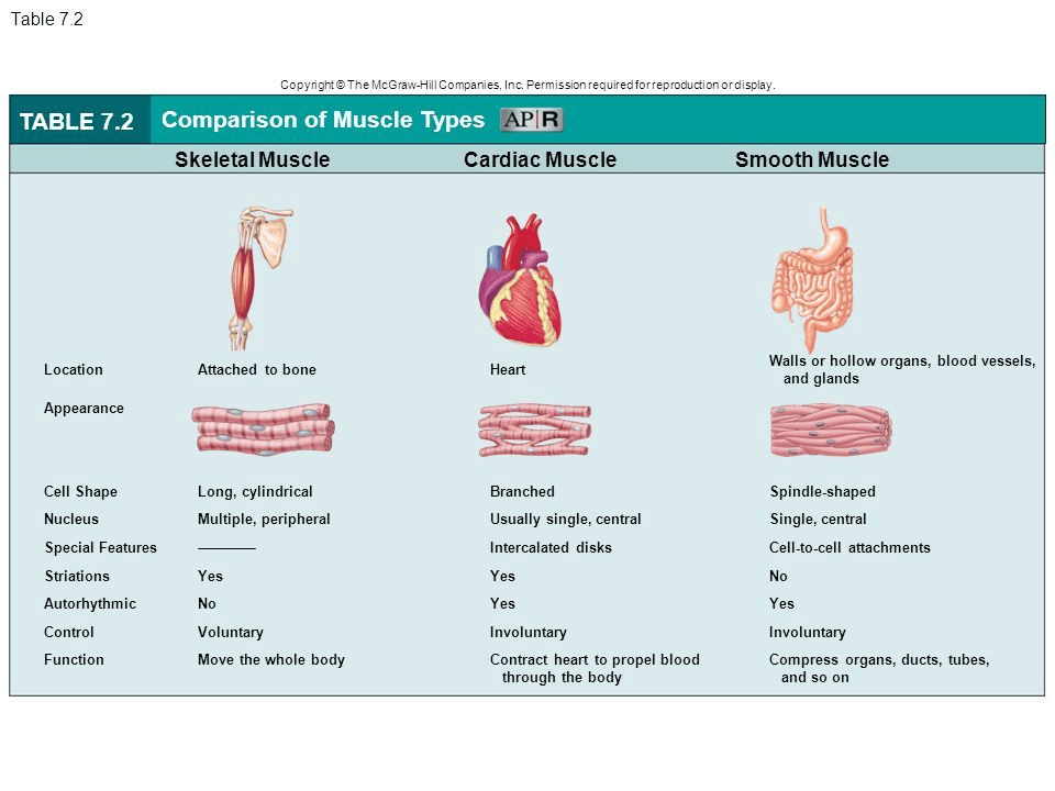
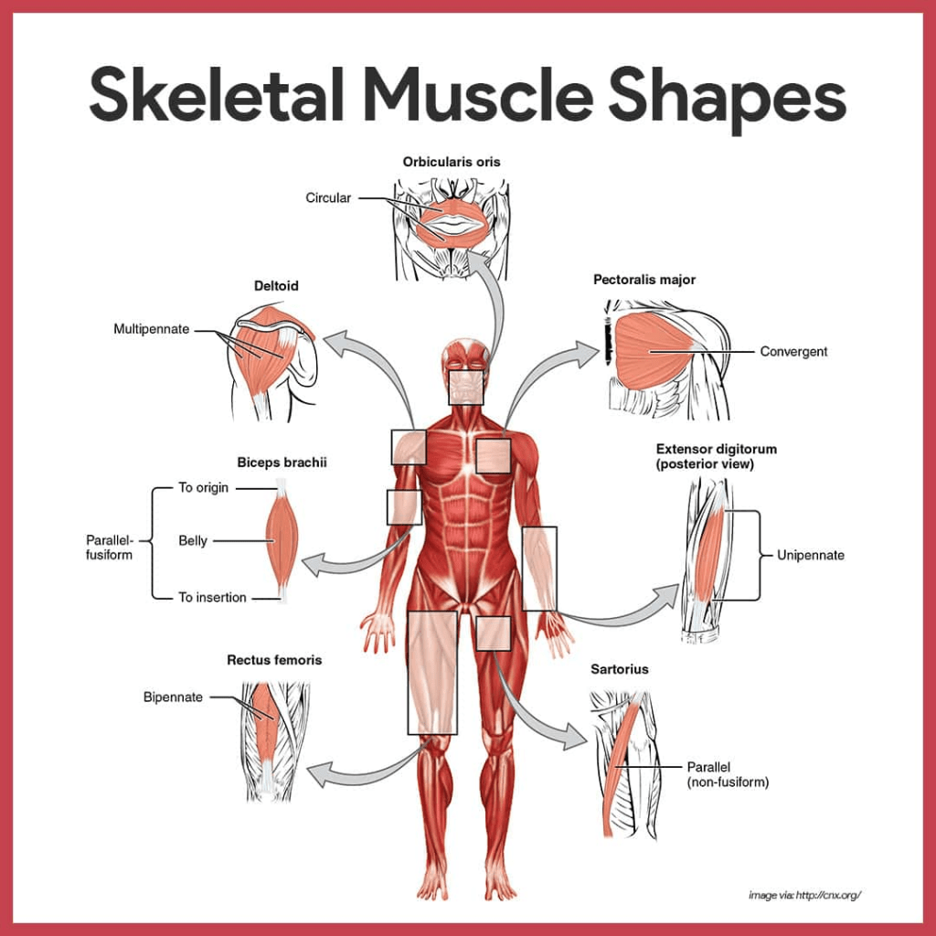
The structure of the three main types of blood vessels (transverse view)The structure and components found in the skeletal muscleTypes of skeletal musclesThe two types of intercostal muscles found in the ribcage protect the lungs whilst breathing.The dome-shaped muscle called the diaphragm is found under the lungs.The movements of the diaphragm when breathing in (inspiration) and breathing out (expiration. The diaphragm flattens when breathing in but returns to its dome-shaped position when breathing out.A table presenting what happens to parts of the breathing system during ventilation (breathing process)Another illustration that presents what happens to the muscles of the respiratory systemA cross-section of the small intestine of the gastrointestinal tract showing where the smooth muscle is. There are two types: longitudinal and circular muscle. They can also be found in other parts of the gastrointestinal tract, such as the food pipe (oesophagus) and large intestine.The structure of the myocardium (heart muscle). It is made of cardiac tissue, which contains cardiac cells (cardiomyocytes).An overview of the different types of muscles
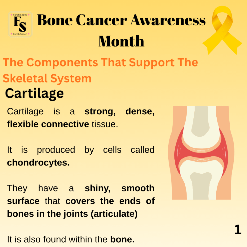
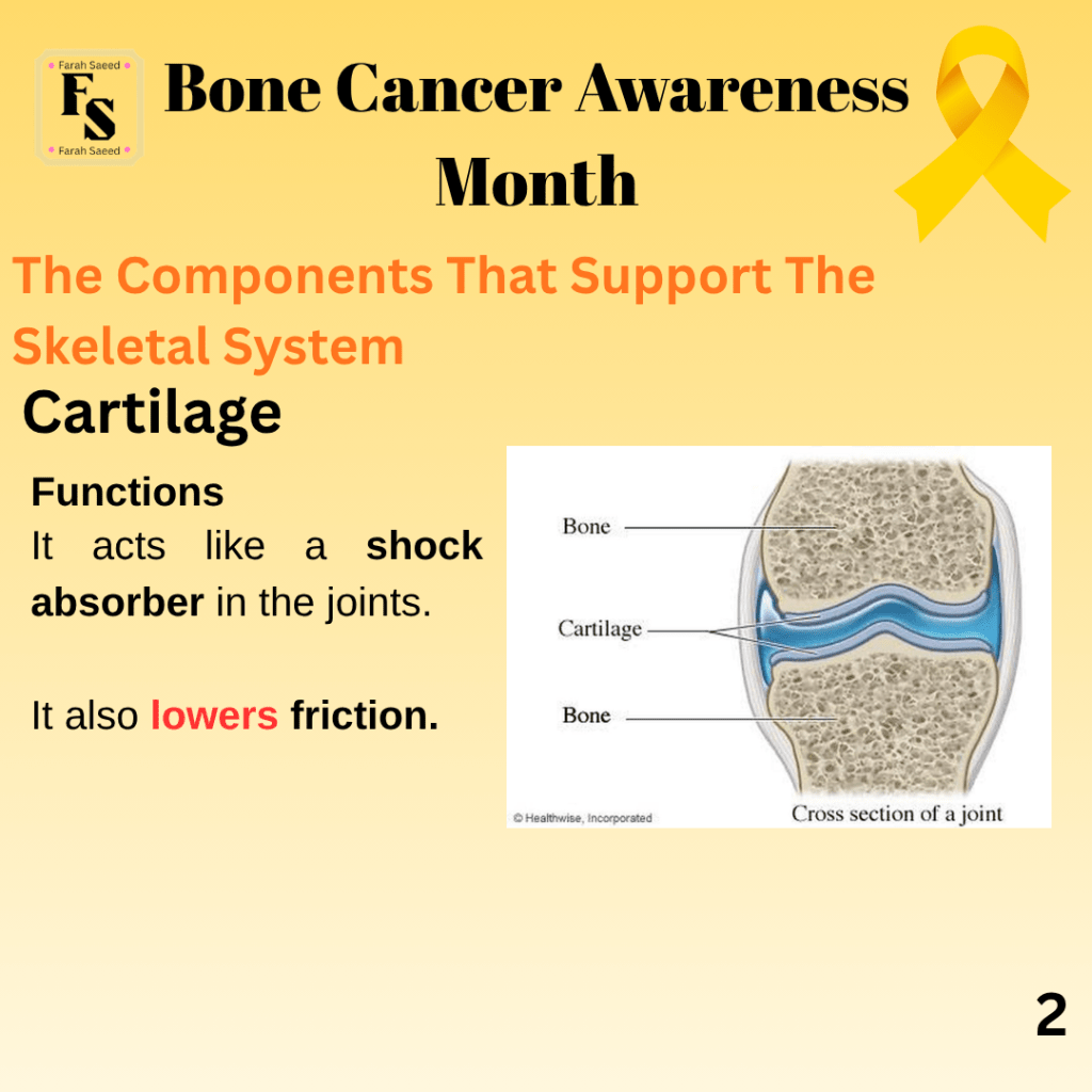
Cartilage
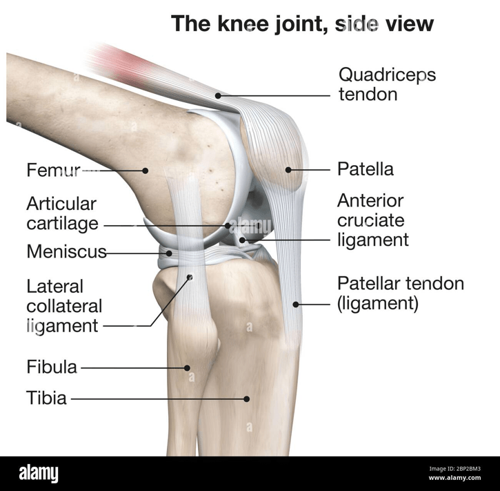
An example of the cartilage present in the knee between the two lower limbs (femur and tibia)
Types Of Cartilage
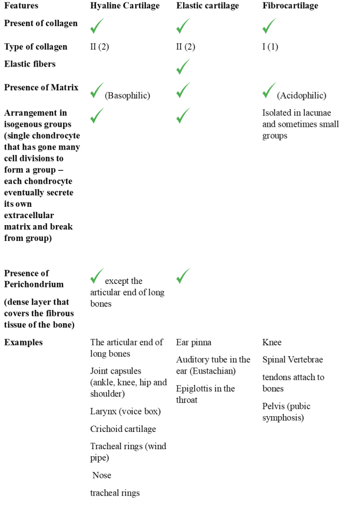
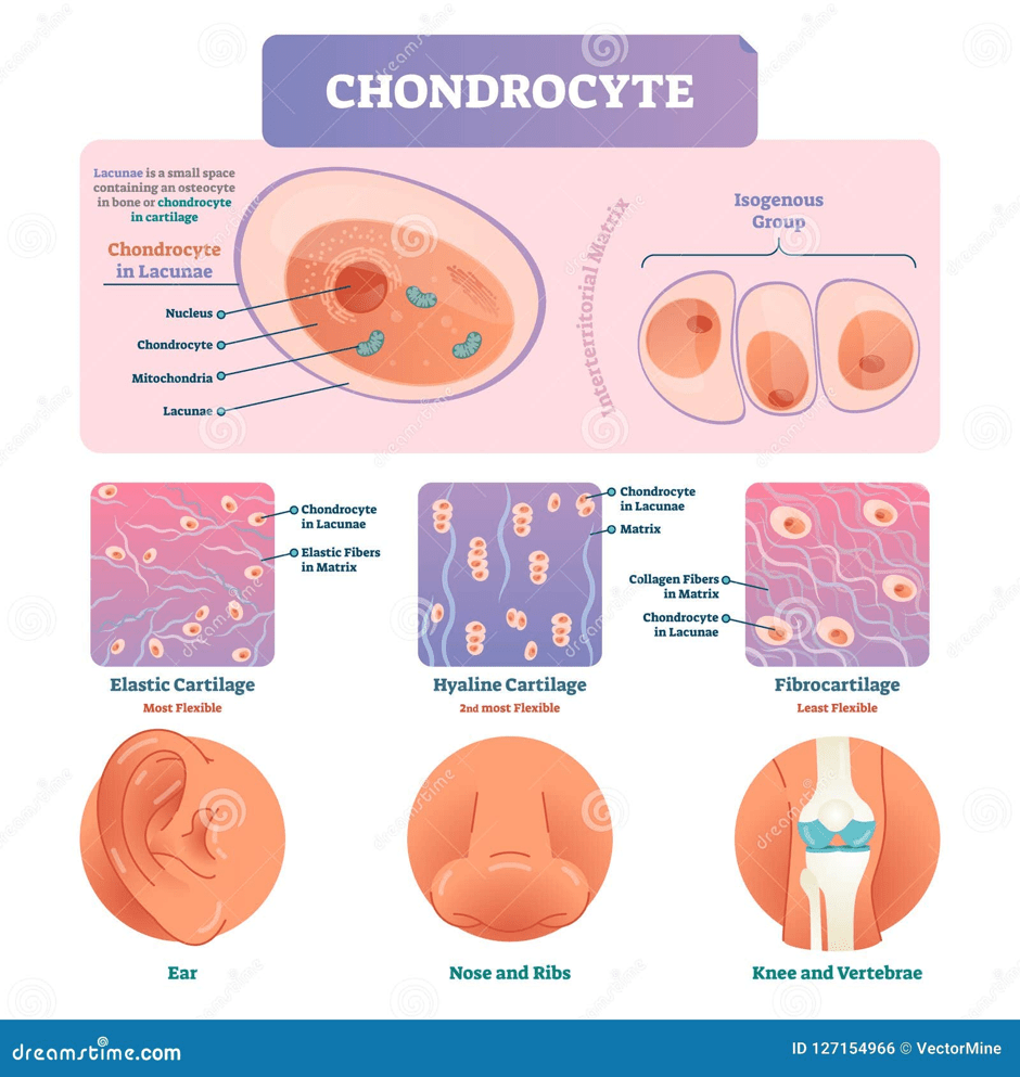
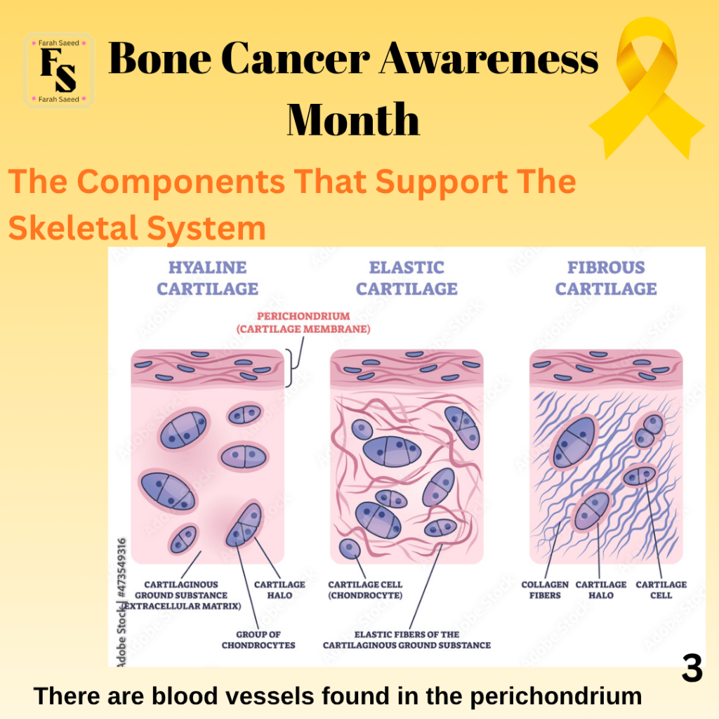
There are three main types of cartilage as presented in the table below:
Supporting Image Resources
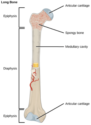
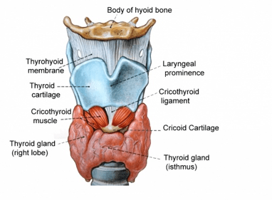
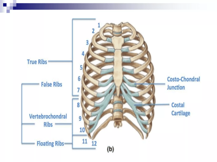
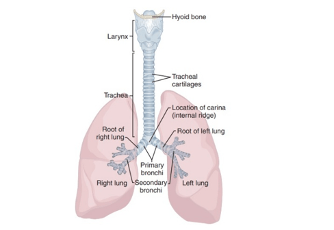
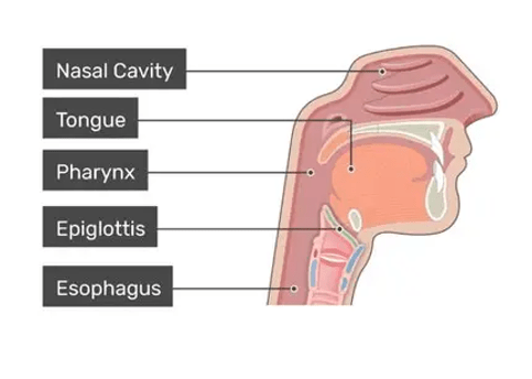
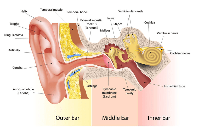
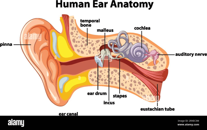
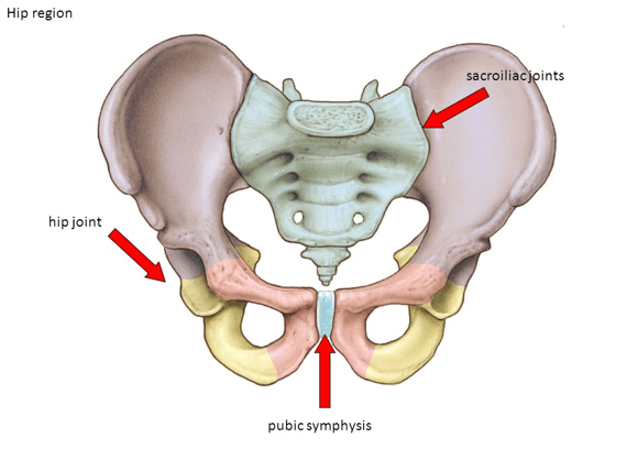
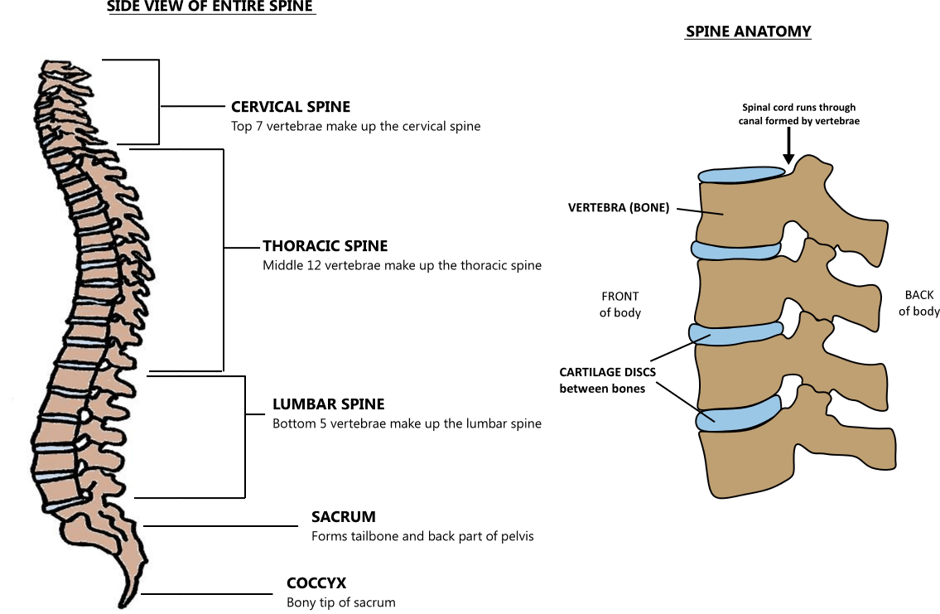
The cellular characteristics of each type of cartilageThe articular cartilage is present at the end of the long bonesThe cricoid cartilage is present near the thyroid gland in the neckThe costal cartilage present in the ribcageThe cartilage present in the windpipe (trachea)The structure of the nasal cavity (nose) and nearby structuresThe cartilage present in the outer earThe structure of the earThe pelvis, where there is cartilage between the jointsThe presence of supporting cartilage between each vertebra of the vertebral column/spine.
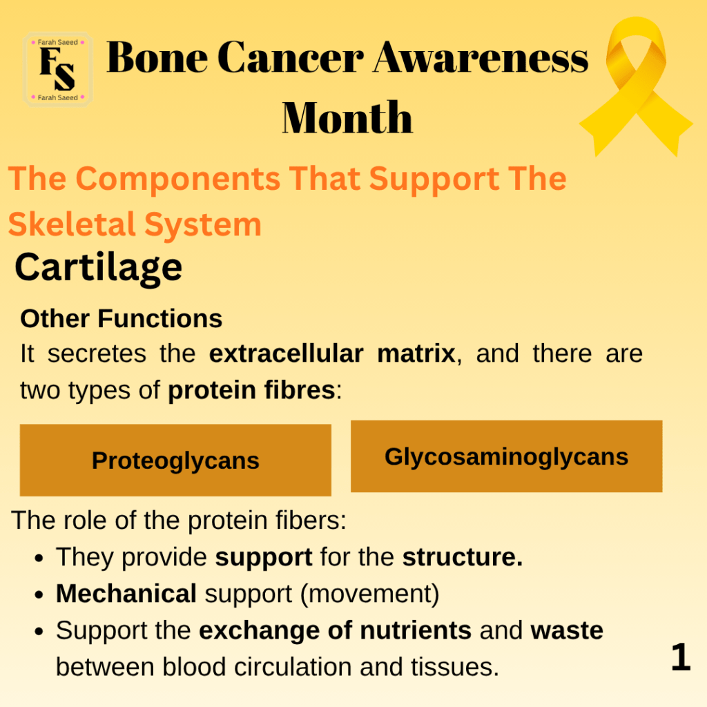
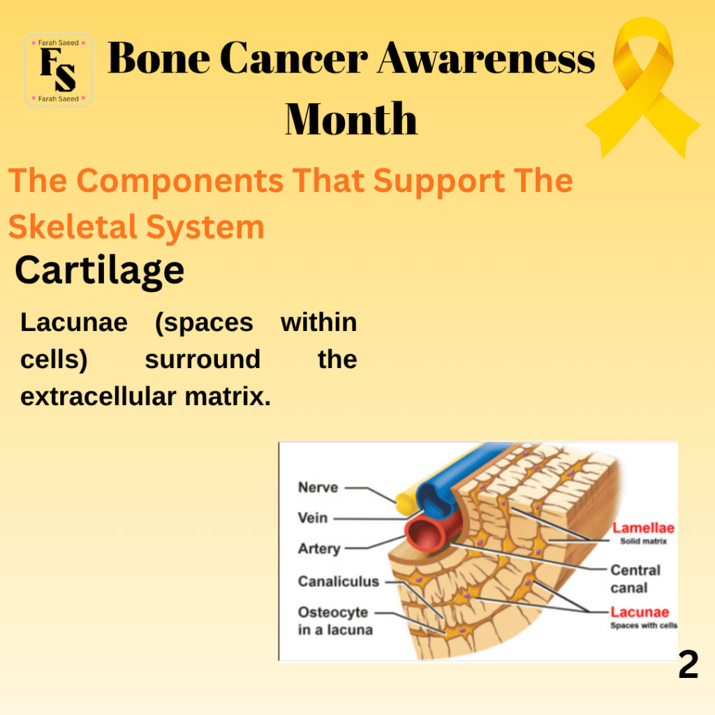
Other Functions Of The Cartilage
Supporting Image Resources
Hyaline Cartilage
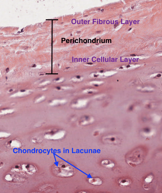
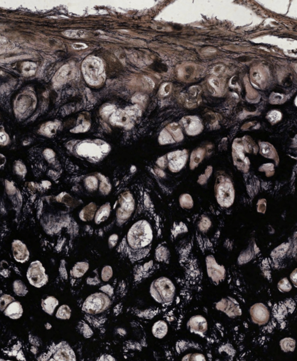
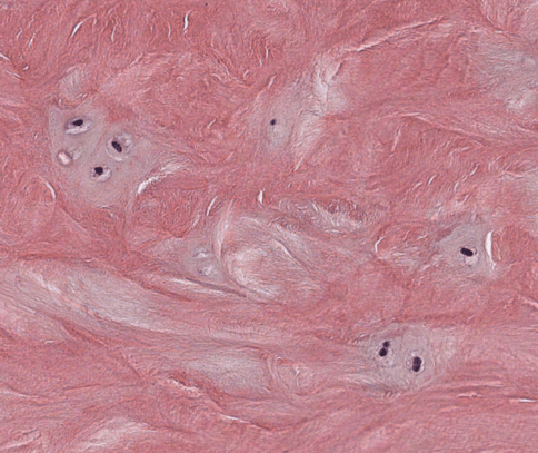
A microscopic image of hyaline cartilage (University of Buffalo, 2026)A microscopic analysis of an elastic cartilage with elastin-containing lacunae and matrix (University of Buffalo, 2026)A microscopic analysis of a fibrocartilage found in the intervertebral disc. There is a pale matrix relative to the eosinophilic staining of the type I collagen fibers found throughout the tissue section.
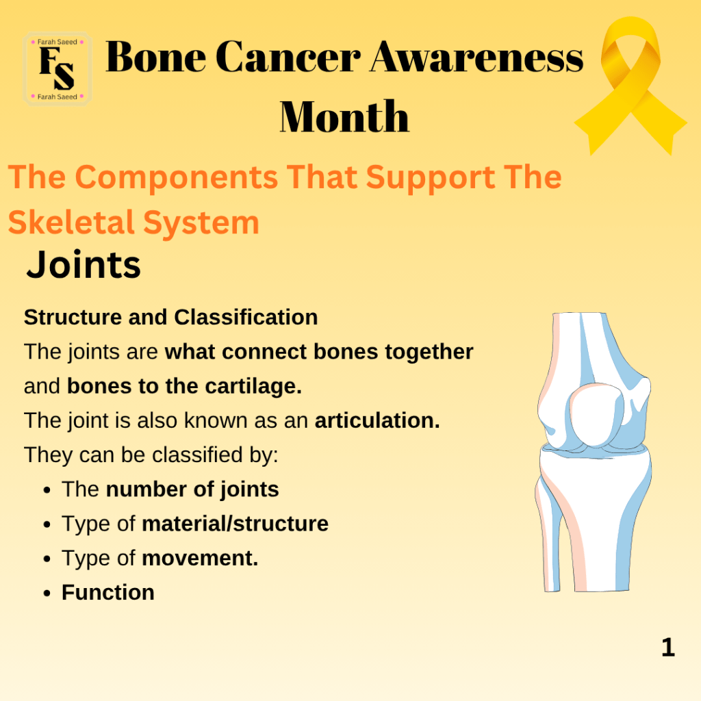
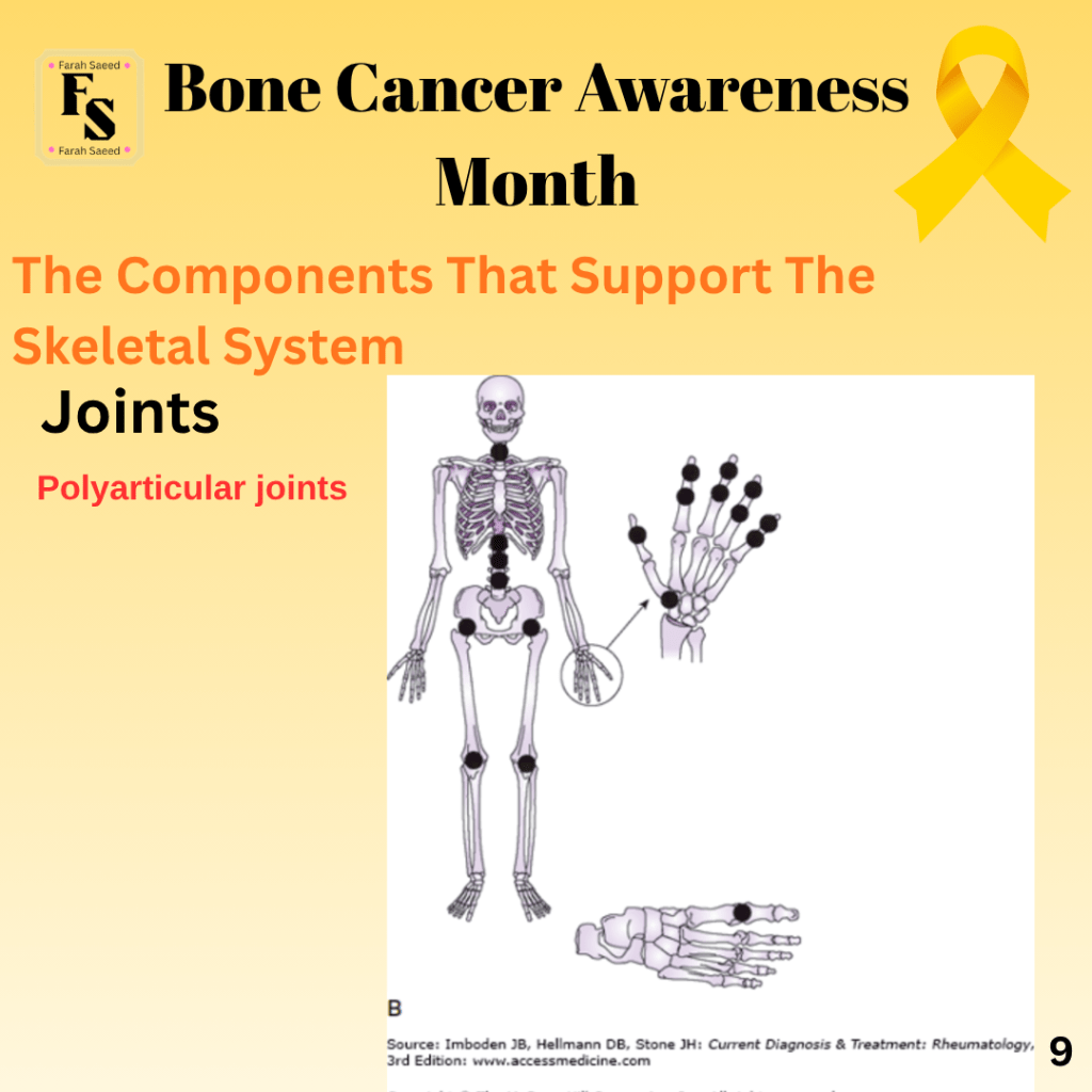
Joints
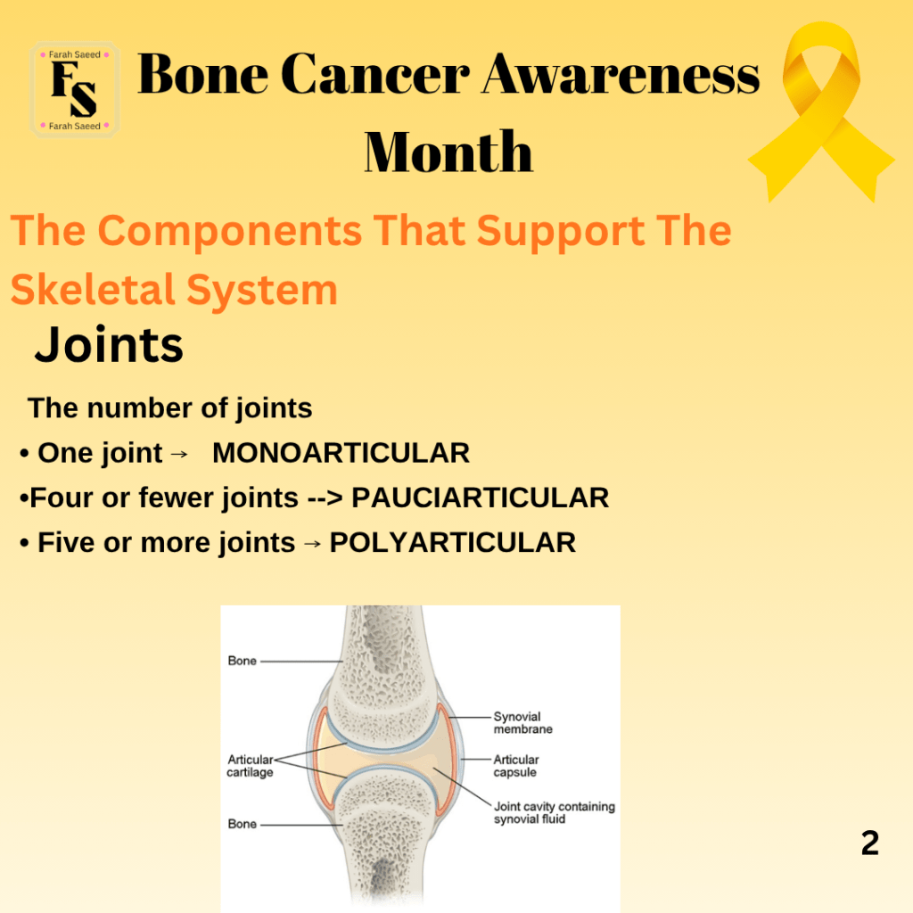
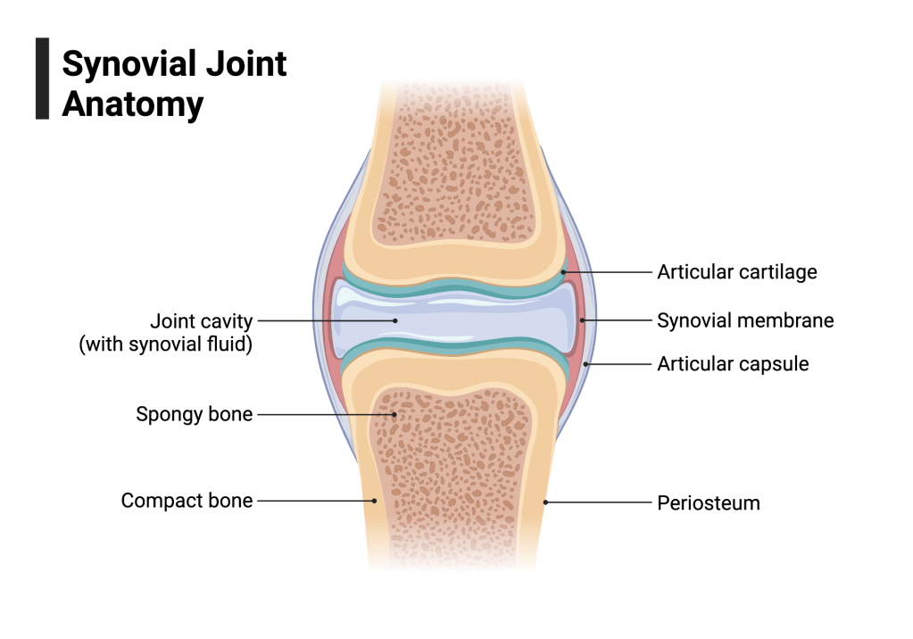
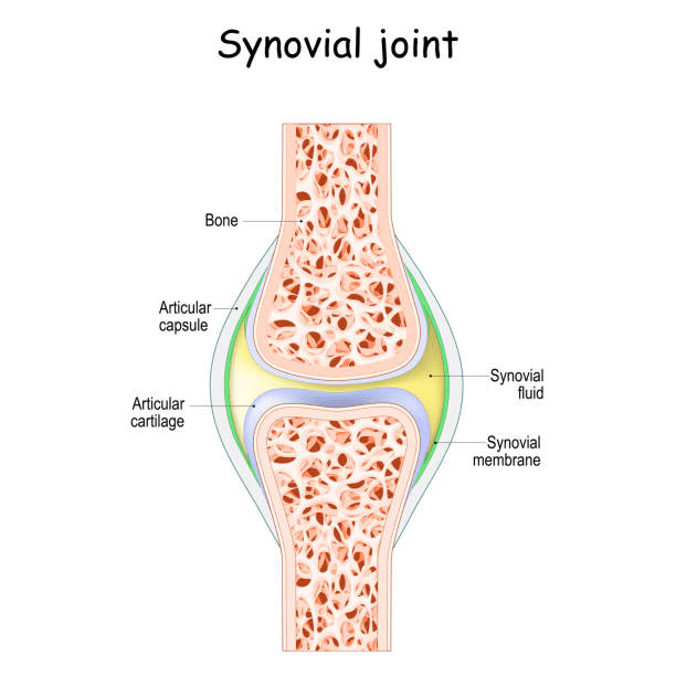
Structure of a joint
Types Of Joints
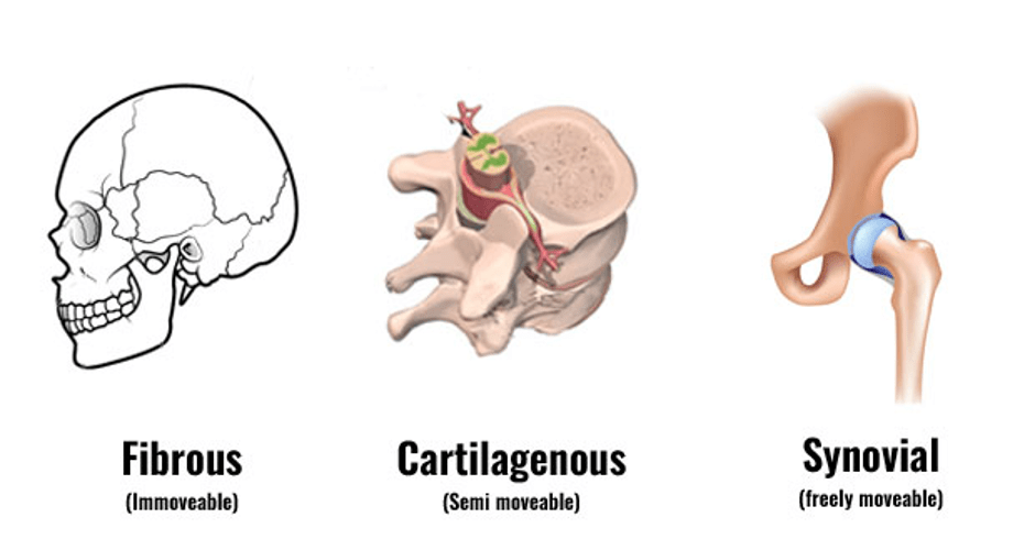
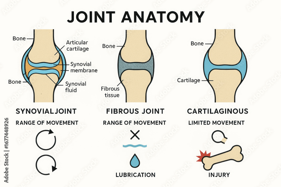
There are three main types of joints: Fibrous Cartilaginous Synovial
Supporting Image Resources
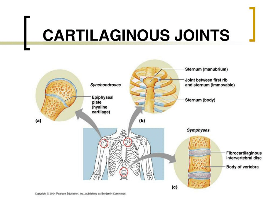
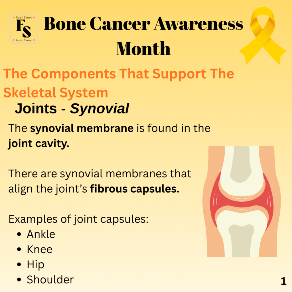
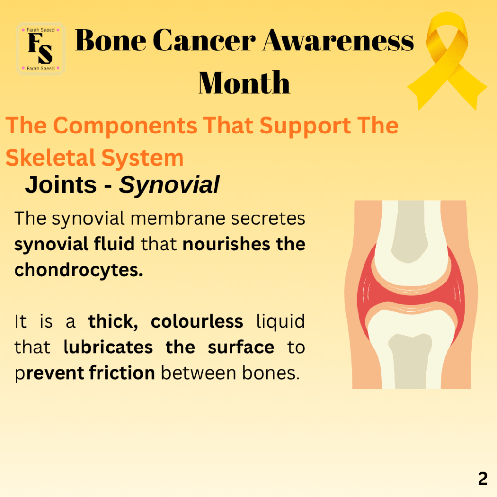
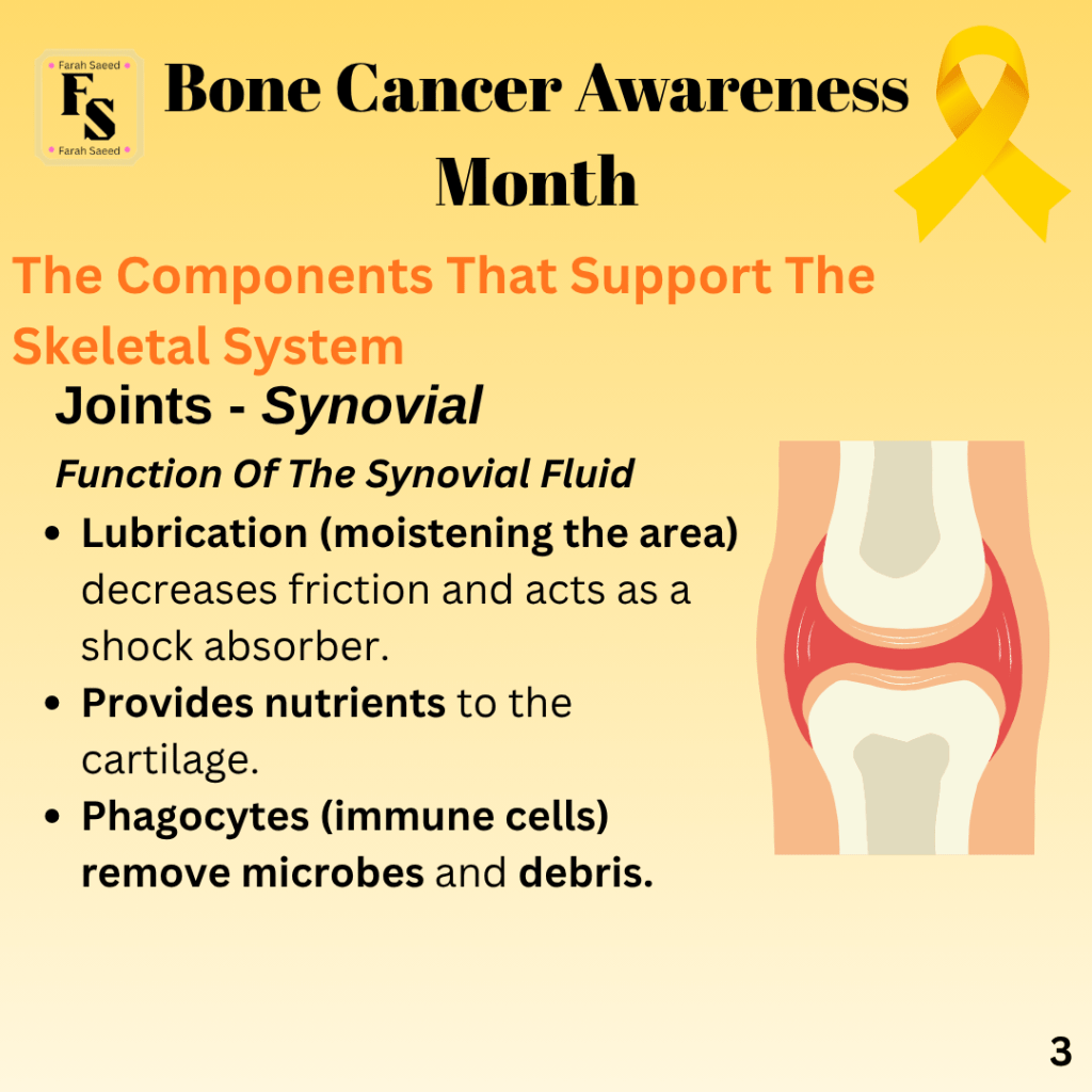
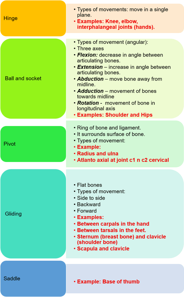
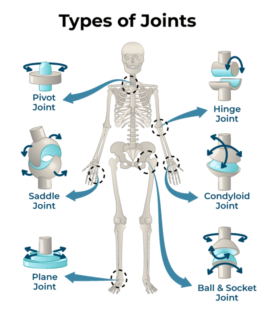
The three main types of jointsCharacteristics and examples of types of movement performed by the three main types of jointsThe location where synovial fluid is produced and stored, protecting the knee jointExamples of each type of cartilaginous joint
Synovial Joints
Types Of Joints
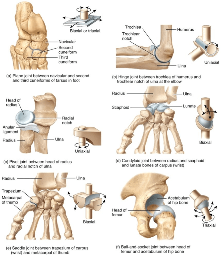
A summary of the main types of joints
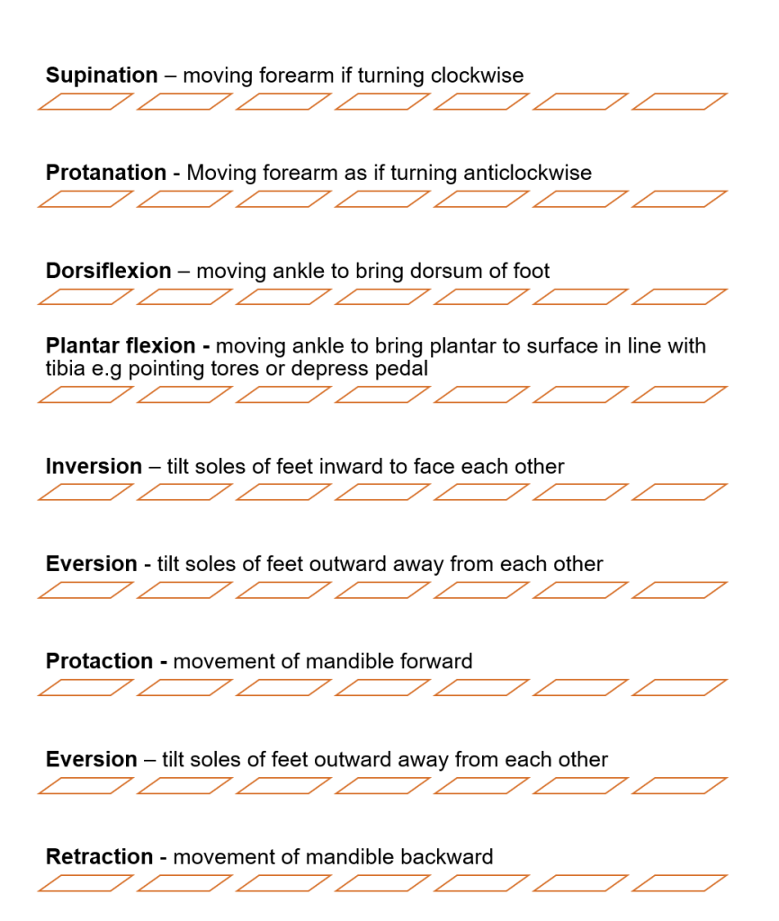
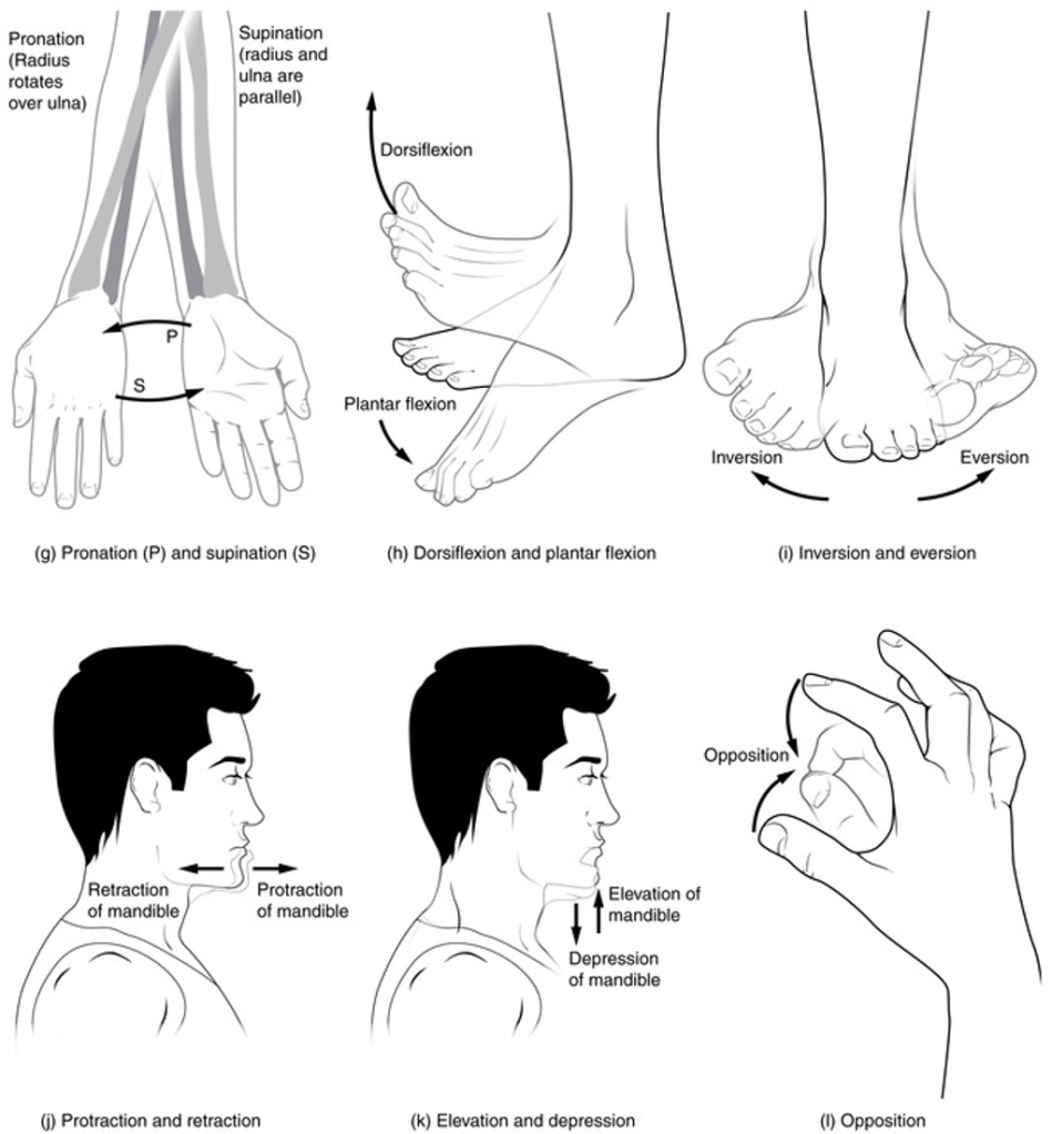
Specialised movements
Supporting Image and Other Resources
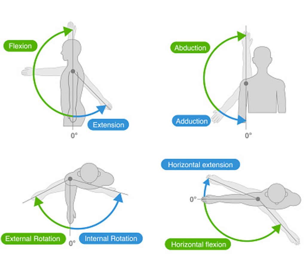
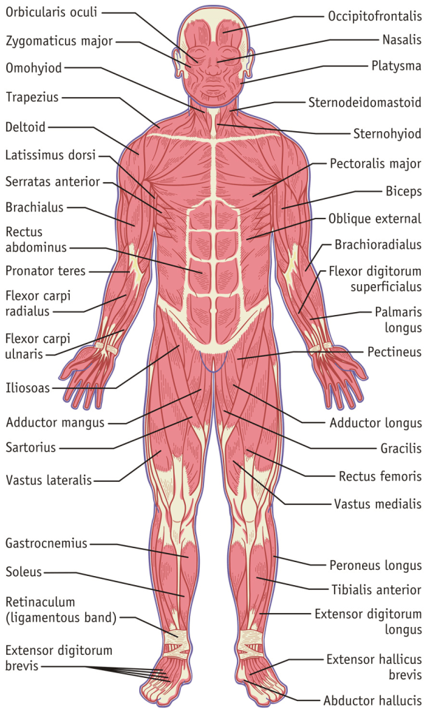
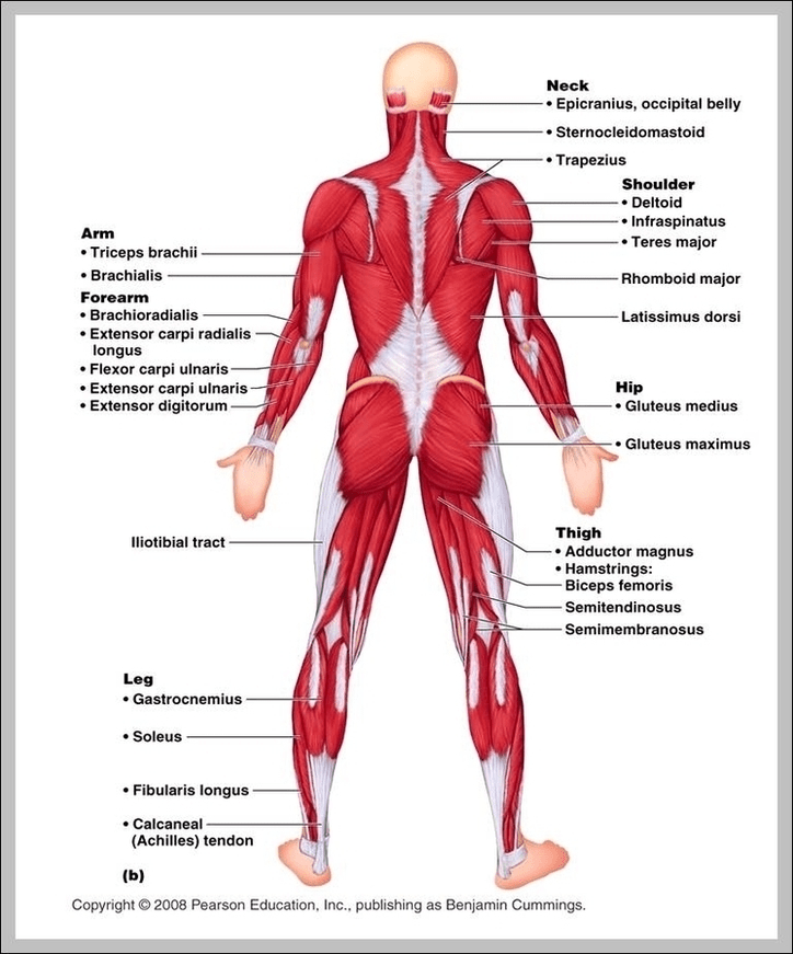
An illustration presenting the main types of movementAn illustration of the main types of jointsA further insight with given examples of different types of joints An illustration of other specialised movementsThe skeletal muscles (frontal view)Skeletal muscles (back view)
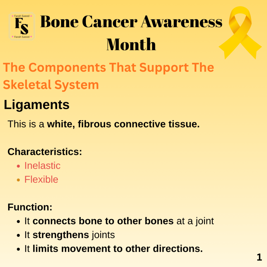
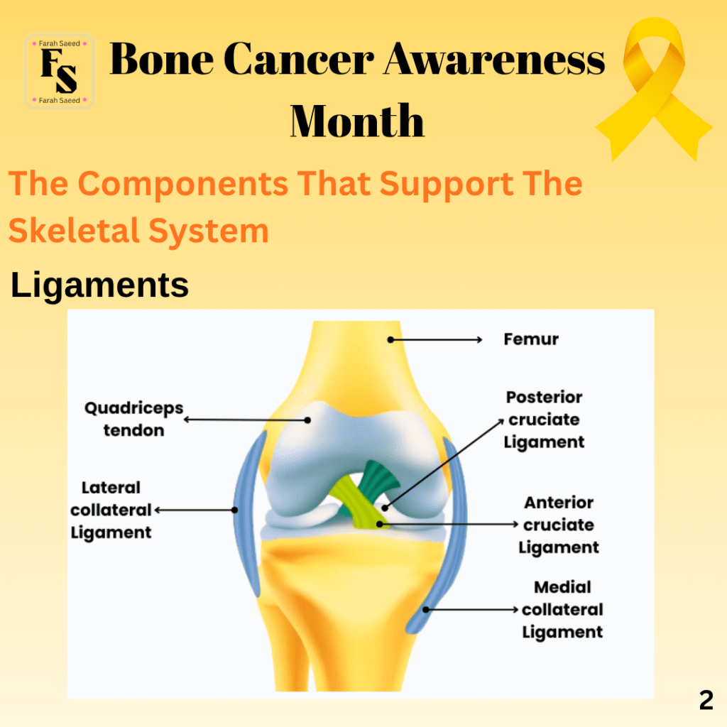
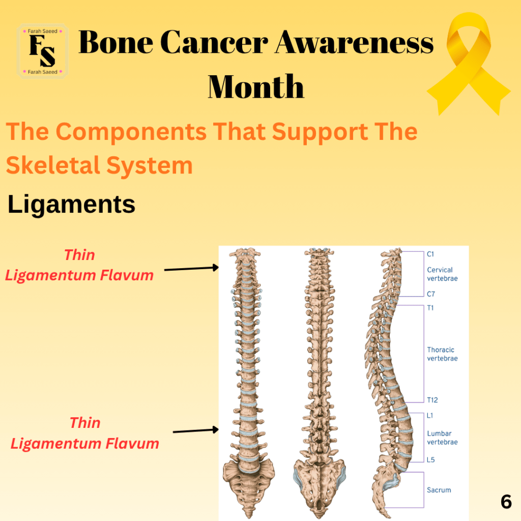
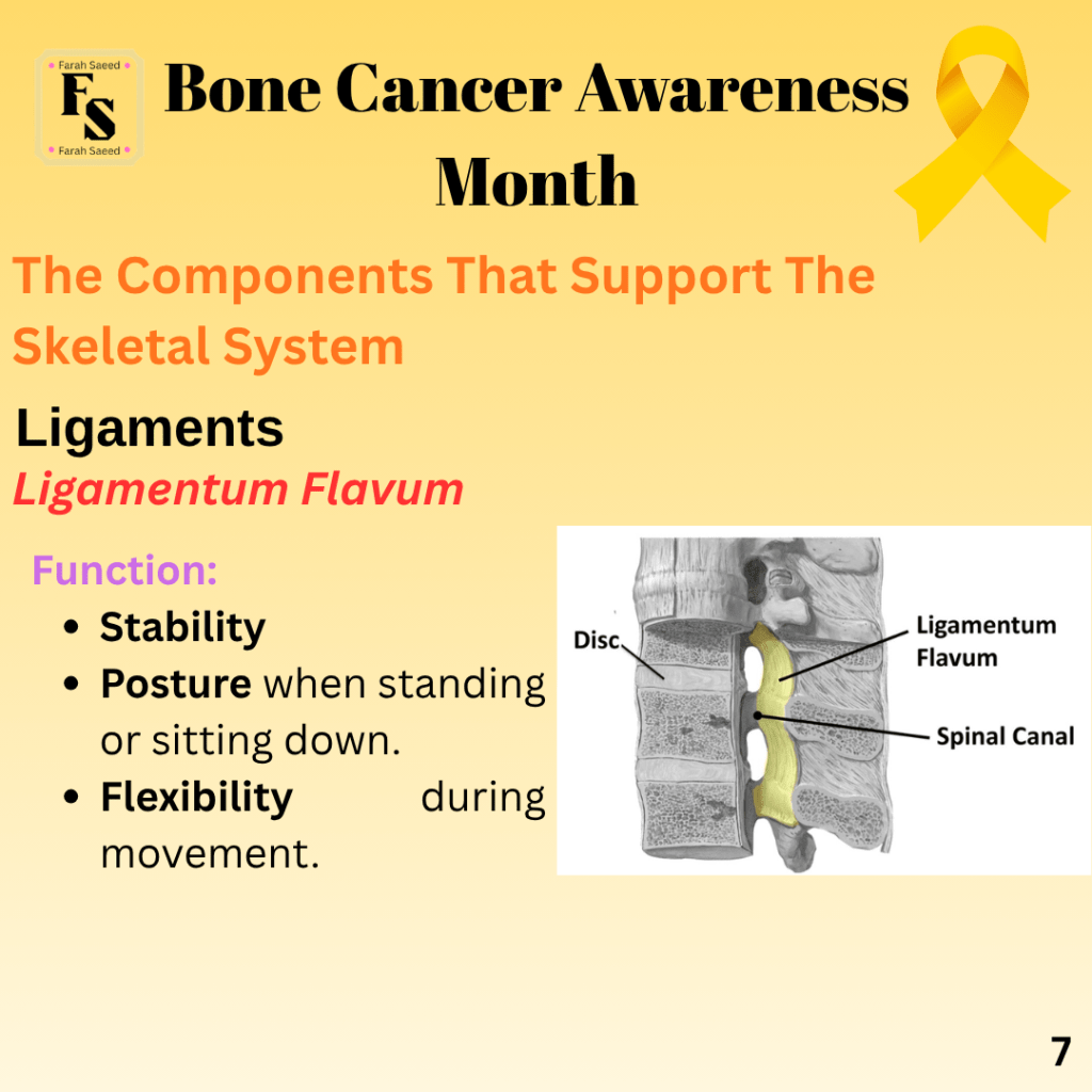
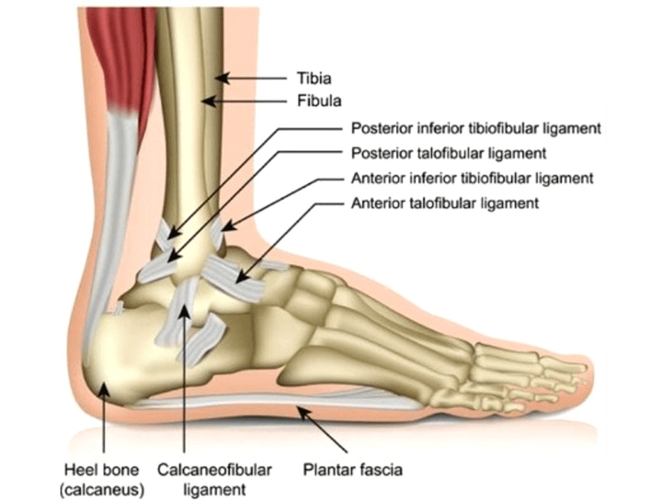
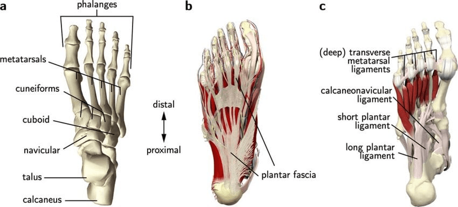
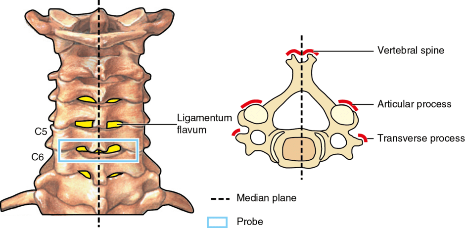
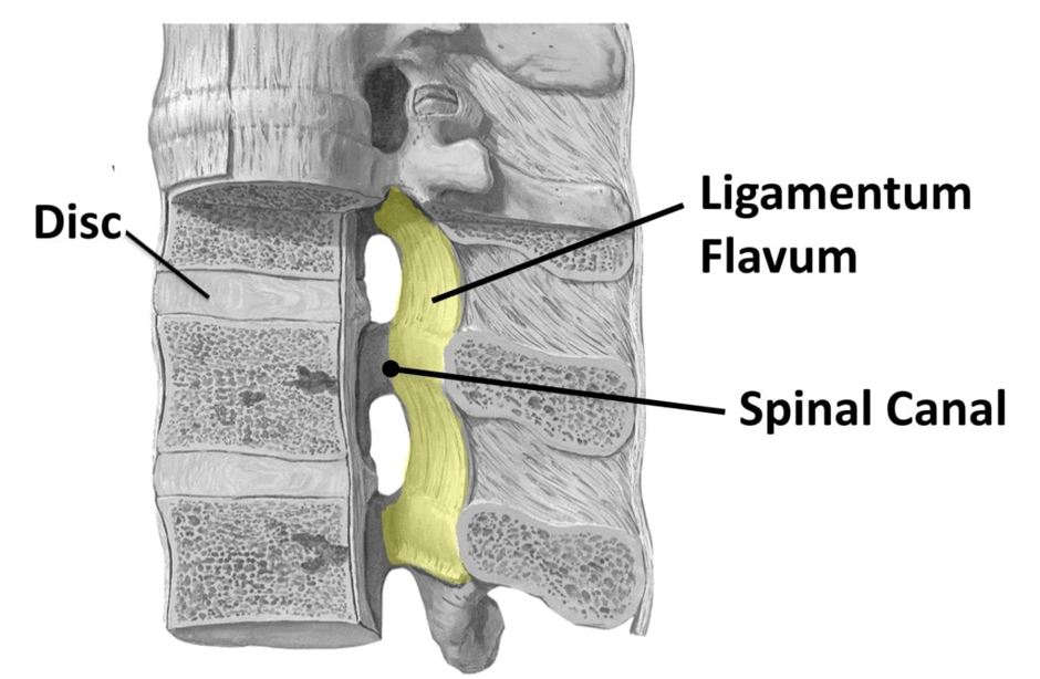
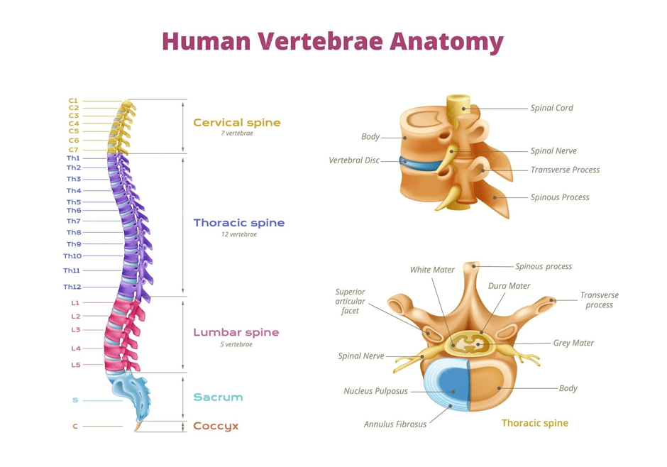
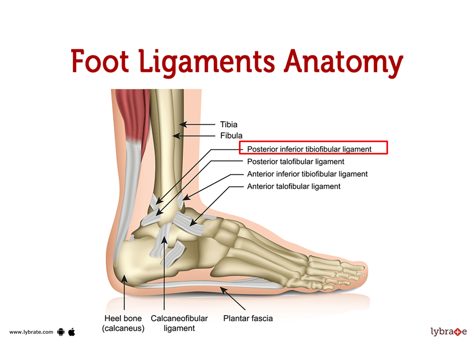
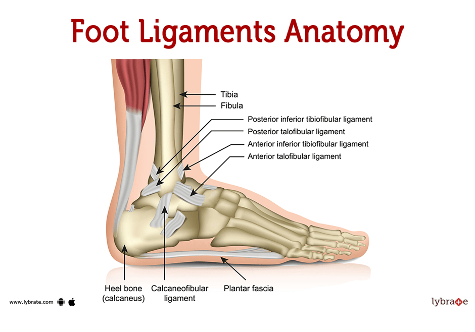
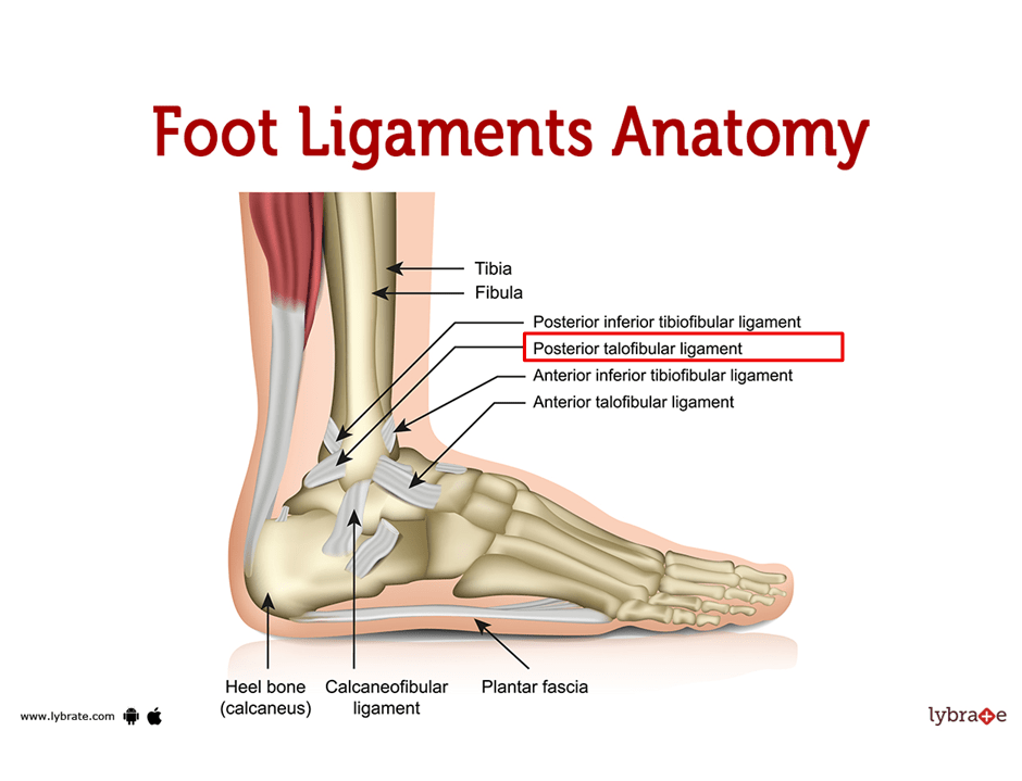
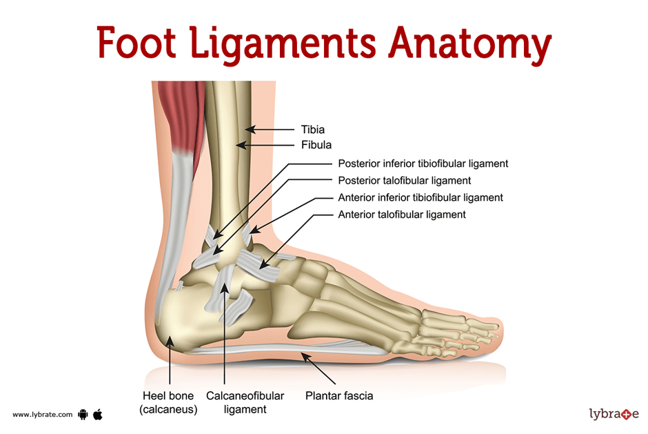
A presentation of the ligaments that support the bones in the foot (medial view)A presentation of the ligaments found in the foot (ventral view)The location of where the ligamentum flavum is situated in the vertebral columnA closer view of the Ligamentum FlavumA recap on the structure of the human vertebral column (spine)
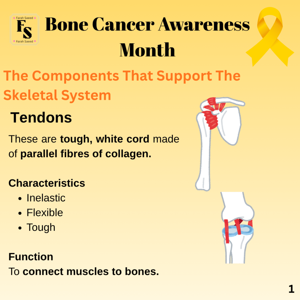
Tendons
Supporting Image Resources
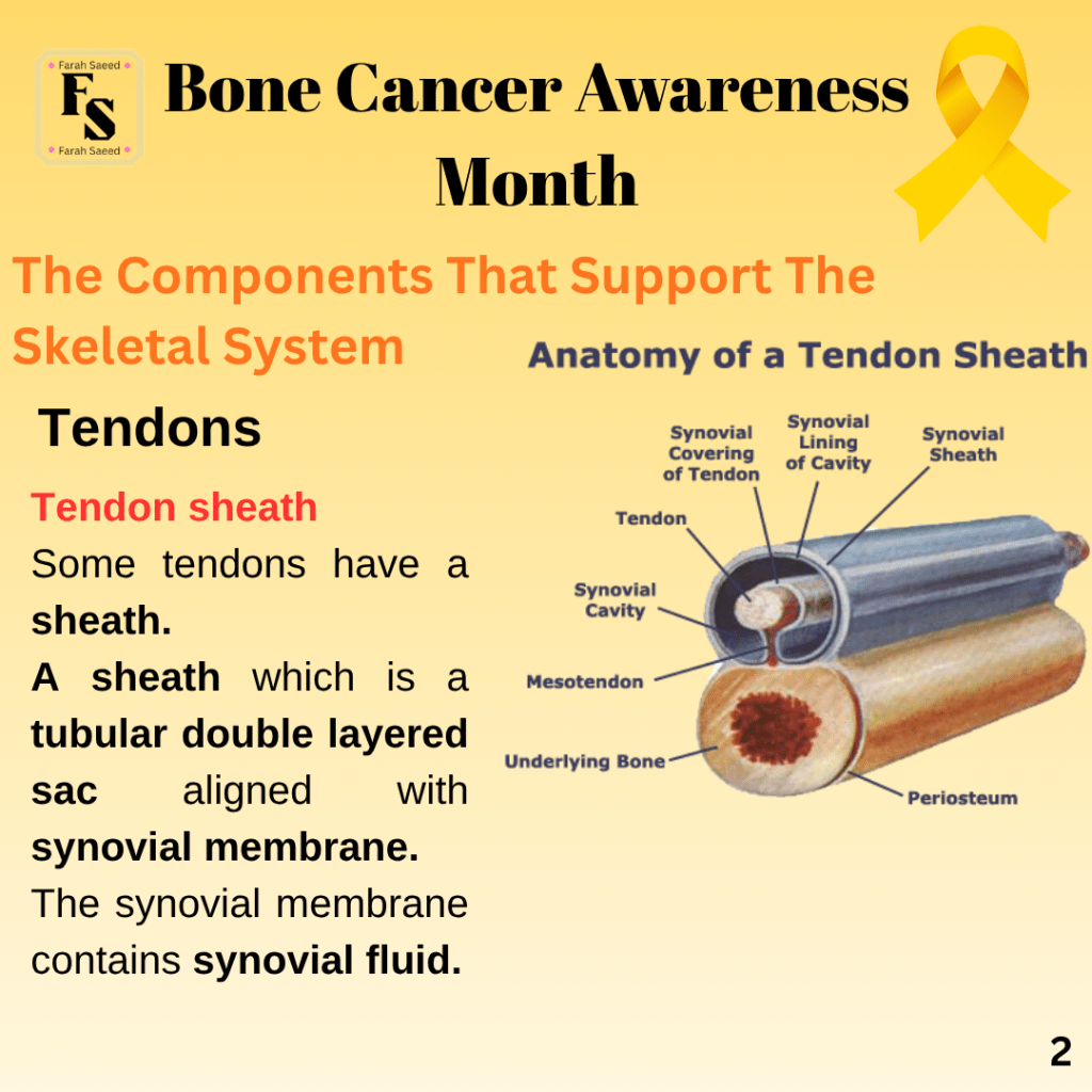
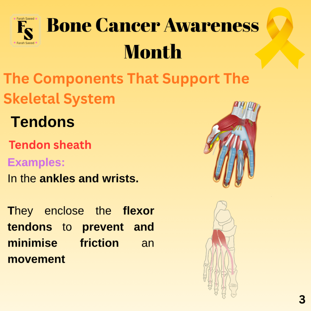
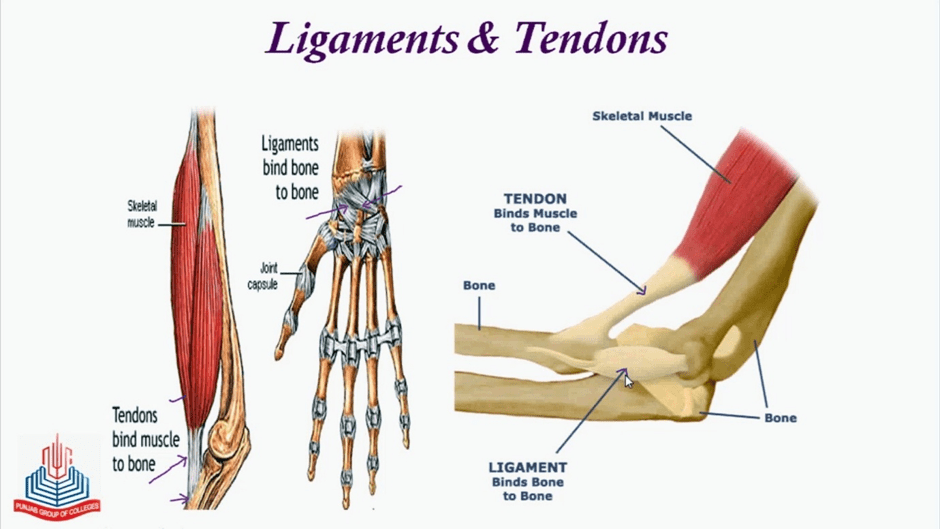
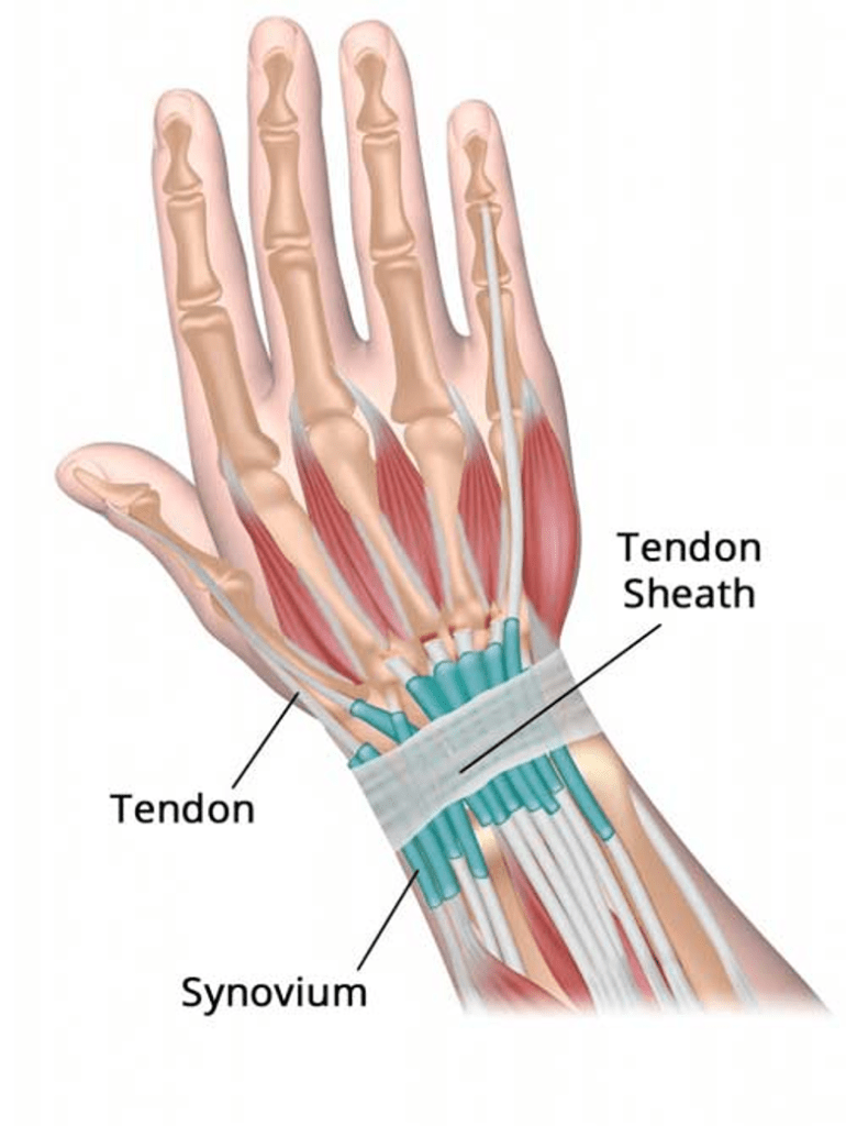
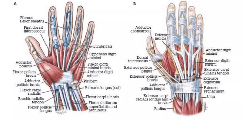
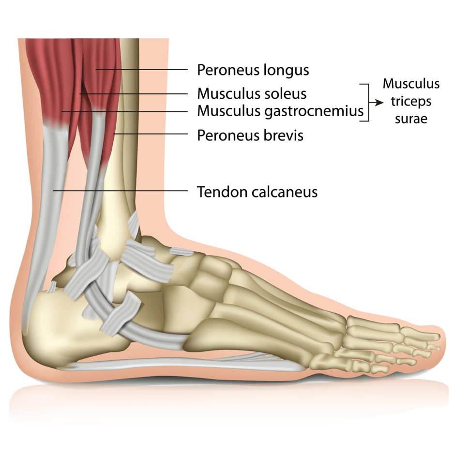
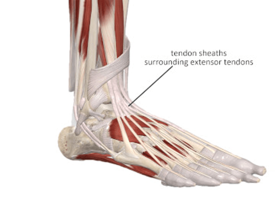
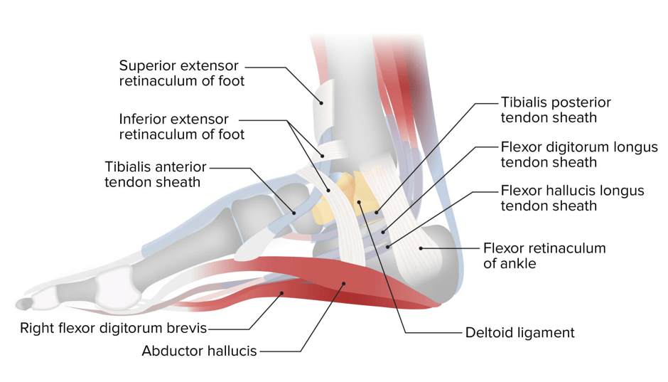
The difference between tendons and ligamentsA presentation of the tendon and tendon sheath on the hand (dorsal view)A closer view of the different supporting tissues found in the hand (dorsal view)An illustration of the tendon calcaneus that connects the muscles in the lower limb to the heel bone (calcaneus) (medial view)A presentation of the tendon sheaths in the foot (medial view)A closer view of the tendon sheath in the foot (lateral view)
The Process of How Ligaments And Tendons’ Names Are Made.
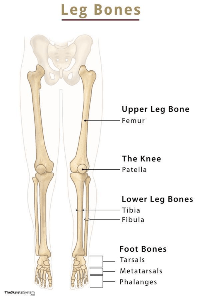
Most ligaments and tendons are named based on the bones and muscles they are attached to and their position. For instance: There are three bones in each lower limb: the femur (one long bone), followed by two bones (tibia and fibula).
Four ligaments connect the bones of the foot.
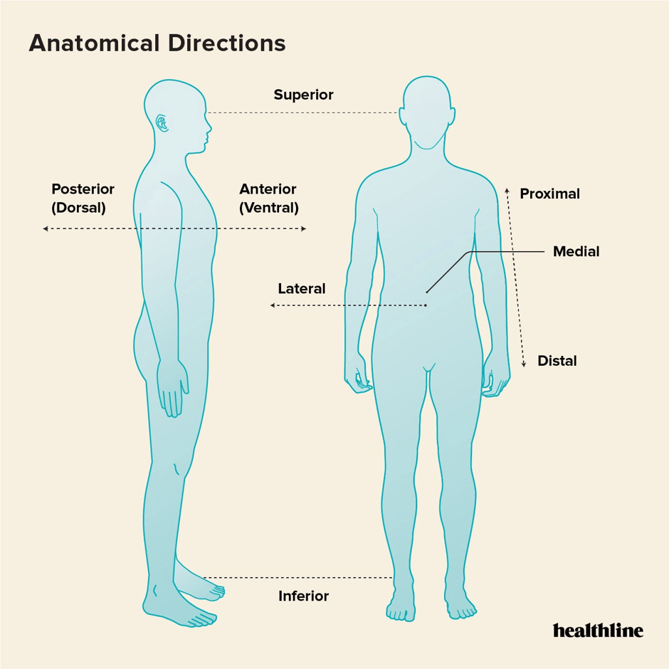
Posterior –> BACK
Anterior –> FRONT
LIGAMENT ONE
Posterior inferior tibofibular ligament
The ligament links the tibia with the fibula – tibiofibular
It is behind –> posterior
It is within –> Inferior
LIGAMENT TWO
Anterior inferior tibiofibular ligament
The ligament links the tibia with the fibula – tibiofibular
It is in front –> anterior
It is within –> INferior
LIGAMENT THREE
Posterior talofibular ligament
The ligament links the talus with the fibula – talofibular
It is in the back –> posterior
LIGAMENT FOUR
Anterior talofibular ligament
The ligament links the talus with the fibula – talofibular
It is in the front –> anterior
Supporting Image Resources
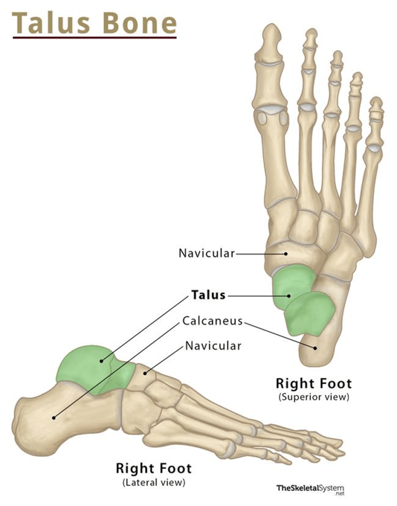
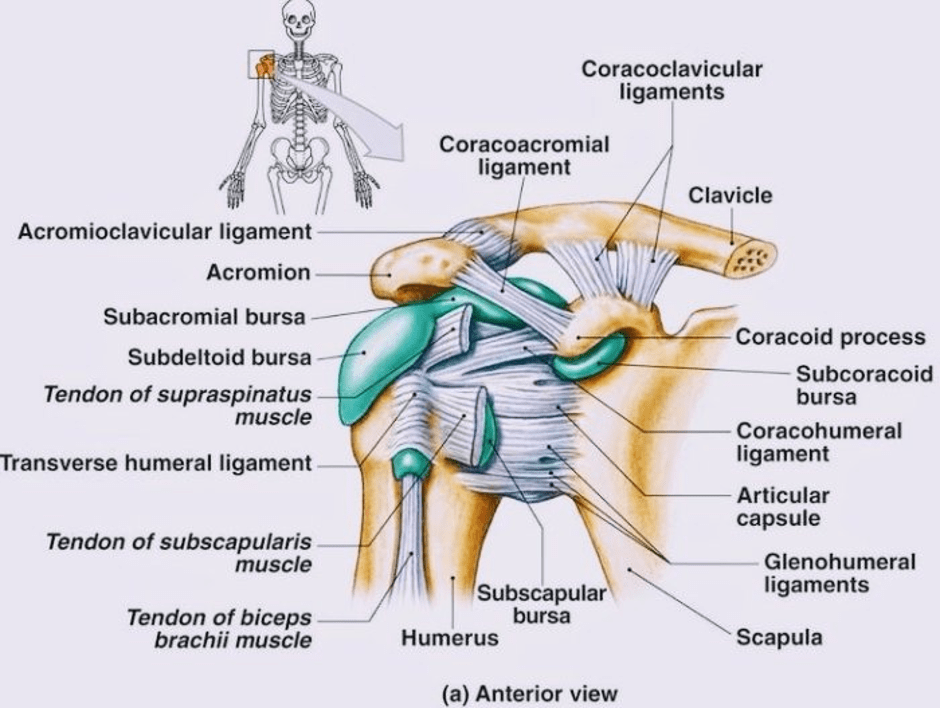
Anatomical planesA presentation of the talus boneA presentation of the bones present in the lower limbsA presentation of the various supporting tissues that connect the shoulder bone (clavicle) with the upper limb (humerus). A bursa is a small sac of fibrous tissue that aligns the synovial membrane (containing synovial fluid). It is present in specific areas where one part moves over another part (overlap) to prevent friction.
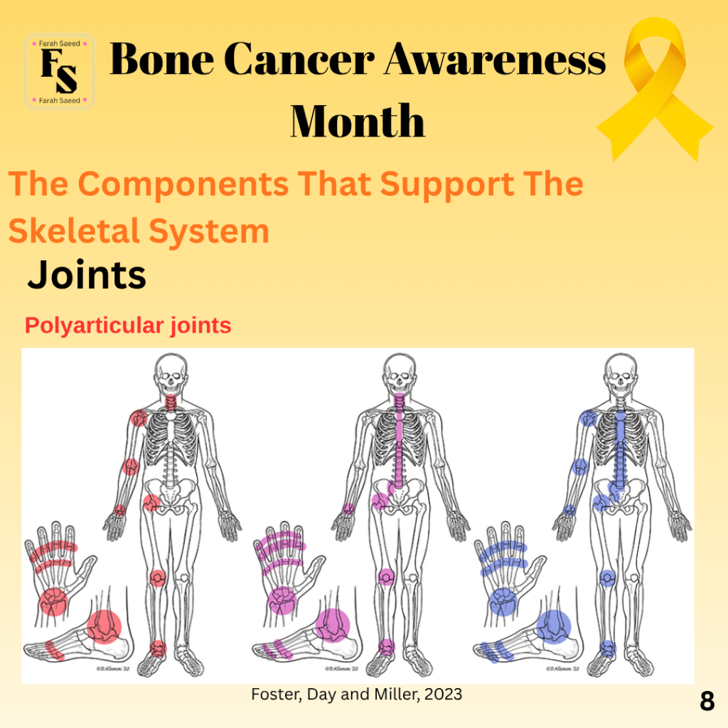
Foster, Z.J., Day, A.L. and Miller, J. (2023) Polyarticular Joint Pain in Adults: Evaluation and Differential Diagnosis. American Family Physician 107(1), pp.42 to 51.