Types Of Vaginal Cancer
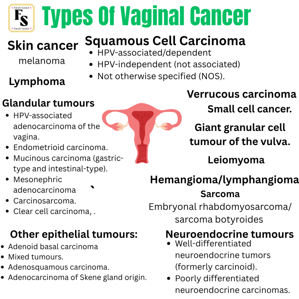
Classification Of Vaginal Cancers
Adams, T.S., Maluleke, J.C. and Cuello, M.A. (2025). Cancer of the vagina: 2025 Update. International Journal of Gynecology & Obstetrics, 171(S1), pp.48–59. doi:10.1002/ijgo.70325.
Höhn, A.K., Brambs, C.E., Hiller, G.G.R., May, D., Schmoeckel, E. and Horn, L.-C. (2021). 2020 WHO classification of female genital tumors. Geburtshilfe und Frauenheilkunde, 81(10), pp.1145–1153. doi:10.1055/a-1545-4279.
WHO Classification of Tumours Editorial Board. Female genital tumours (WHO classification of tumours series). Vol 4. 5th ed. International Agency for Research on Cancer; 2020.
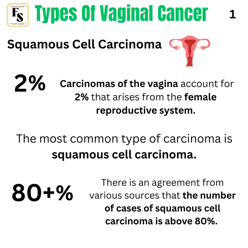
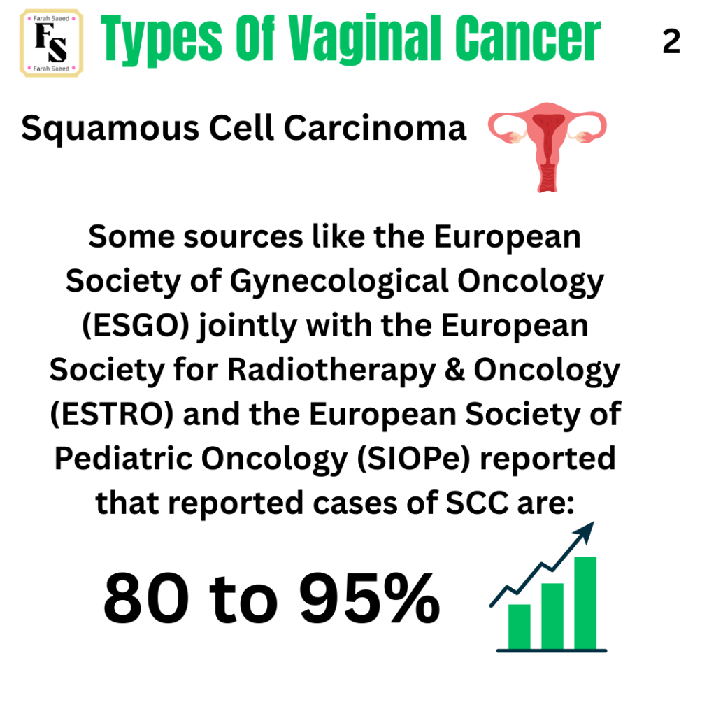
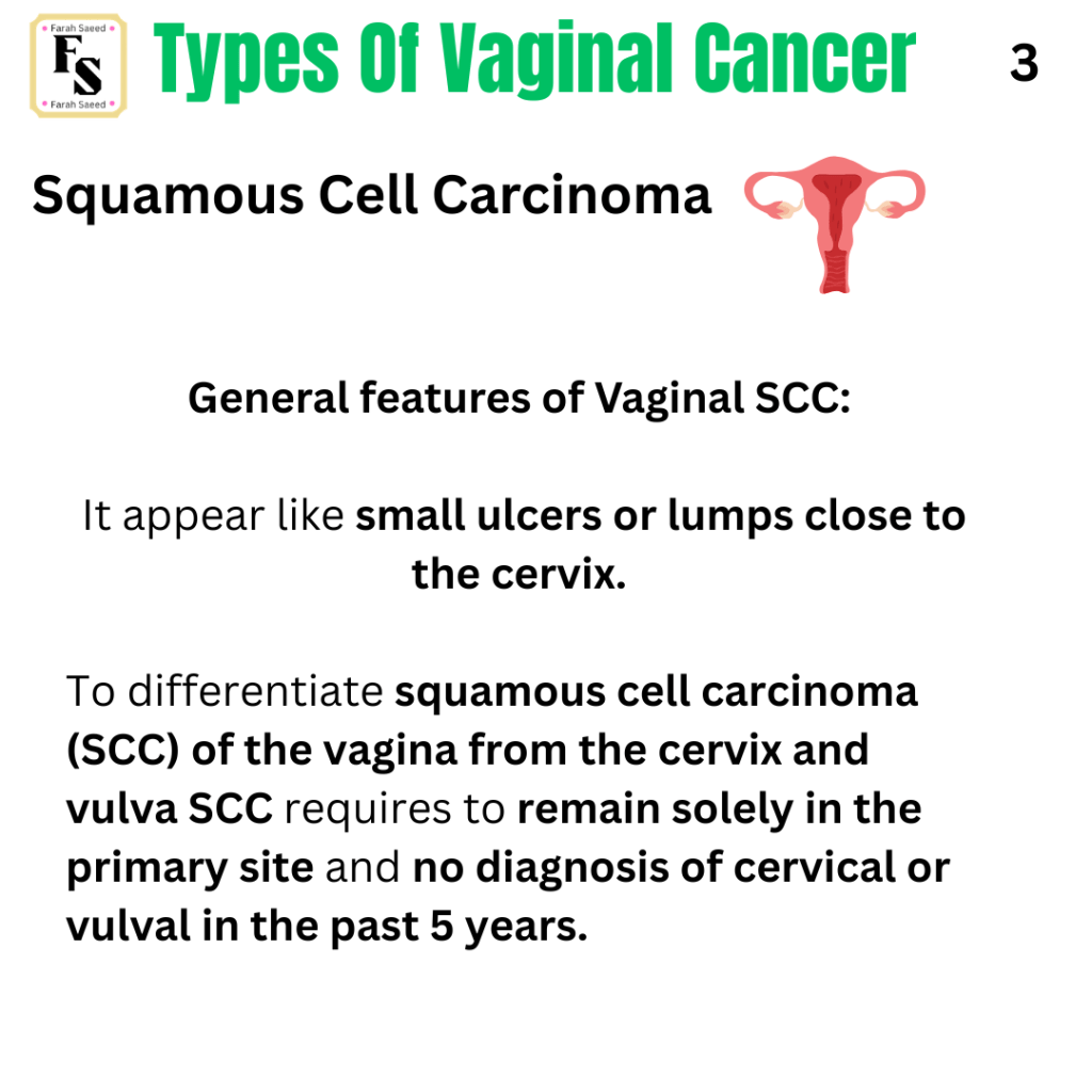
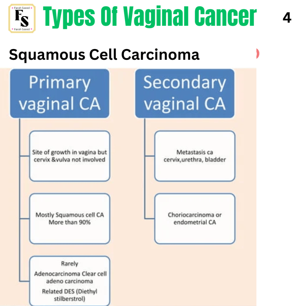
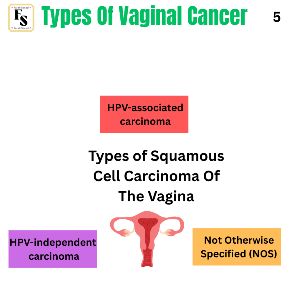
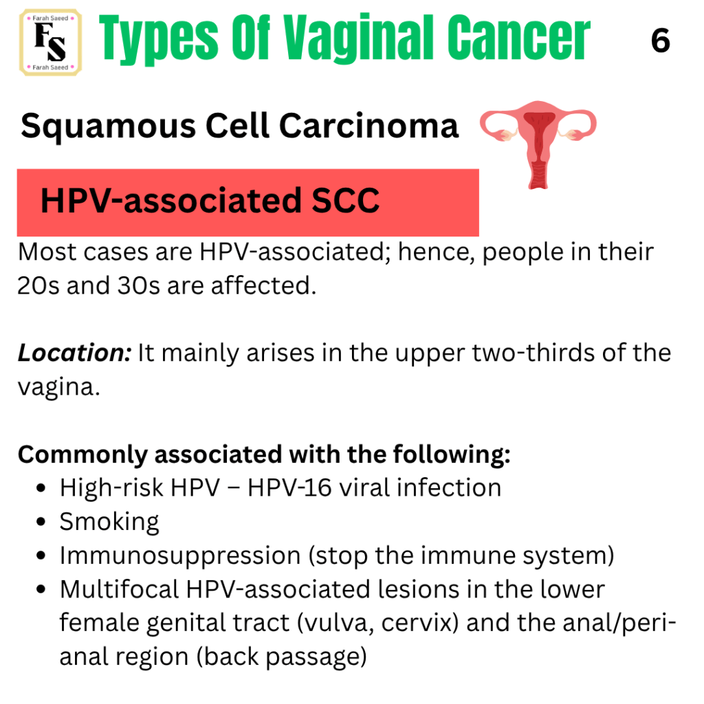
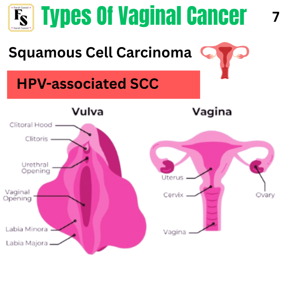
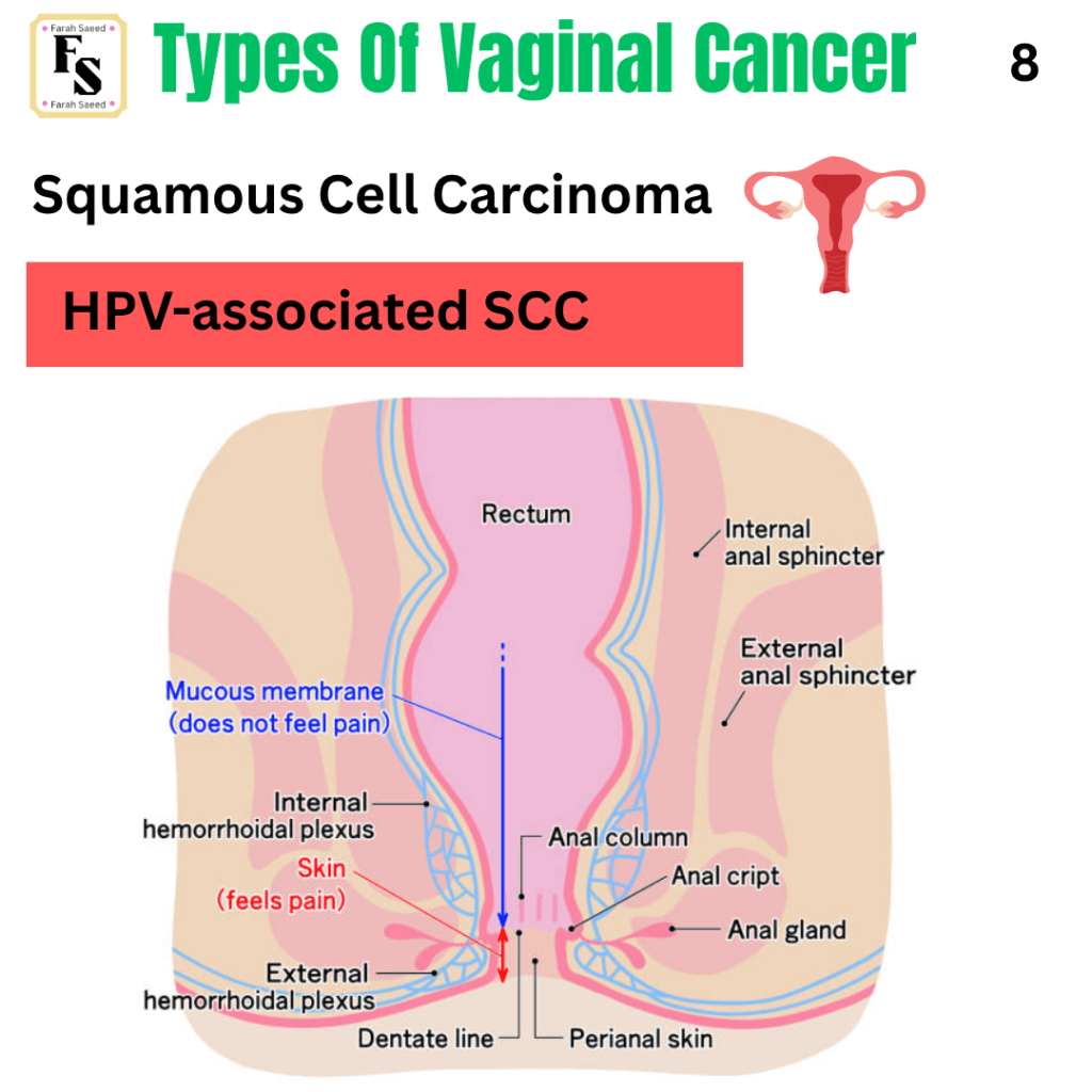
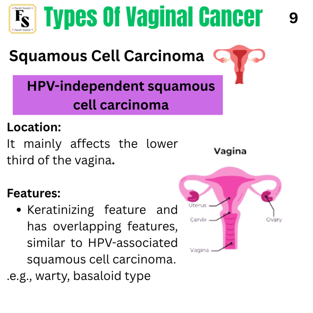
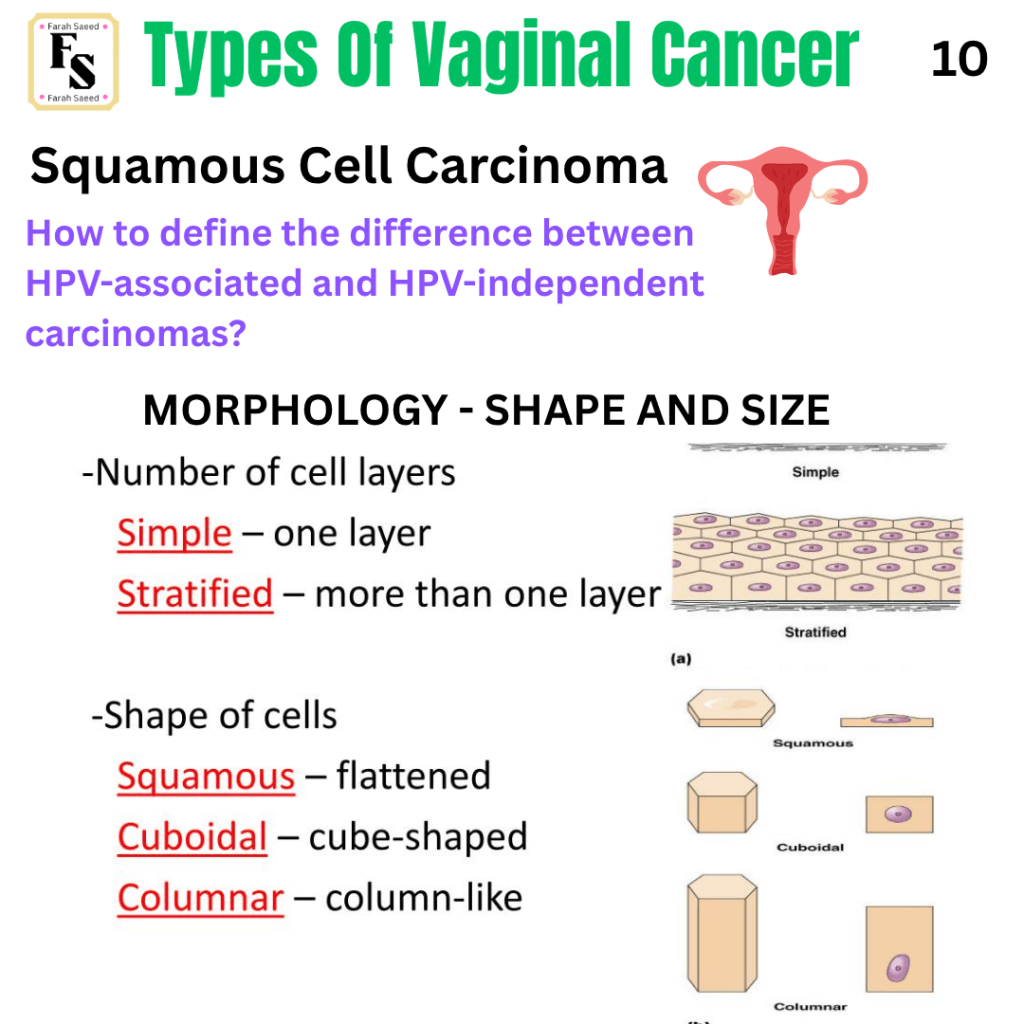
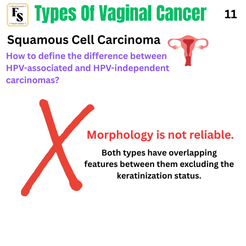
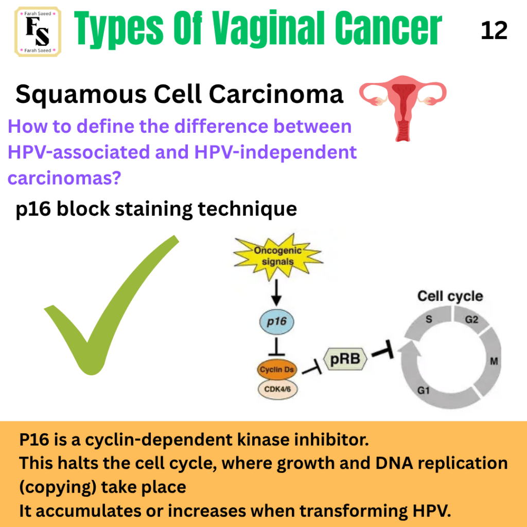
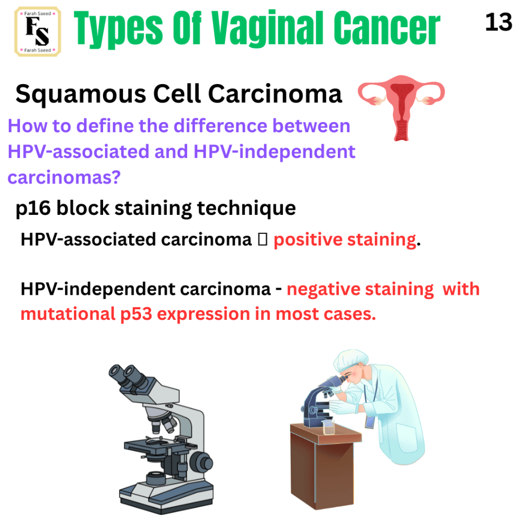
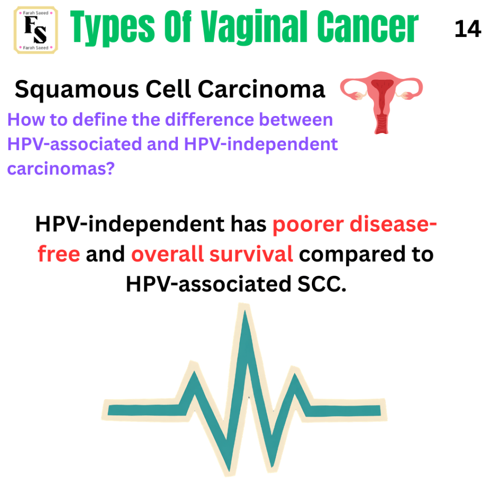
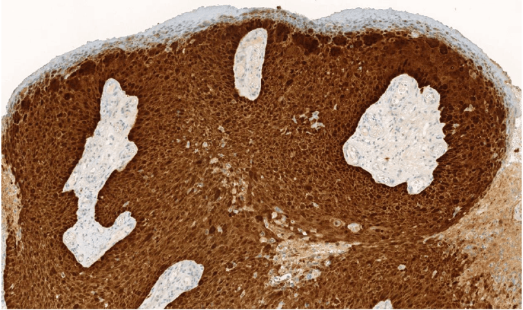
Squamous Cell Carcinoma Of The Vagina
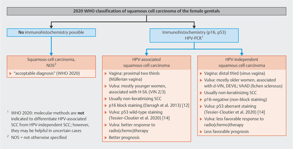
Source: Höhn, A.K., Brambs, C.E., Hiller, G.G.R., May, D., Schmoeckel, E. and Horn, L.-C. (2021). 2020 WHO classification of female genital tumors. Geburtshilfe und Frauenheilkunde, 81(10), pp.1145–1153. doi:10.1055/a-1545-4279
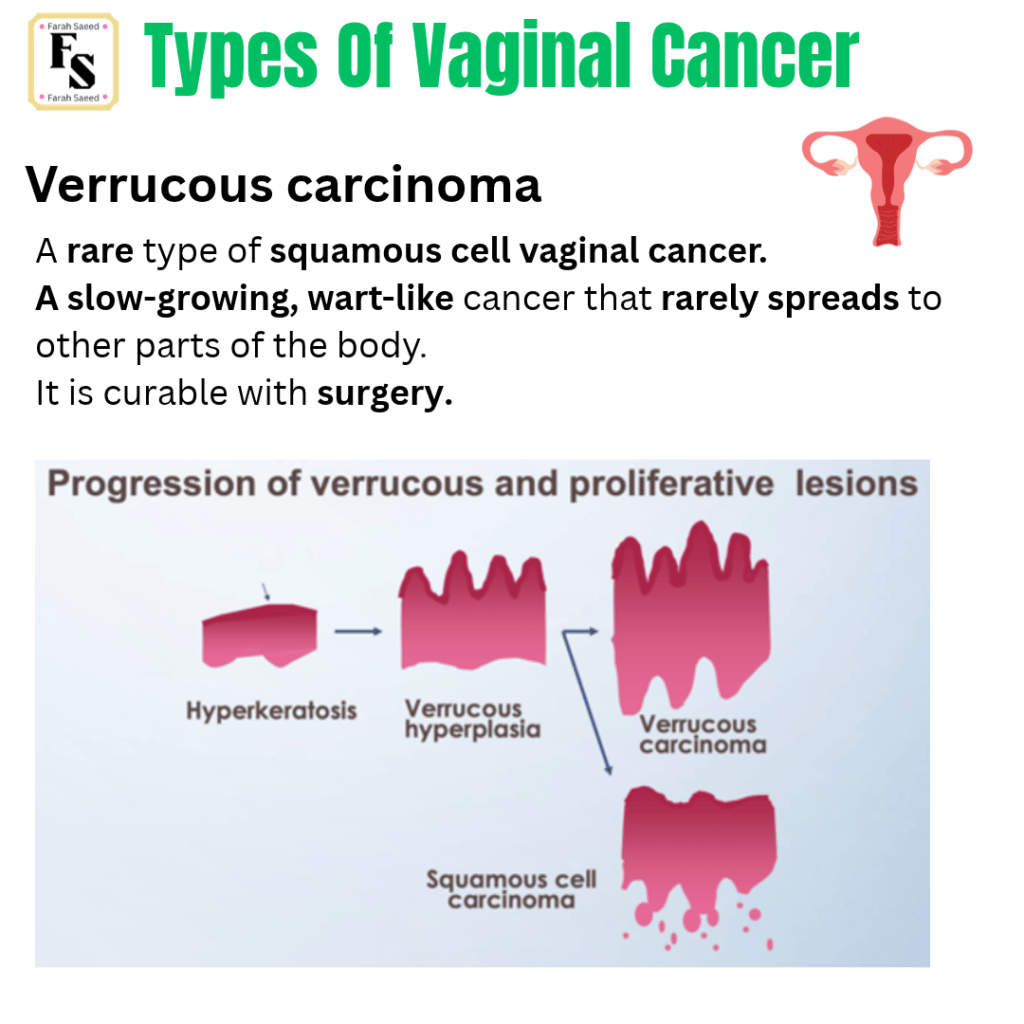
Verrucous Carcinoma
Primary Small Cell Carcinoma Of The Vagina
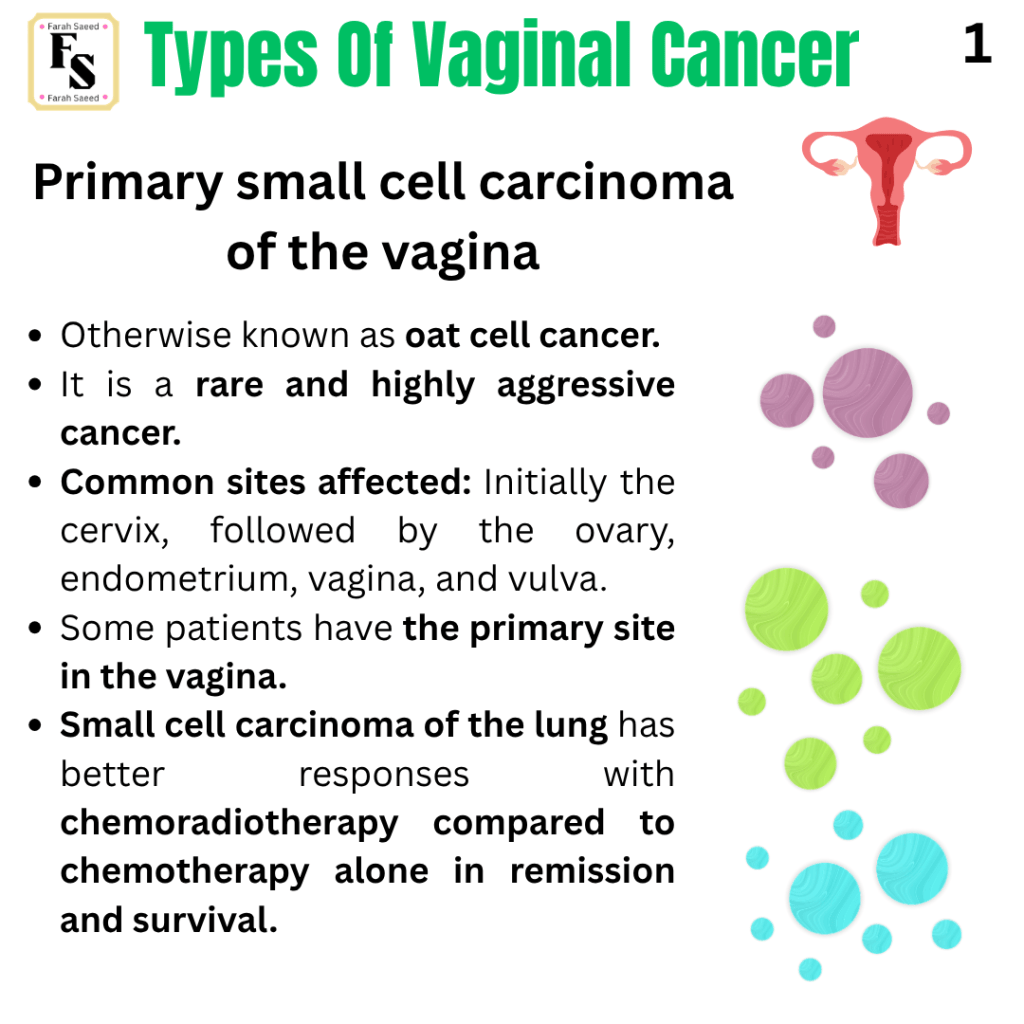
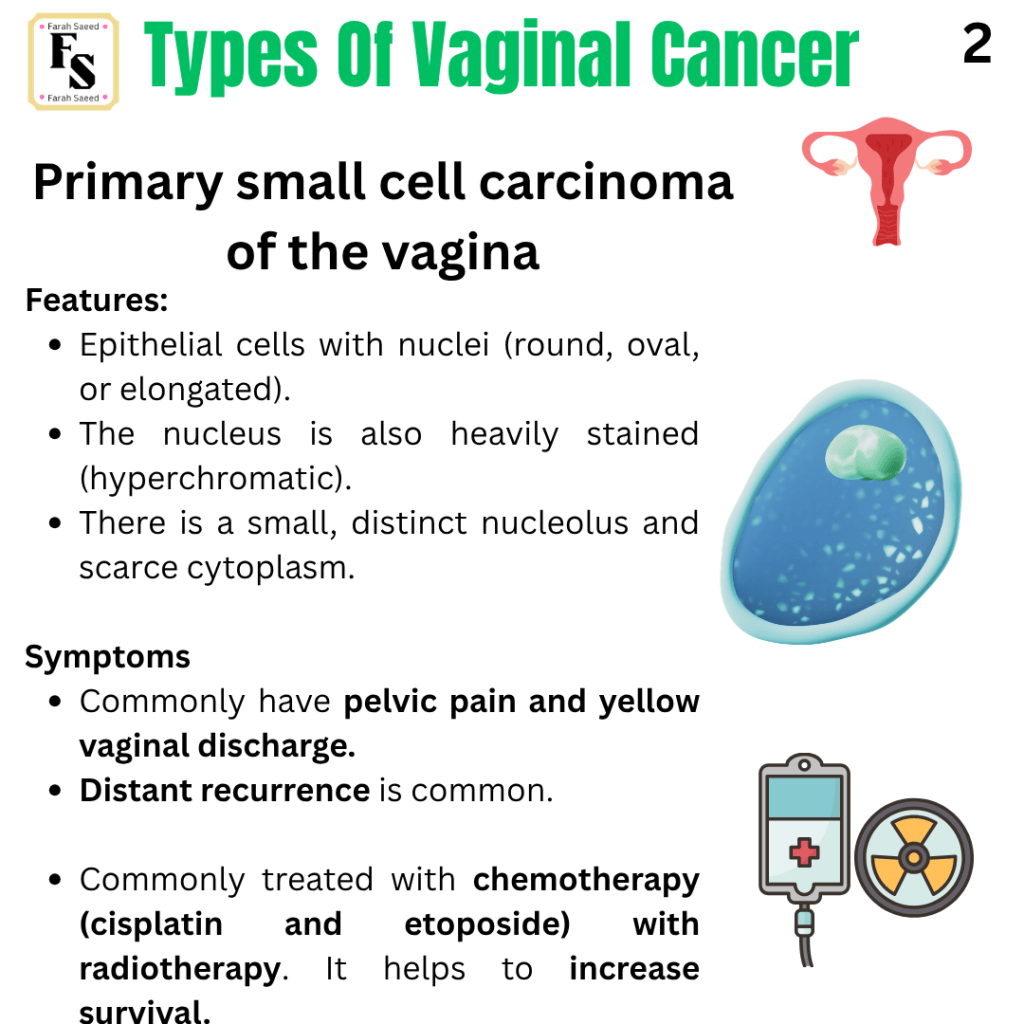
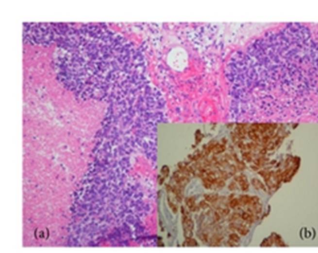
Oliveira, R., Bócoli, M.C., Saldanha, J.C., Murta, E.F.C. and Nomelini, R.S. (2013). Primary small cell carcinoma of the vagina. Case Reports in Obstetrics and Gynecology, 2013, pp.1–4. doi:10.1155/2013/827037.
Giant Granula Cell Tumour Of The Vagina
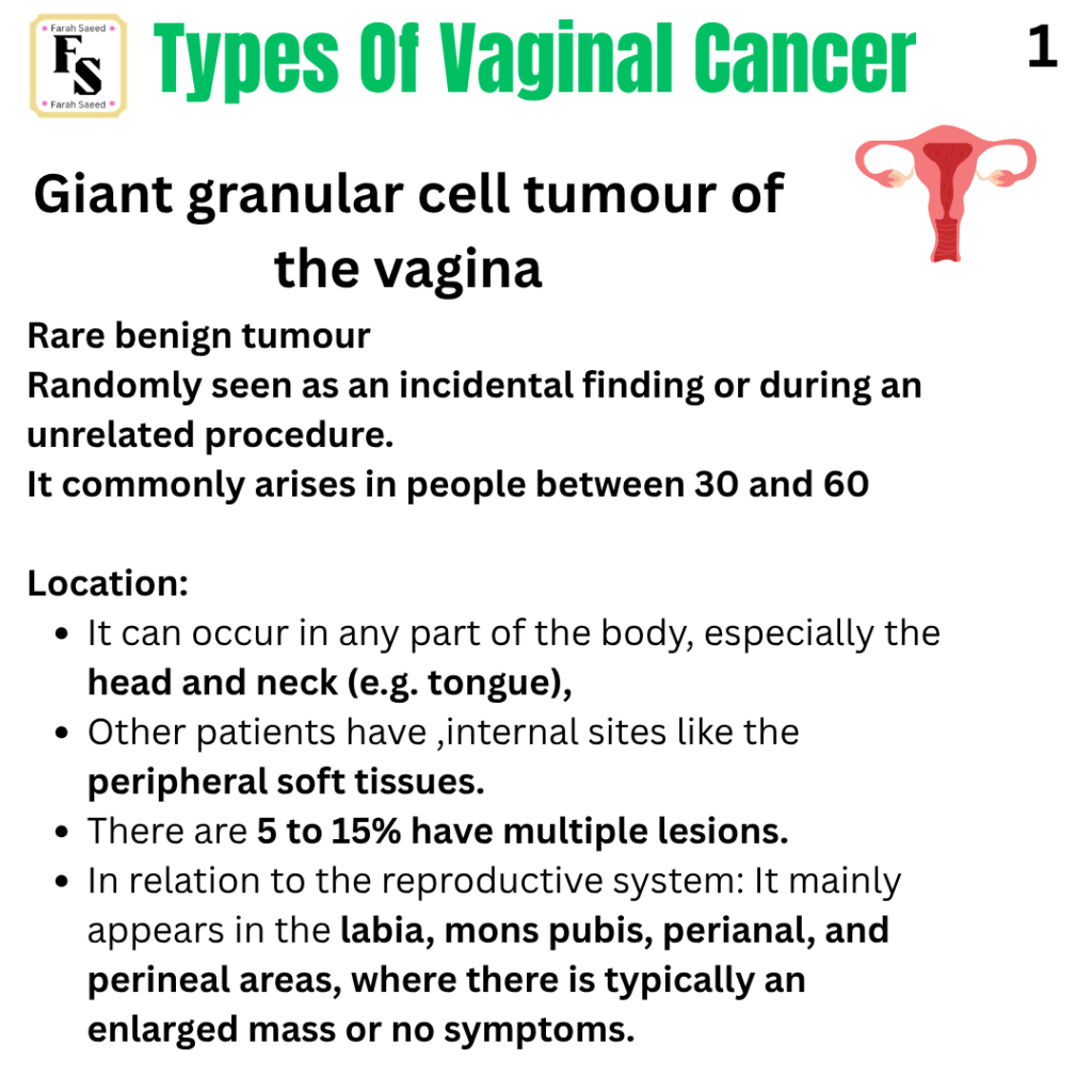
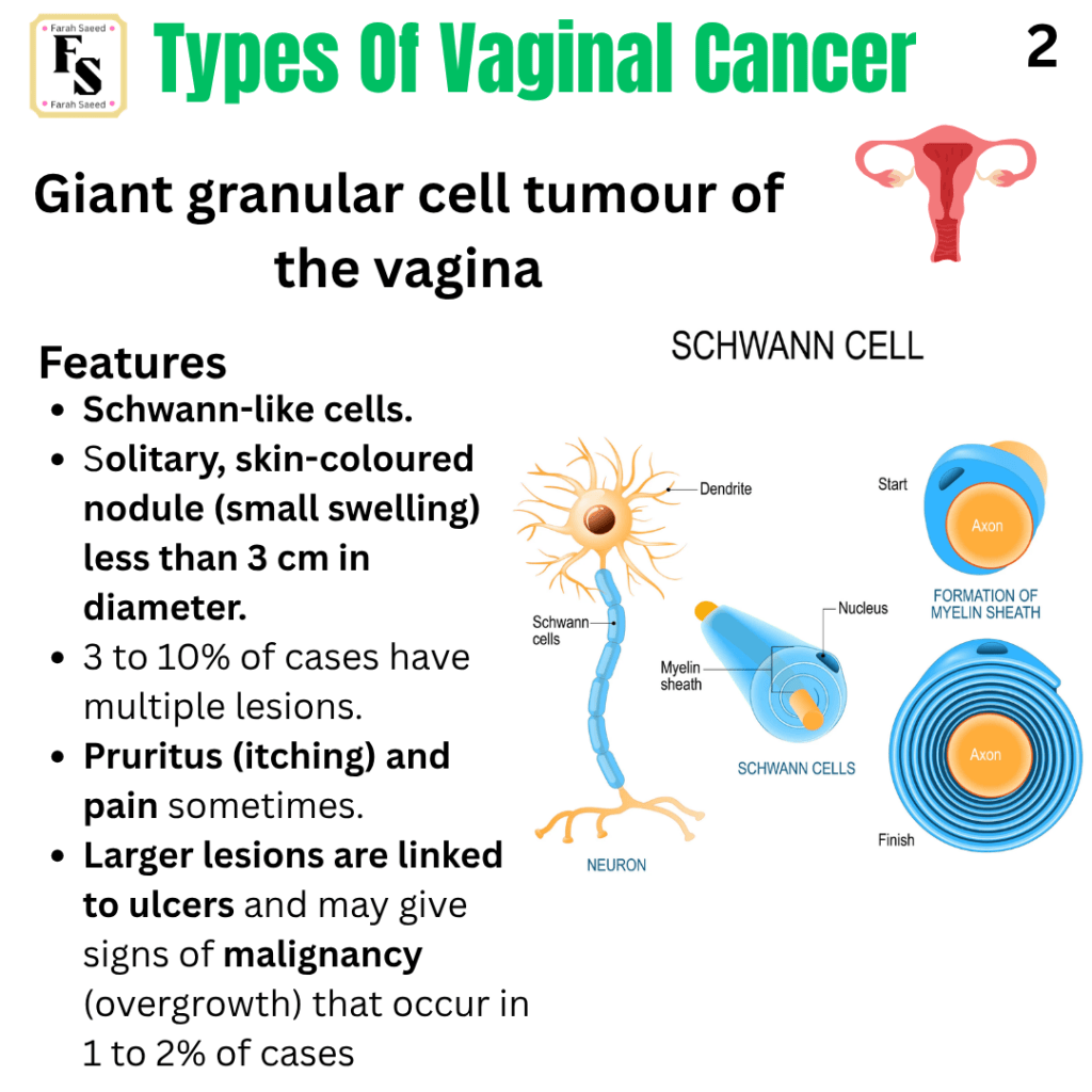
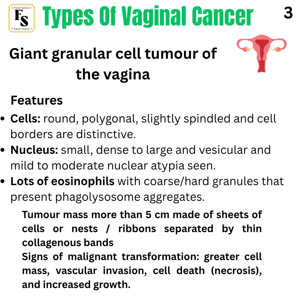
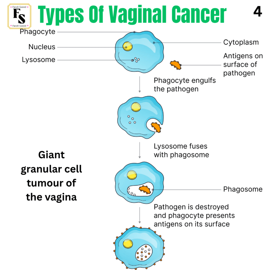
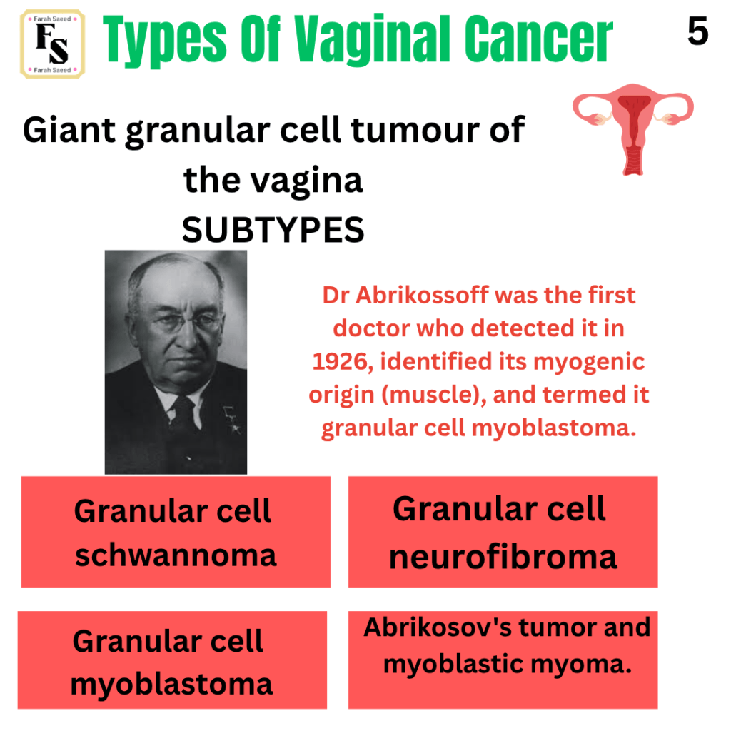
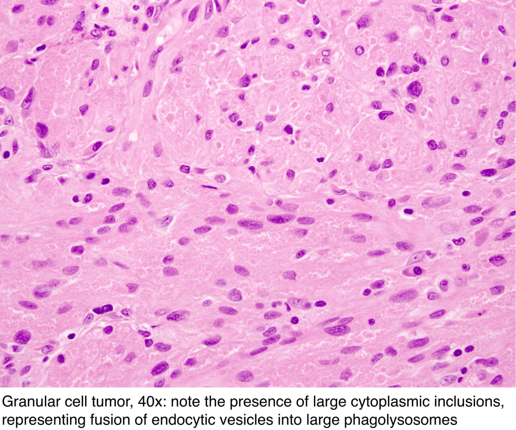
Obeidin, F. and Alexiev, B. (2026) Soft tissue. Available at: https://www.pathologyoutlines.com/topic/softtissuegranularcell.html (Accessed: 17th October 2026)
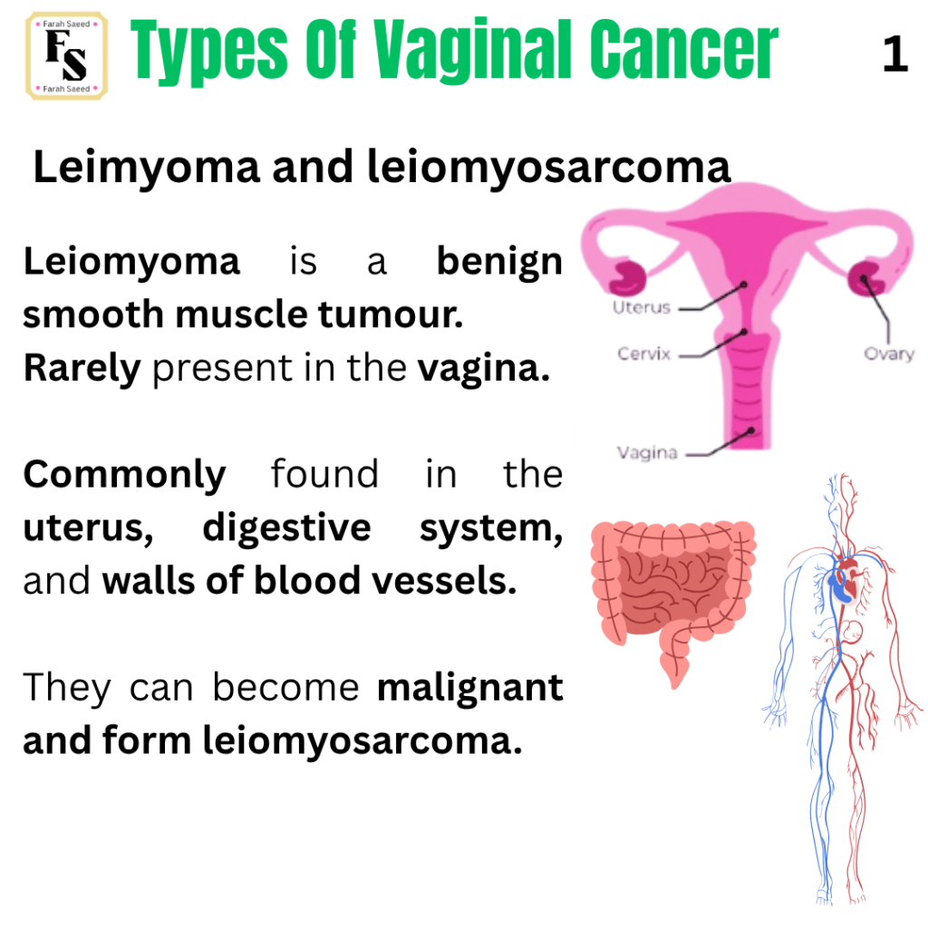
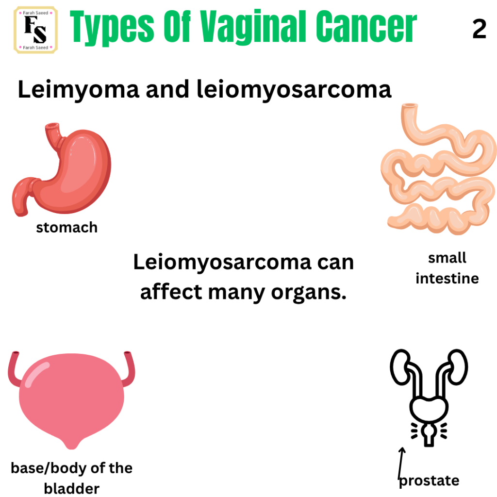
Leiomyoma and Leiomyosarcoma
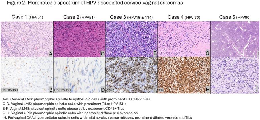
This image is by Archila et al. (2026), who investigated HPV-associated sarcoma of the cervix and vagina: leiomyosarcoma (LMS), undifferentiated pleomorphic sarcoma (UPS), and deep aggressive angiomyxoma (DAA). The data provided above indicate there are distinctive features that differentiate between all three subtypes: histological features, the HPV subtypes, common (16, 51) and rare (30, 90, 114), the type of genetic mutation, necrosis (cell death), mitotic activity, size of mass, type of borders, the presence of nuclear atypia, which influences the area of recurrence, overall survival (OS), and progression-free survival (PFS). However, all have tumour-infiltrating lymphocytes (TIL). The vaginal LMS presents as spindled to epithelioid cells with eosinophilic to amphophilic cytoplasm. Eosinophils are a type of immune cell released to help prevent allergy and ingest pathogens. Amphophilic cytoplasm can stain with haemotoxylin and eosin (HE). Haematoxylin is basic, whereas eosin is acidic. There is also a presence of several abnormally shaped nuclei (severe nuclear atypia) and hyalinized stroma (a form of connective tissue). Here, the researchers investigated cervical LMS and two vaginal LMS cases.
Cervical LMS is associated with subtype HPV51. It has the smallest mass size, 3 cm, among all the subtypes but does not have necrosis. It has an infiltrative border and a high mitotic count (high-power field) of 26-40. It commonly recurs in bone, gluteus (muscle on back/buttocks), abdomen, scalp, and chest. Cervical LMS is commonly associated with genetic mutations like the chromatin remodelling gene ATRX.
Similarly, vaginal LMS is commonly linked with ATRX mutation but lacks mutations of tumour suppressor genes TP53/RB1. Vaginal LMS has a large mass that varies between 10.5 and 13. It has a pushing border. It is commonly associated with HPV51, HPV16, and HPV114. The mitotic count per HPF varies; some have 6-9, whereas others have 15-20 HPF. They vary with recurrence, some in the pelvis, others in the lung.
Perivaginal DAA has variable cellularity, spindled to epithelioid, with mild atypia. It does not have necrosis but has infiltrative borders. It is commonly linked with HPV90. It has low mitotic activity with 1 -2 mitotic counts per HPF. Its mass size is 6cm. Mostly expresses ER or PR (oestrogen and progesterone). Perivaginal DAA does not have any genetic mutations. Recurrences commonly appear in the perianal region (back passage). It also has high overall survival.
Vaginal UPS has a mass size of 3.9 cm with the presence of pushing borders and severe cellular atypia. It is commonly associated with HPV30. It has mitotic activity of 9 HPF; it is commonly associated with ATRX mutations but has no gene fusions. There is also necrosis, but it has no recurrences.
Archila, L.R., Balakrishnan, R., Boroujeni, A.M., Chiang, S., Ellenson, L., Connelly, C., Bowman, A., Vanderbilt, C., Agaram, N., Abu-Rustum, N., Hensley, M. and Park, K. (2026). 1048 HPV-Associated sarcomas of the cervix and vagina – detection of common (16, 51) and rare (30, 90, 114) HPV types, recurrent ATRX alterations and wild-type TP53/RB1 By next-generation sequencing (NGS). Laboratory Investigation, 106(3), p.105338. doi:10.1016/j.labinv.2025.105338.
Hemangioma
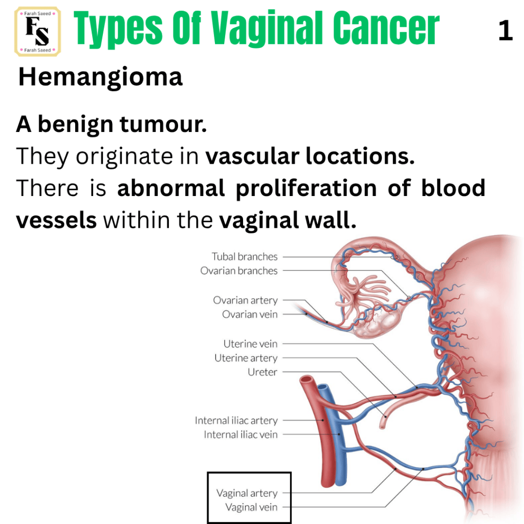
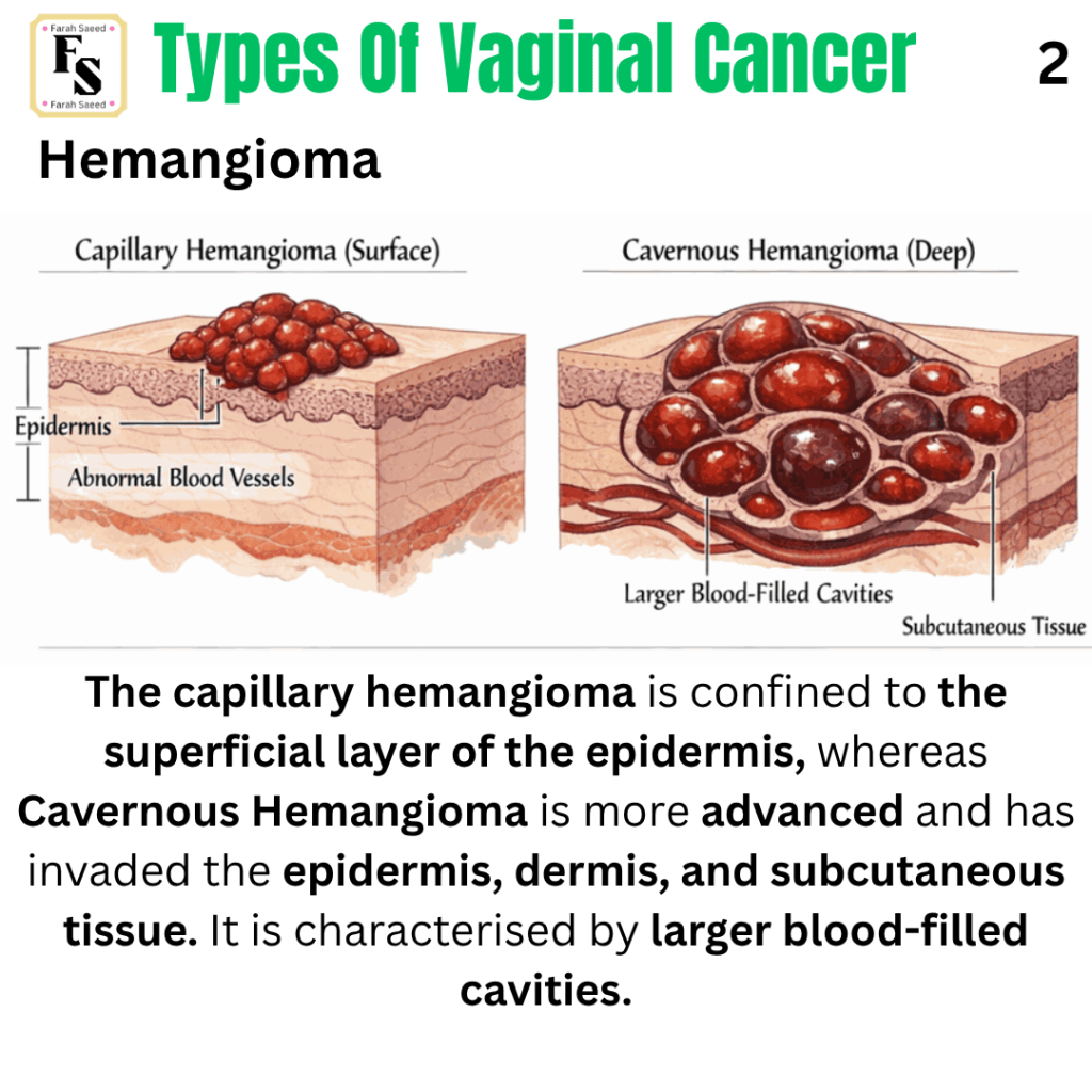
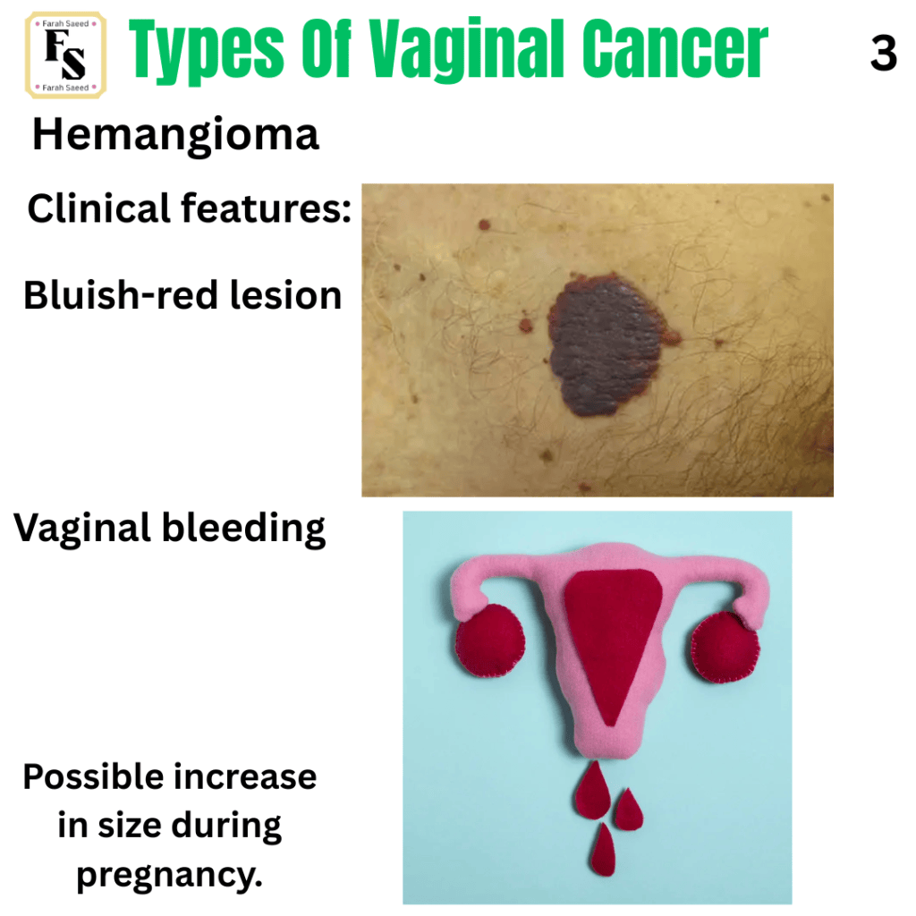
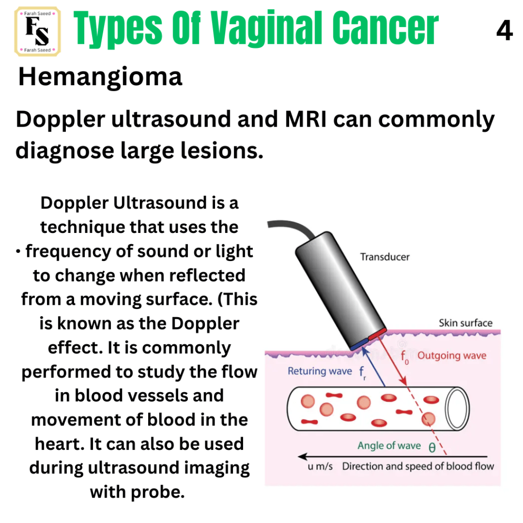
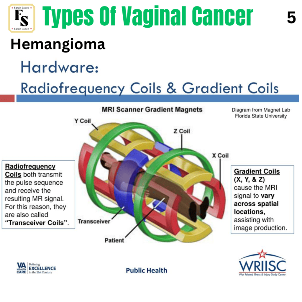
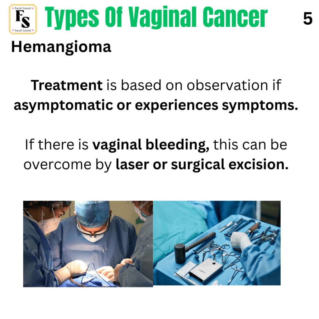
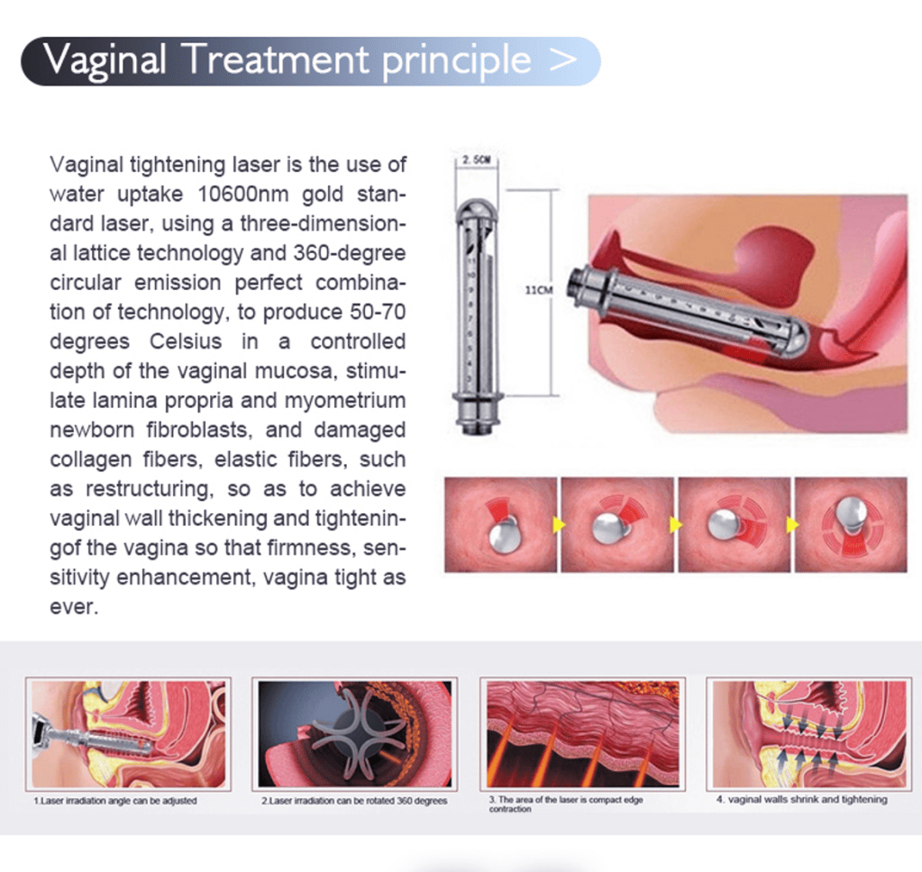
Supporting Images For Hemangioma



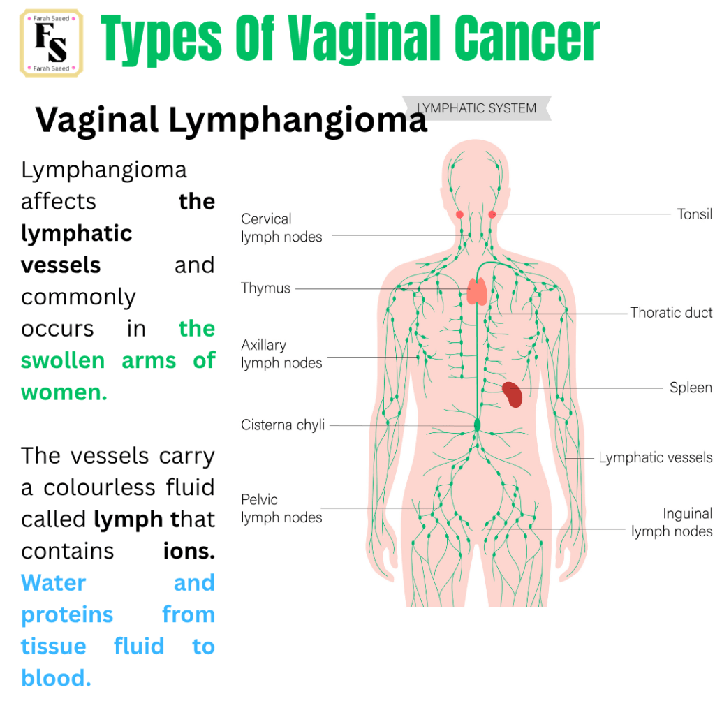
Vaginal Lymphangioma
Recommended Link For Lymphangioma detection and diagnosis
https://www.ultrasoundmedicvn.com/2012/12/case-161vaginal-mass-suspected.html
Recommended Reading Resource For Lymphangioma of the vulva that shares similarities with vaginal lymphangioma
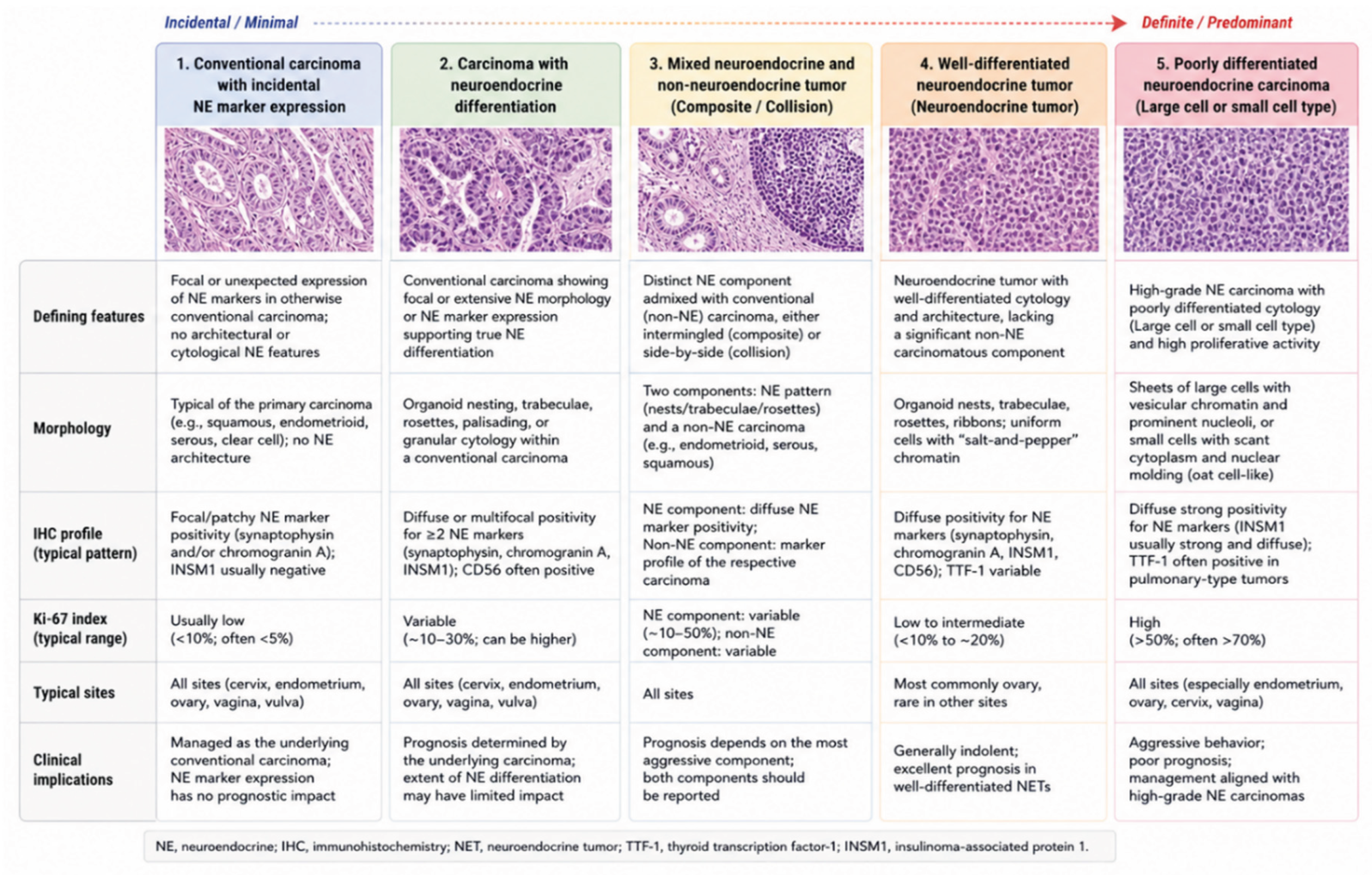
Neuroendocrine Tumours
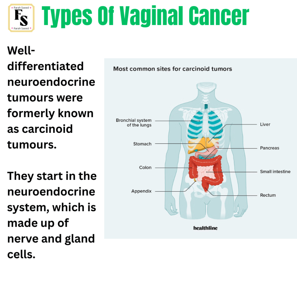
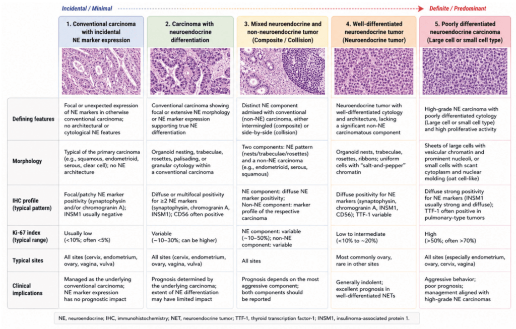
Source of table.
Glandular Tumours
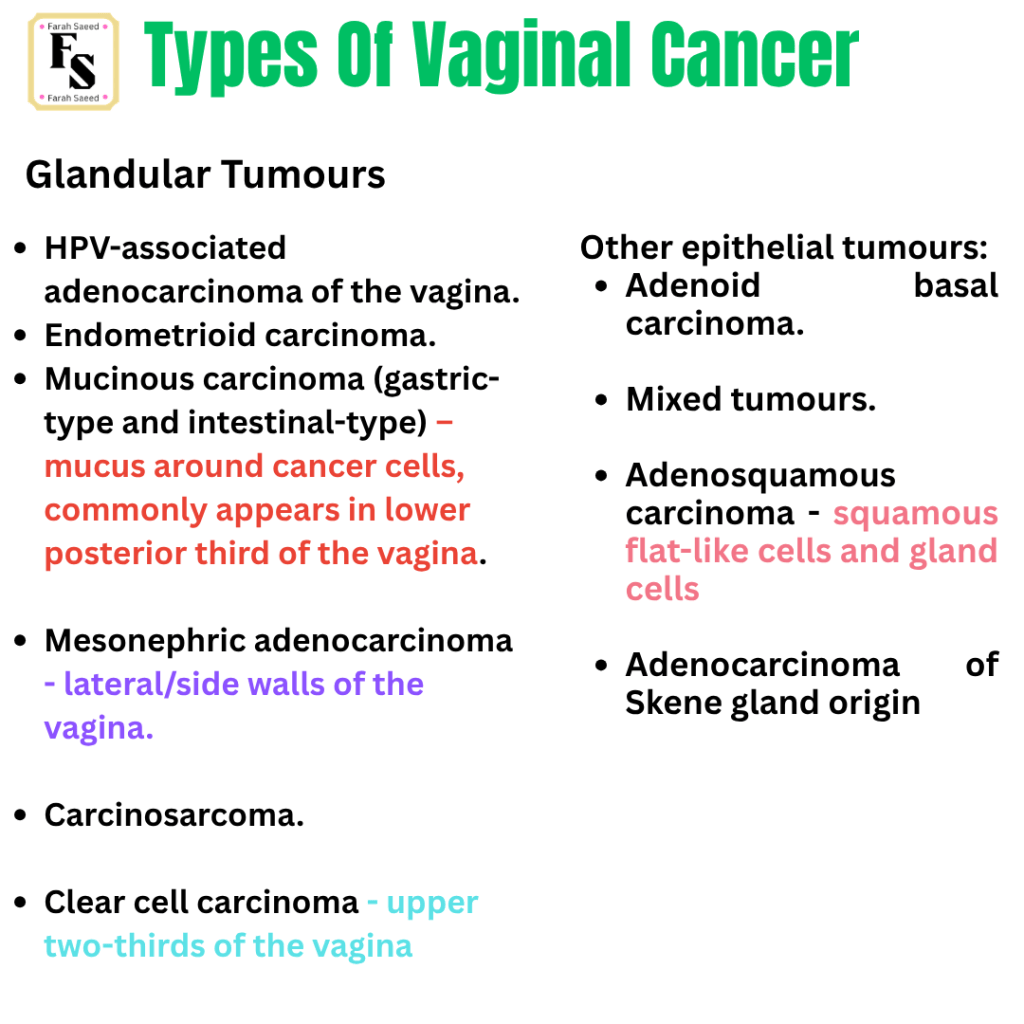
Overview Of Adenocarcinoma And Clear Cell Carcinoma
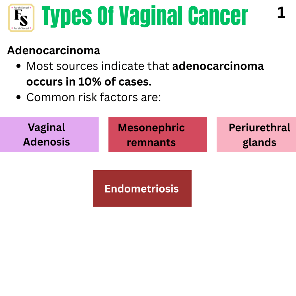
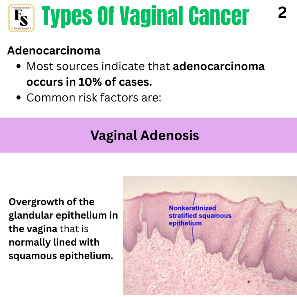
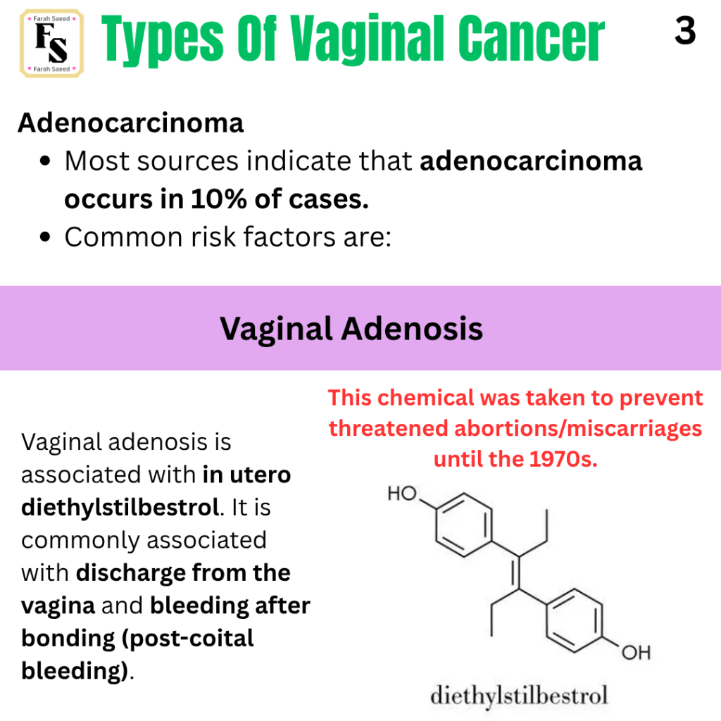
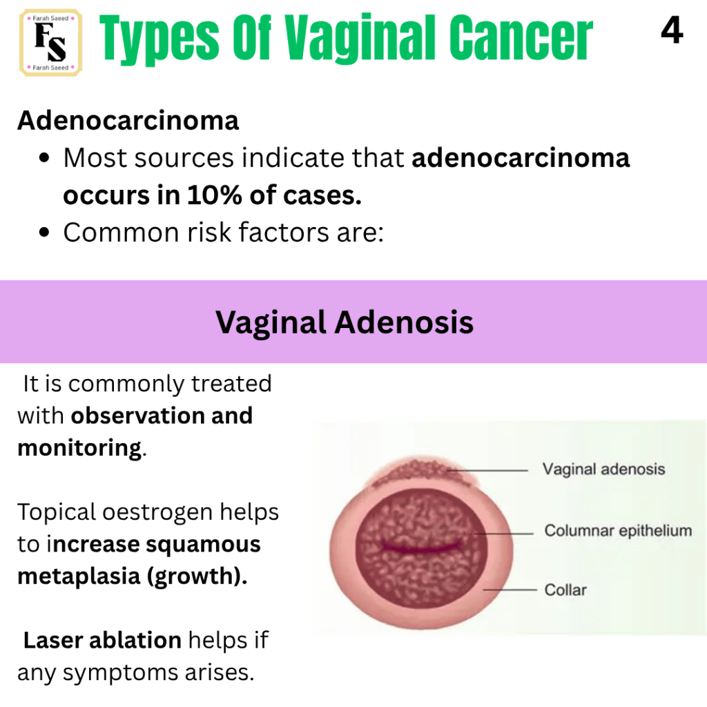
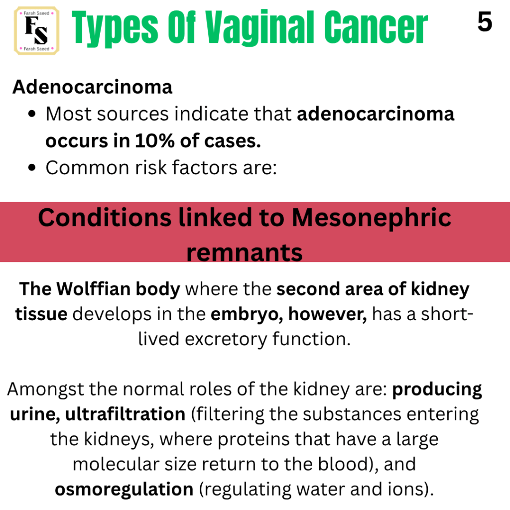
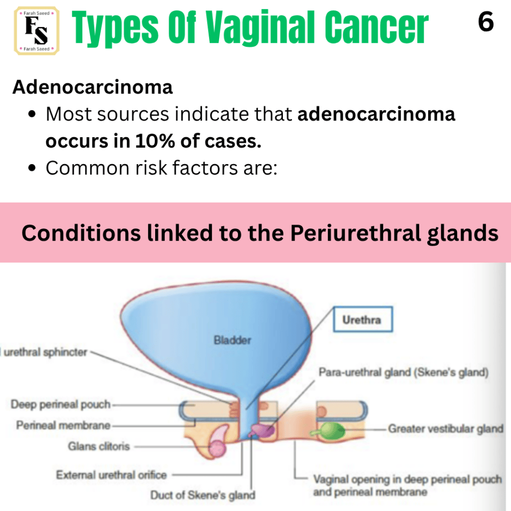
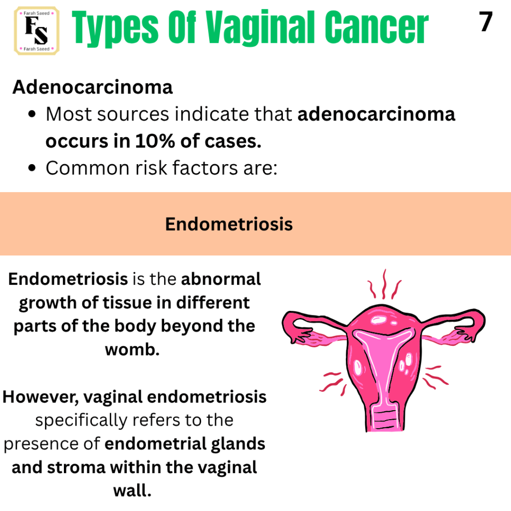
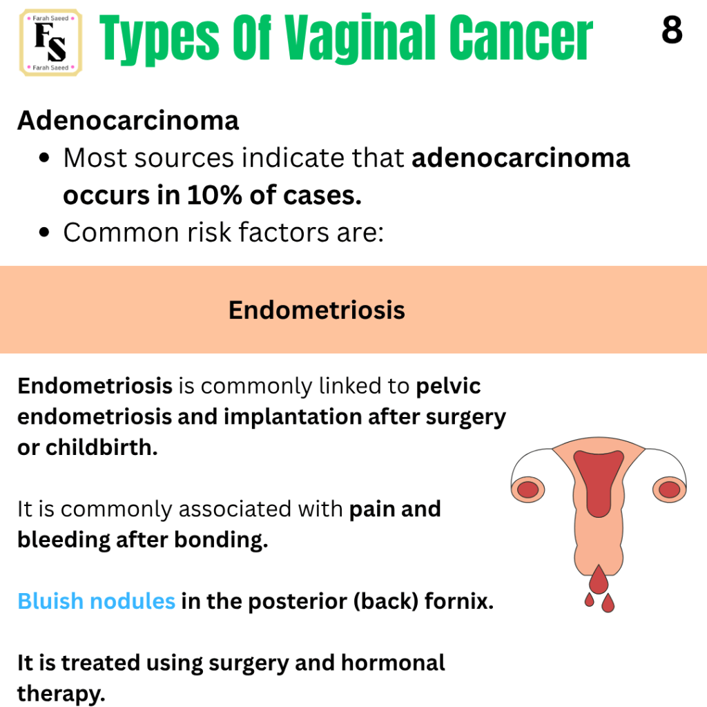
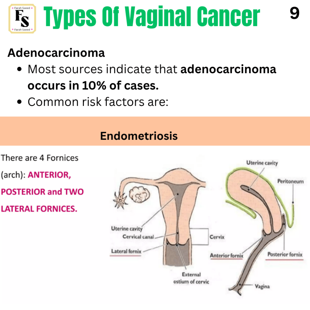
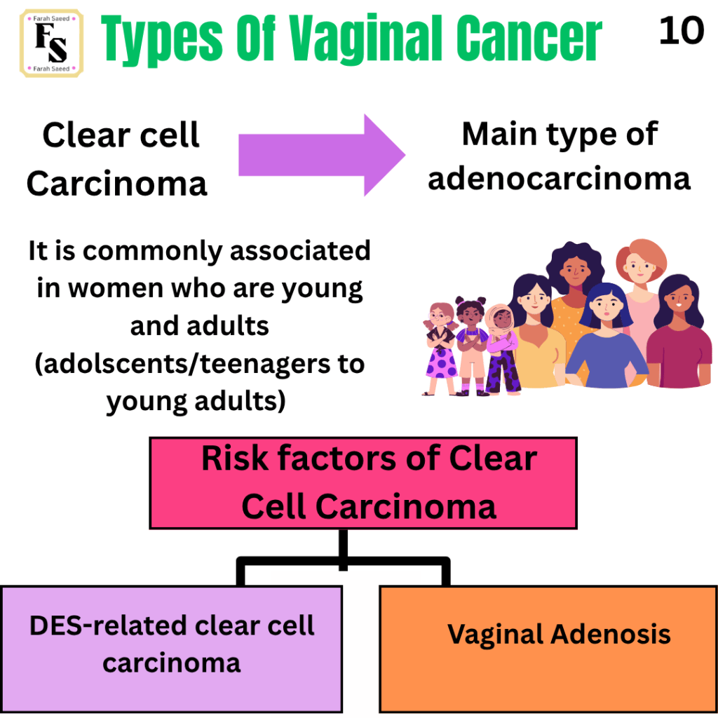
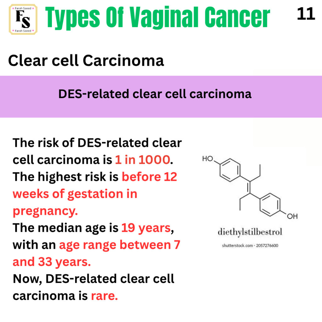
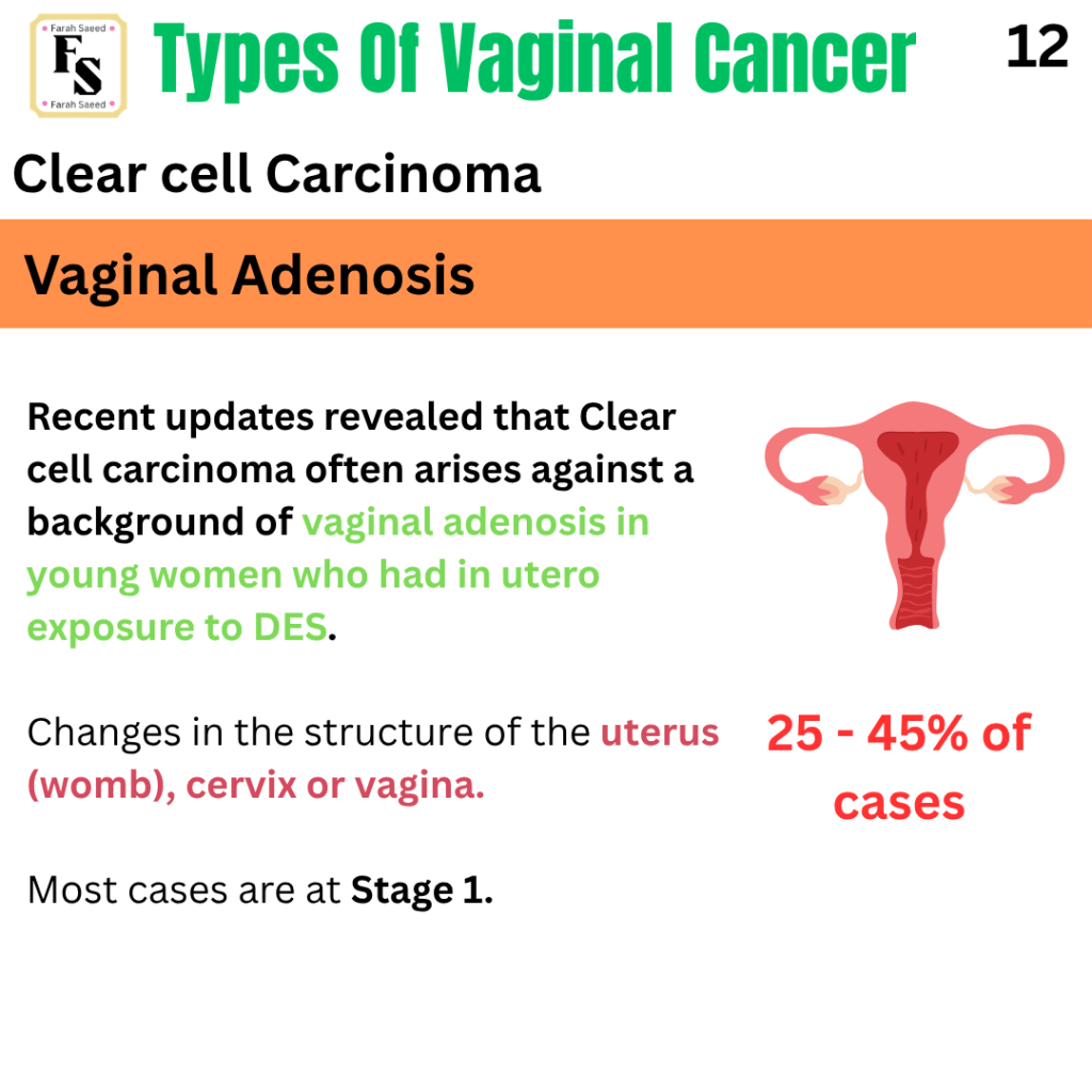
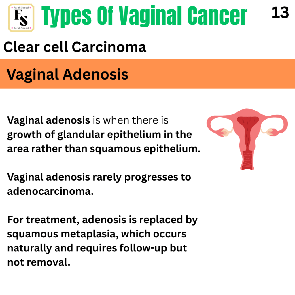

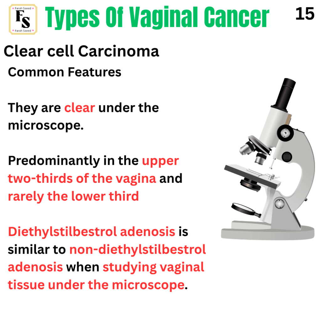
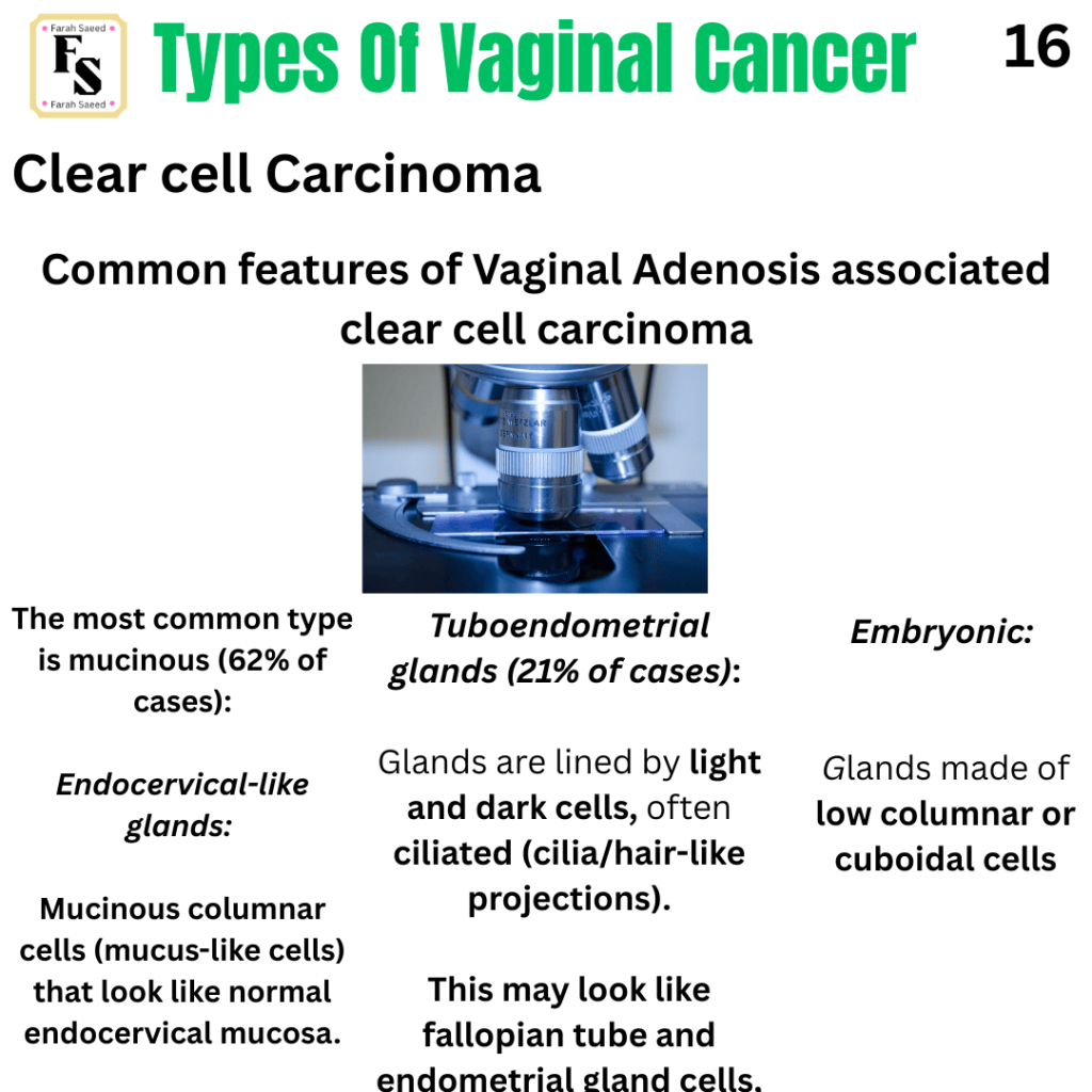
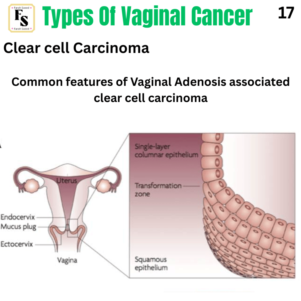
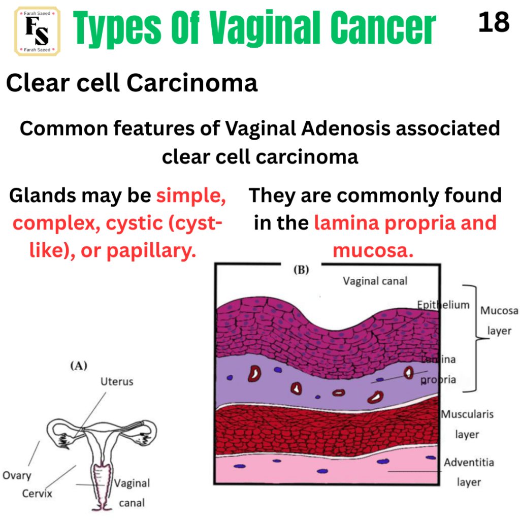
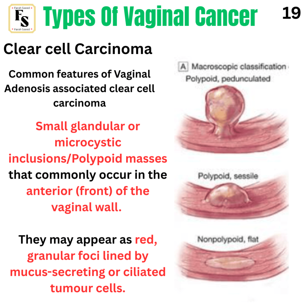
Supporting Images Of Adenocarcinoma
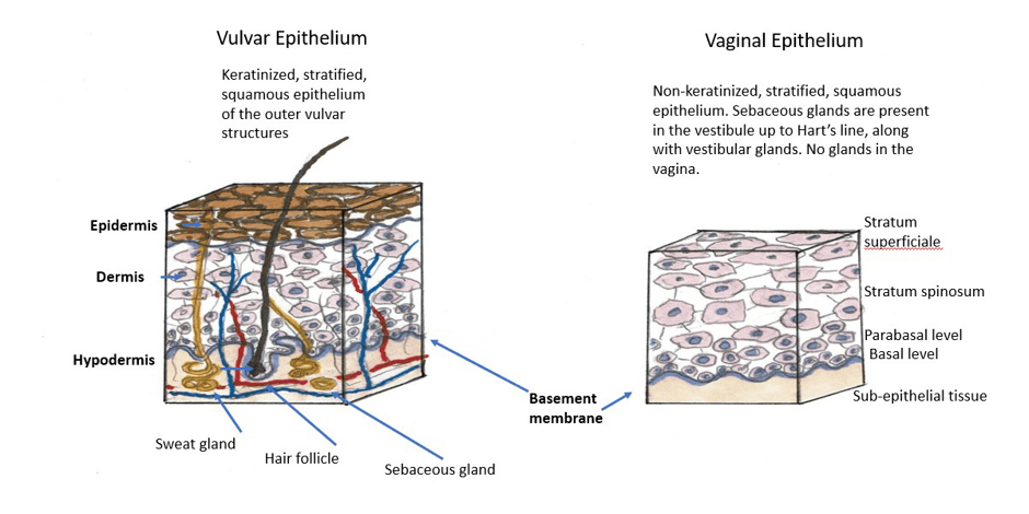
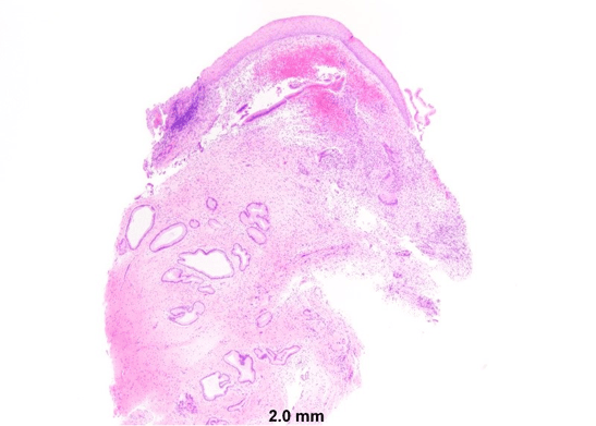
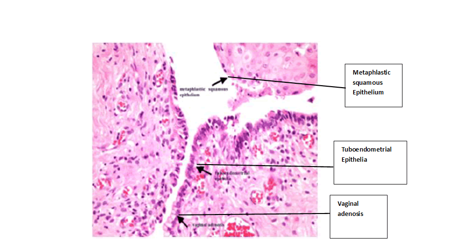
A microscopic presentation of vaginal tissue. The black arrows present the following: metaphlastic squamous epithelium, tuboendometrial epithelia and Vaginal adenosis.
Dagdeviren, H., Helvacioglu, C., Kanawati, A., Kaya, C. and Cengiz, H. (2017). Posterior vaginal wall cyst mimicking rectocele. Journal of Pregnancy and Child Health, 04(02). doi:10.4172/2376-127x.1000306.
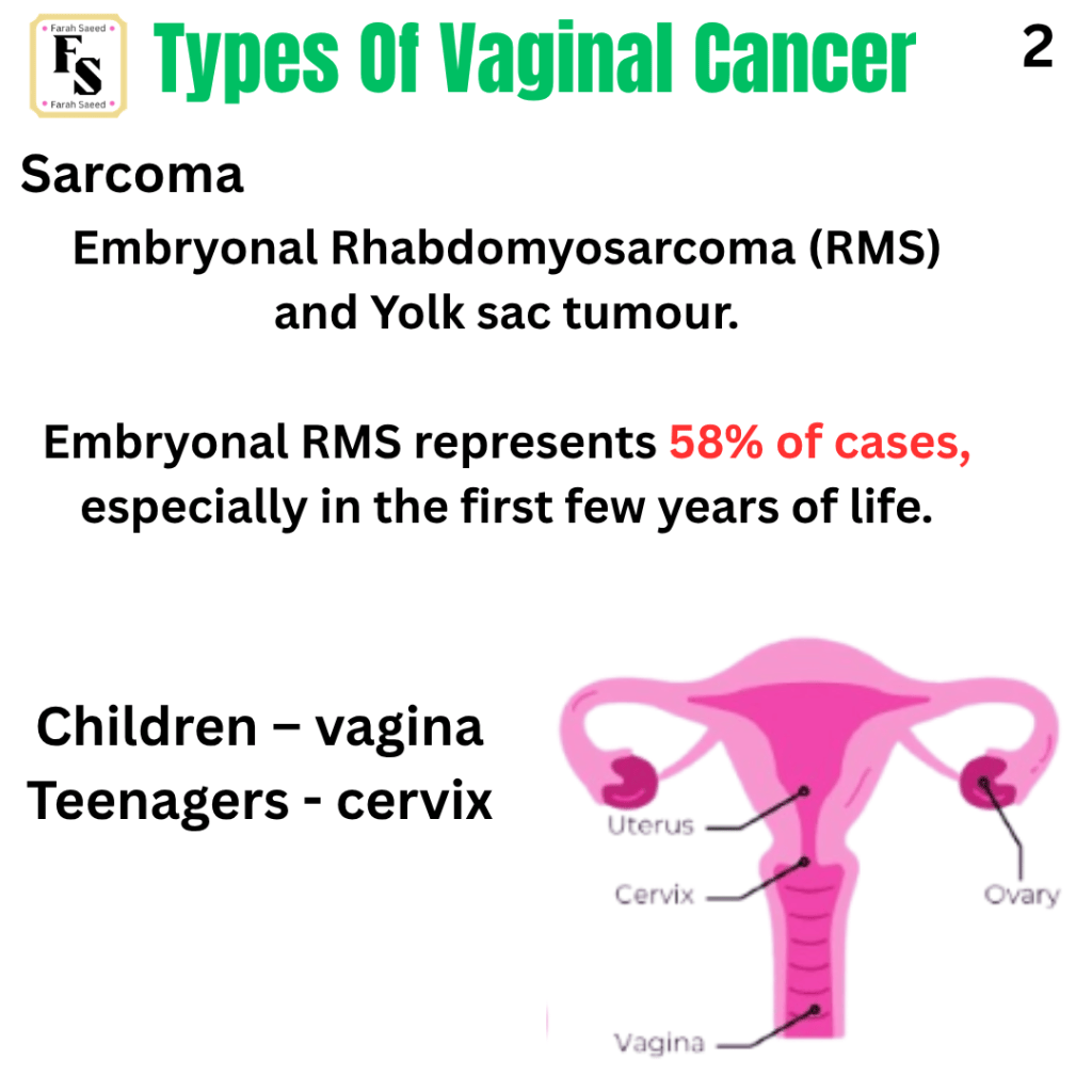
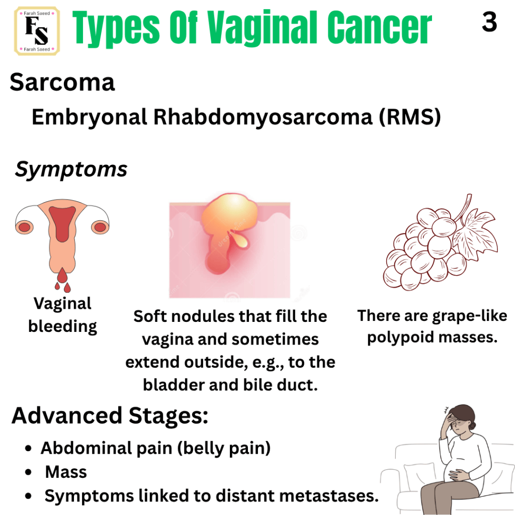
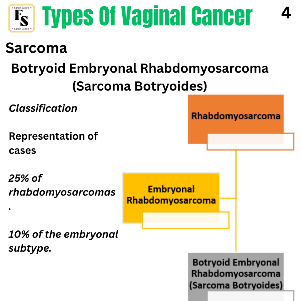
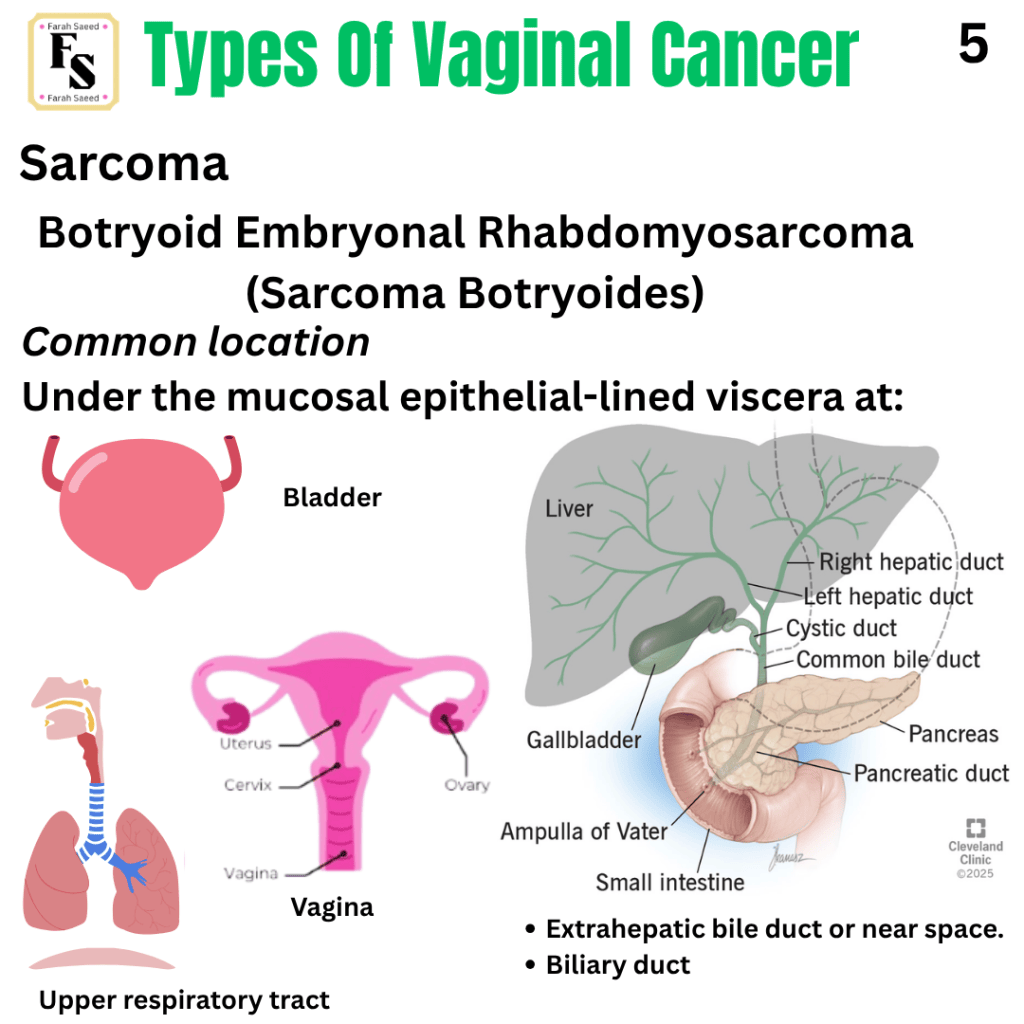
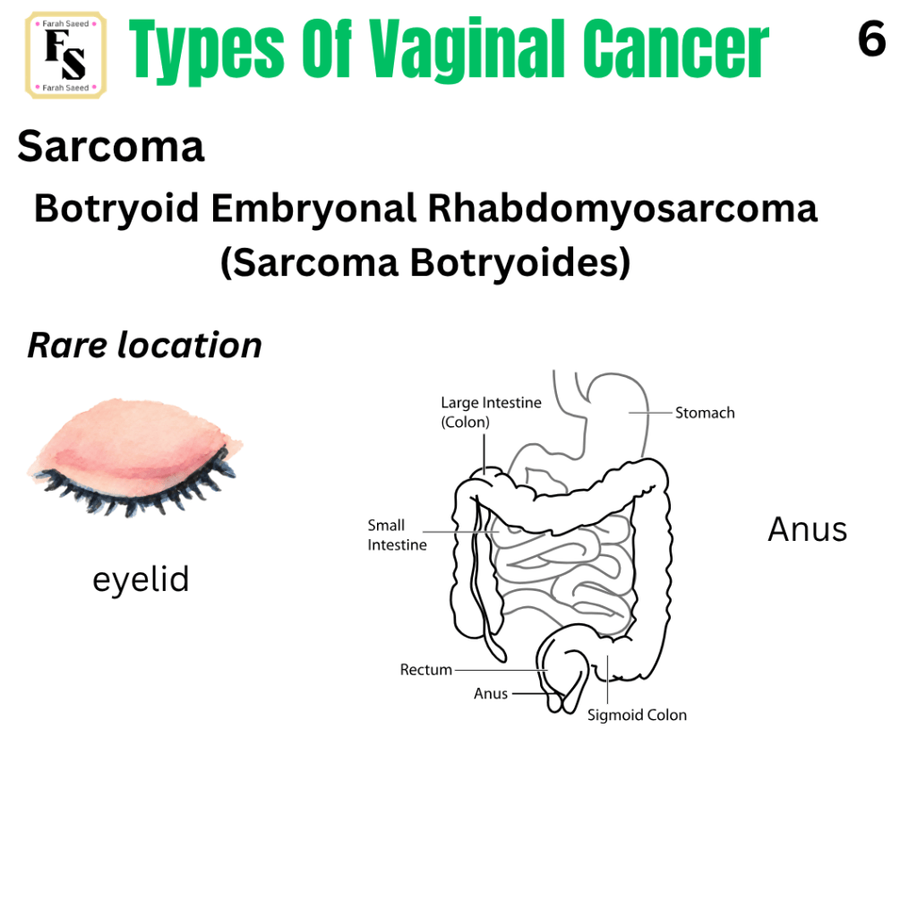
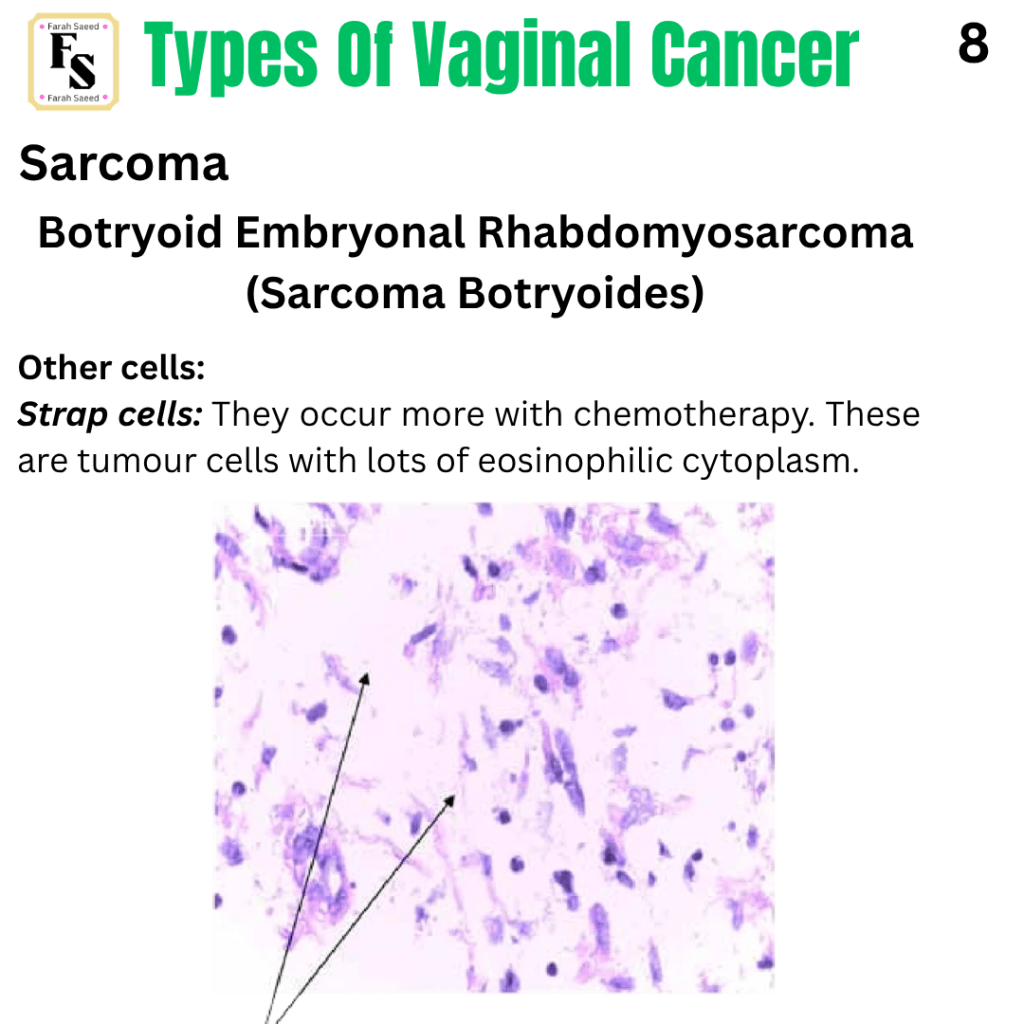
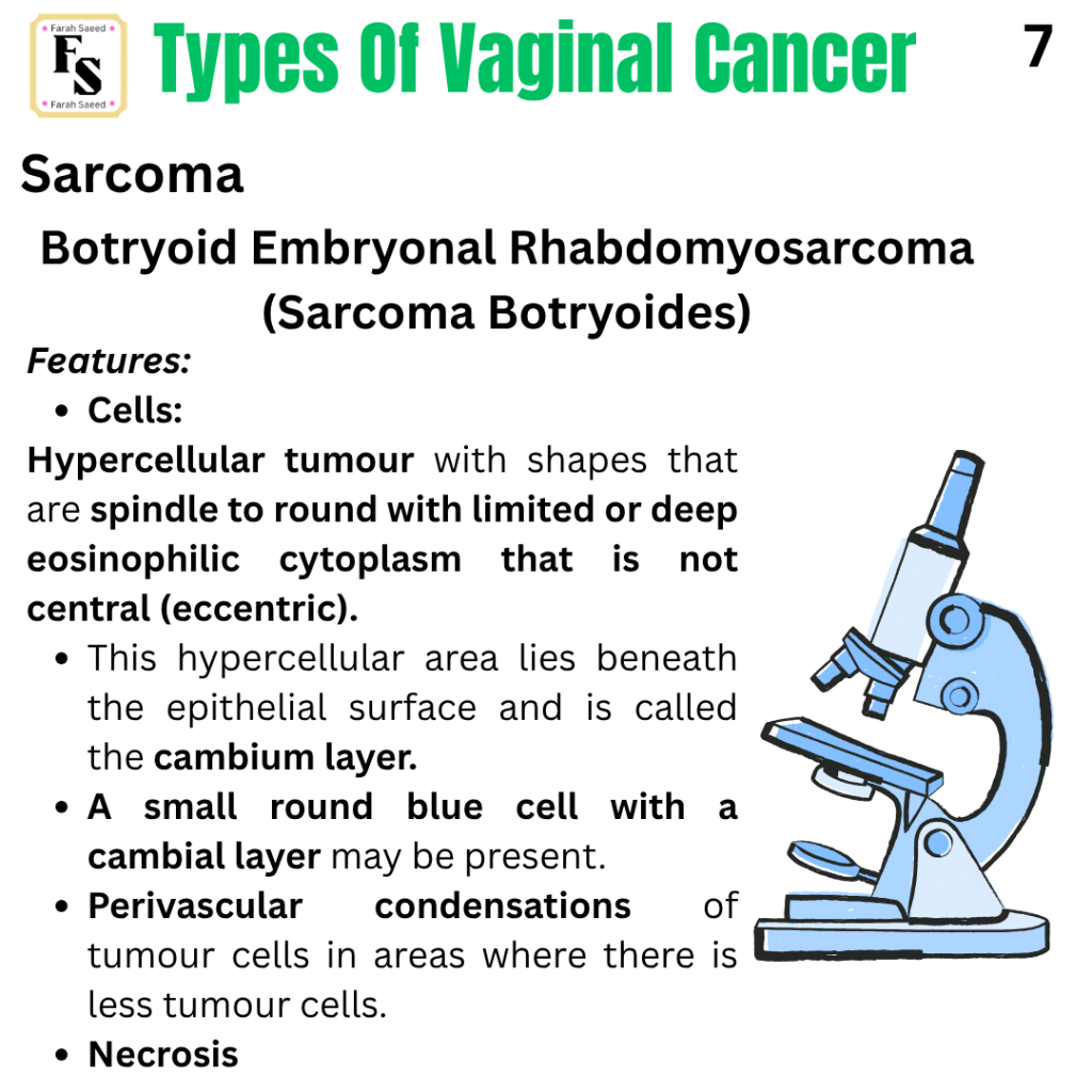
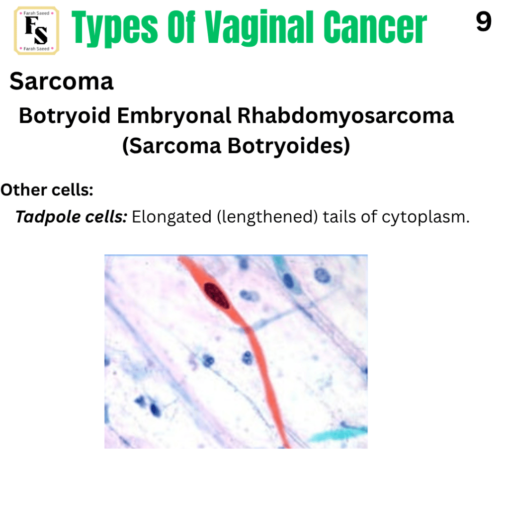
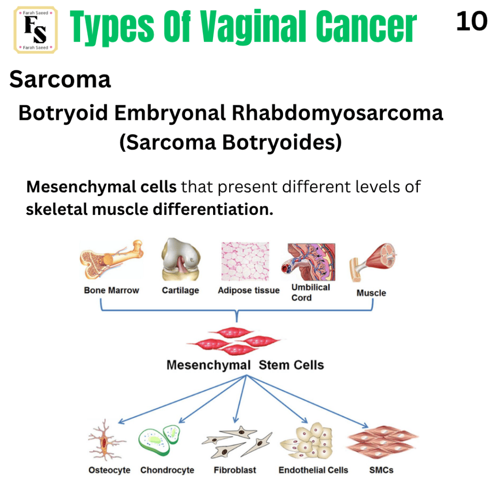
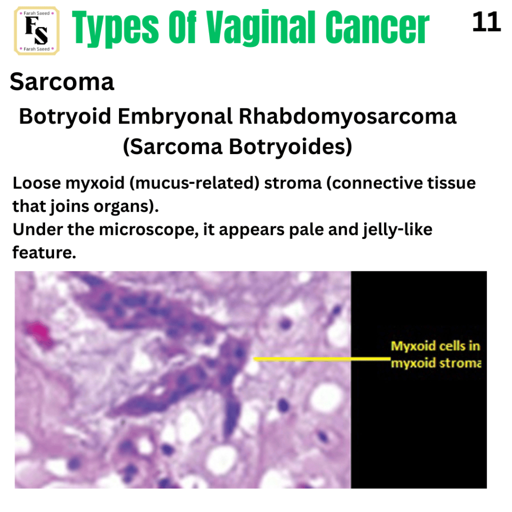
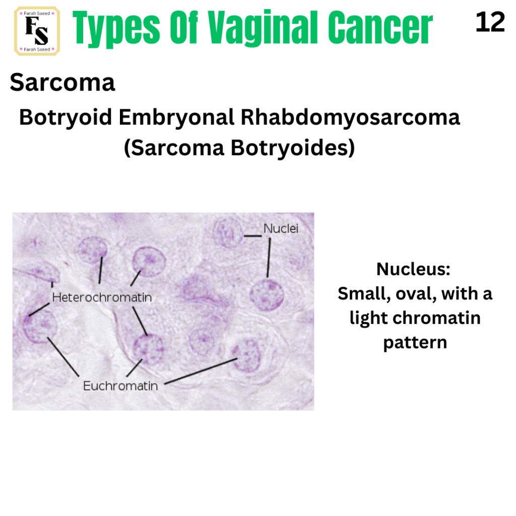
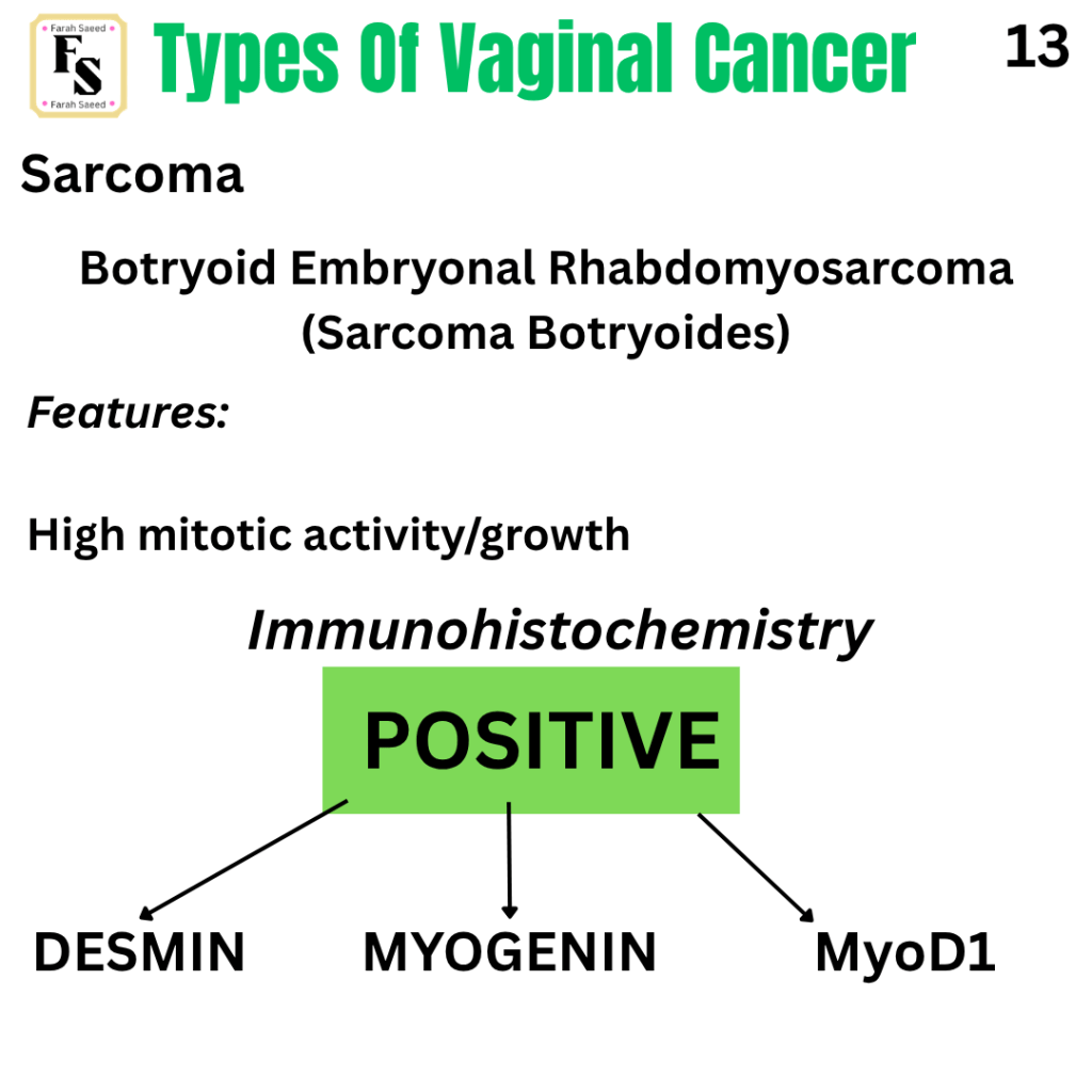
Sarcoma
An Insight Into The Immunohistochemistry Markers
Desmin
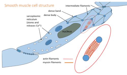
This is a major muscle-specific intermediate filament protein. It is highly expressed in the cardiac tissue (heart tissue) and helps with the development of the heart.
Desmin represents 2% of cellular protein in myocytes (muscle cells).
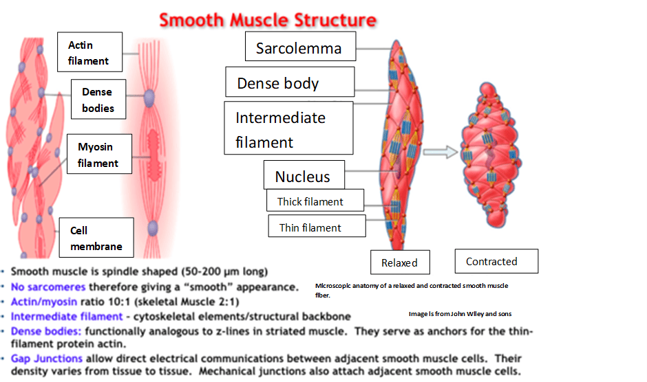
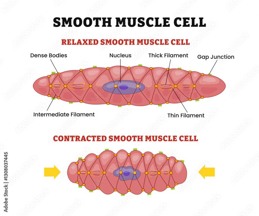
Smooth muscle cells, which are commonly found in the gastrointestinal tract, blood vessels, and other areas, contains desmin with the cytoplasmic dense bodies and subplasmalemmal dense plaques.
Skeletal/striated muscles have desmin filaments associated with sarcomeric Z disks.

Myogenin
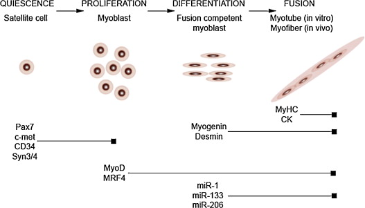
Myogenin is a transcription factor that is expressed in myotubes. It helps in the differentiation stages of making muscle cells (myogenesis), muscle regeneration, and activation of genes involved in myogenesis. The procedure involves mononuclear muscle precursor cells leaving the cell cycle, differentiating, and connecting with existing myofibers or with each other to make new myofibers.
MYOD1
This is a gene that encodes a transcription factor that is important for the production of muscles (myogenesis).
The MYOD1 transcription factor activates its own transcription, muscle-related genes, and helps to leave the cell cycle. It is also involved in the differentiation of the skeletal muscle.
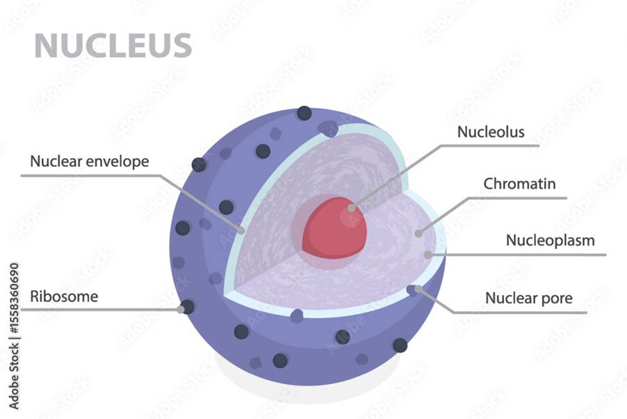
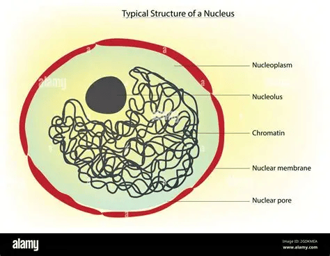
In the nucleus, more specifically the nucleoplasm and euchromatin, MYOD1 connects with MYF5 and MYOF to begin the myogenesis programme and reprogram fibroblasts into myoblasts. The upregulation of the CDKN1A gene helps to stop the cell cycle so myogenesis can begin.
Mutations in MYOD1 can lead to diseases in the muscles referred to as myopathy. It is characterised by weakness in the muscles, pain, and tenderness. This can be born with (congenital), such as foetal akinesia (loss of muscle tone or response) and arthrogryposis (limited movement due to contractures in two or more joints.
Mutations in MYOD1 can also be inherited from other family members who have the condition.
Acquired-related myopathies may be associated with other health conditions, for instance, endocrine/hormone disorders, drug therapy, and cancers like rhabdomyosarcoma.
Important Reminder
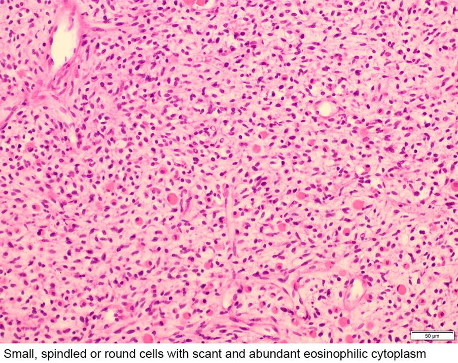
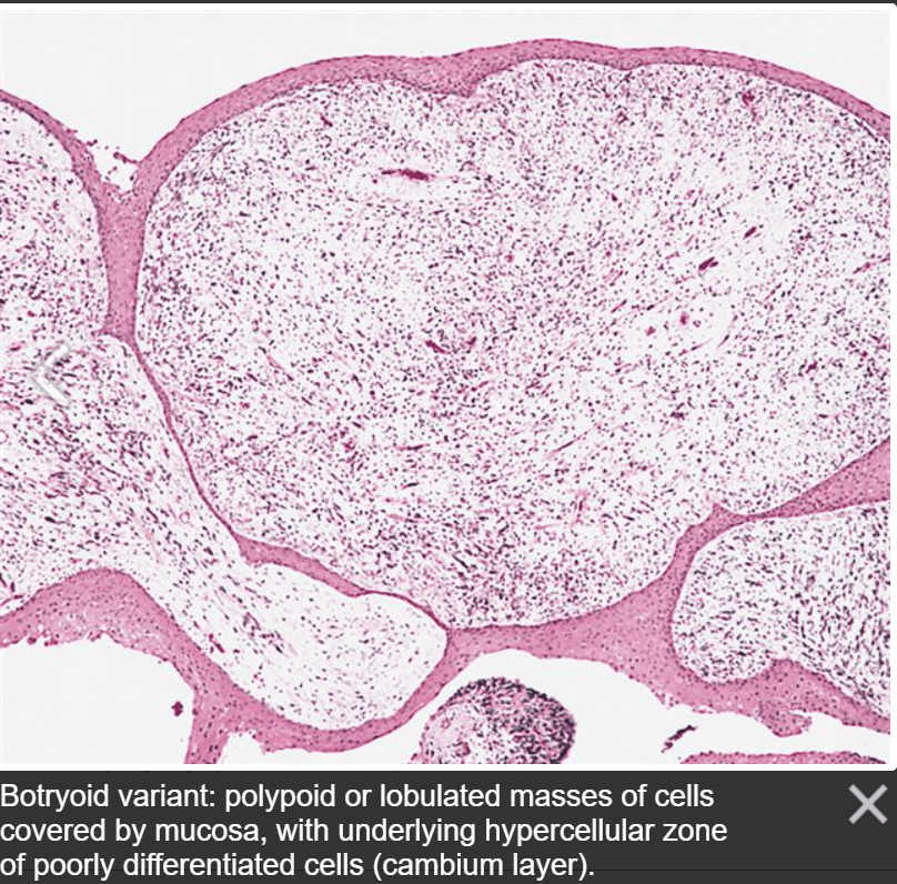
MyoD1/myogenin positive marker. It appears in a linear pattern. There is staining of the muscle and not the tumour.
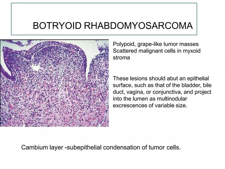
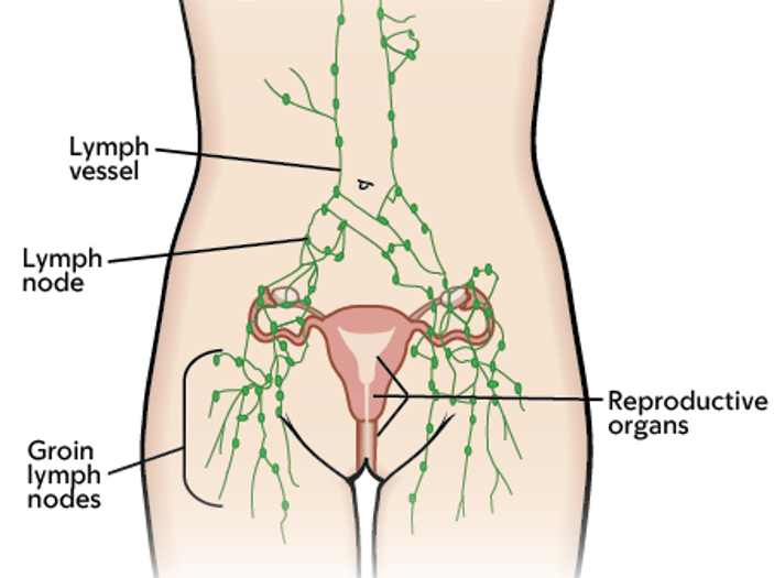
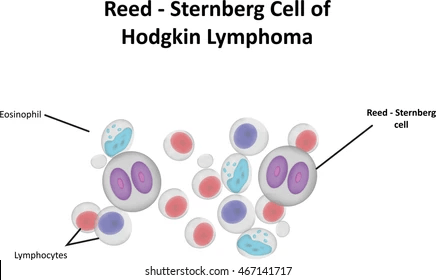
Vaginal Lymphoma
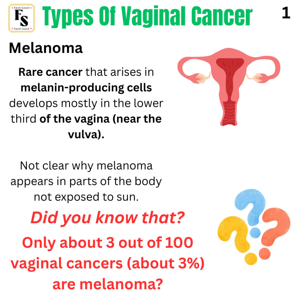
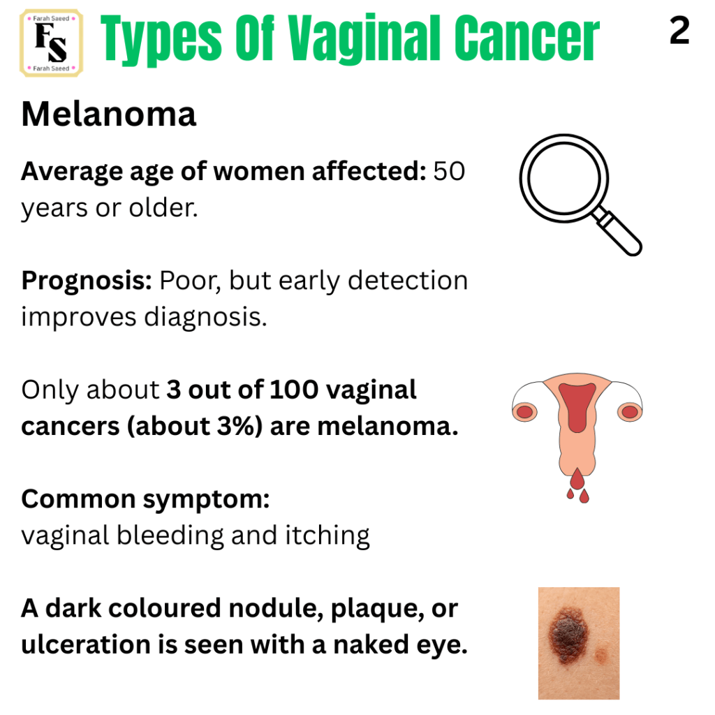
Melanoma
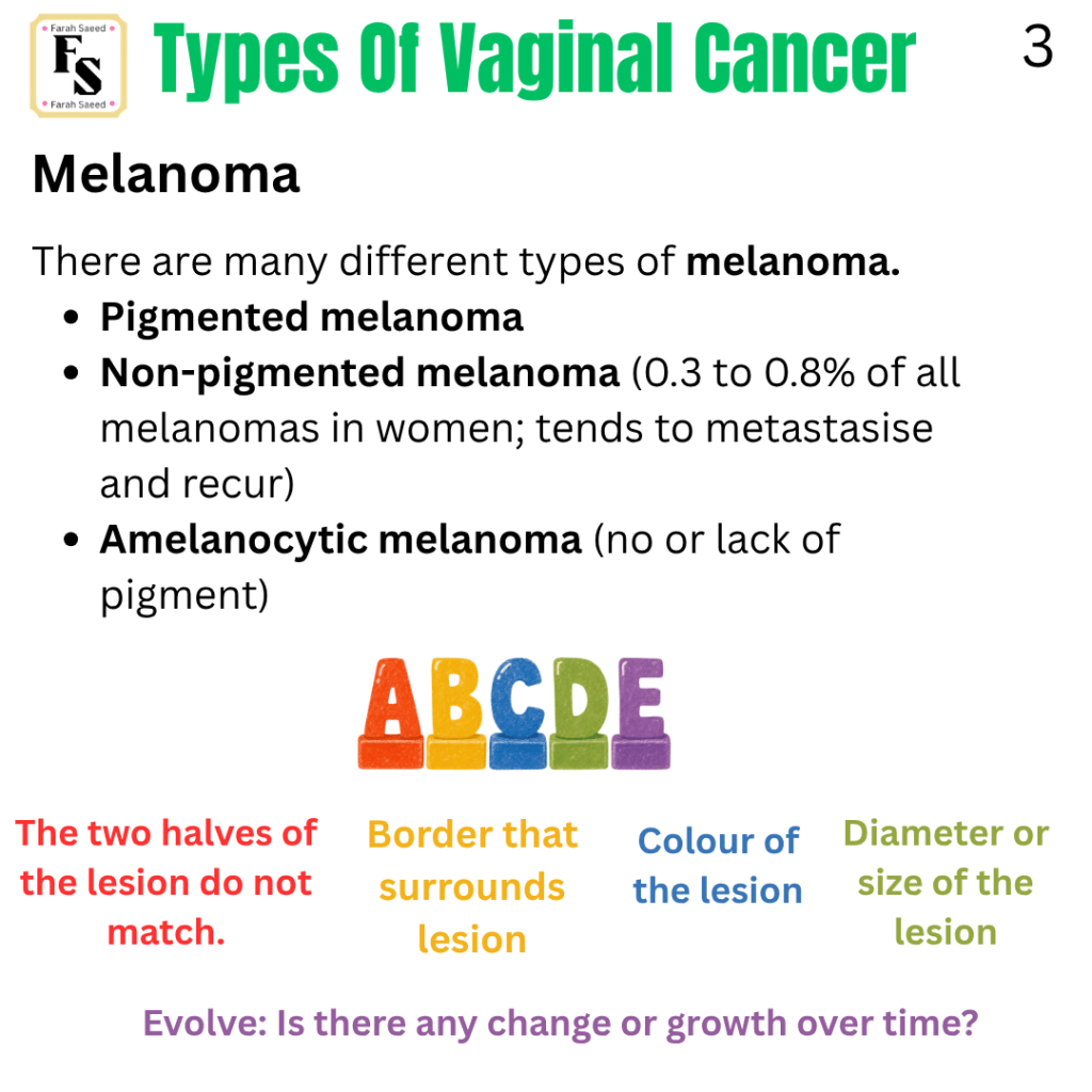
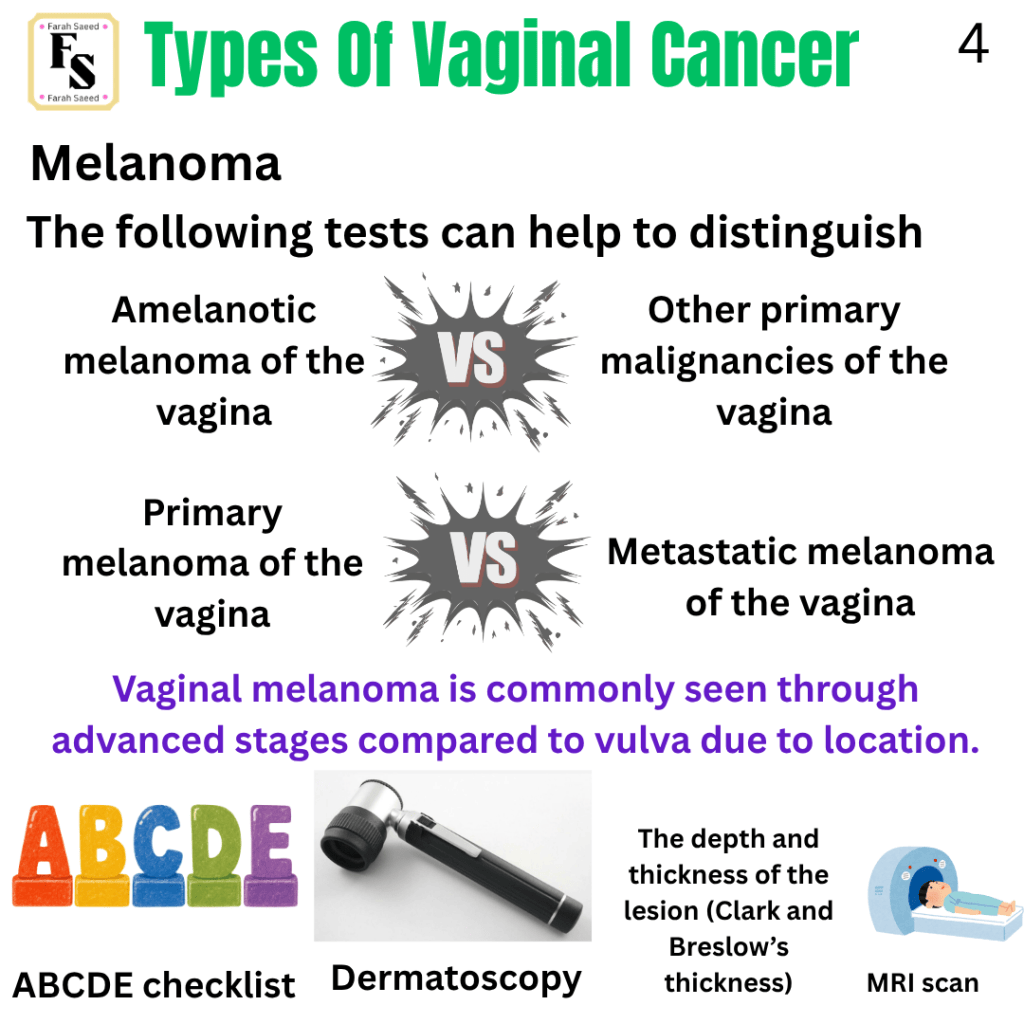
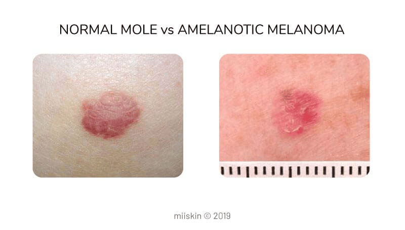
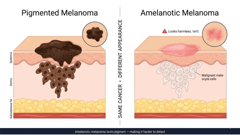
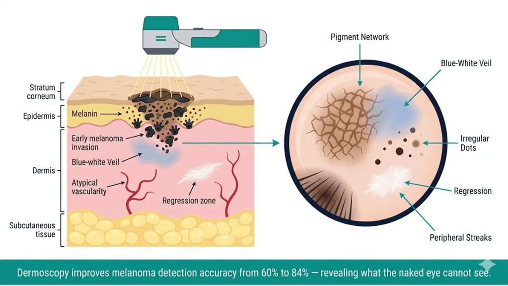
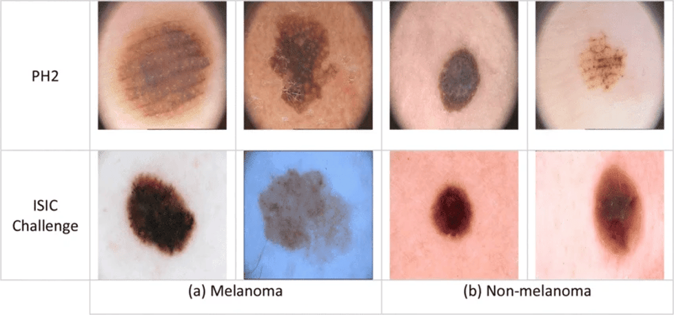
Here, this image presents features that distinguish the difference between melanoma and non-melanoma using two types of databases: PH2 and ISIC. PH2 is a dermoscopic Image Database for Research and Benchmarking to help detect cases and classify melanomas based on the following factors: segmentation of the lesion, clinical diagnosis, diagnosis under the microscope (histology), and evaluation of different dermoscopic criteria: colour, pigment, dots/globules, streaks, regression areas and whether there is a blue-whitish veil. The other data were generated by the International Skin Imaging Collaboration (ISIC). It aims to improve the diagnosis and treatment of melanoma by setting standards in skin imaging, studying dermatologic images, which increases communication between healthcare professionals and those involved in computer science, artificial intelligence, and medical engineering.
Further information on PH dataset can be found on the following link:
Further information on ISIC dataset can be found on the following link:
Reference
Adams, T.S., Maluleke, J.C. and Cuello, M.A. (2025). Cancer of the vagina: 2025 Update. International Journal of Gynecology & Obstetrics, 171(S1), pp.48–59. doi:10.1002/ijgo.70325.
Archila, L.R., Balakrishnan, R., Boroujeni, A.M., Chiang, S., Ellenson, L., Connelly, C., Bowman, A., Vanderbilt, C., Agaram, N., Abu-Rustum, N., Hensley, M. and Park, K. (2026). 1048 HPV-Associated sarcomas of the cervix and vagina – detection of common (16, 51) and rare (30, 90, 114) HPV types, recurrent ATRX alterations and wild-type TP53/RB1 By next-generation sequencing (NGS). Laboratory Investigation, 106(3), p.105338. doi:10.1016/j.labinv.2025.105338.
Cancer Research UK (2024) Vaginal melanoma. Available at: https://www.cancerresearchuk.org/about-cancer/vaginal-cancer/types-grades/melanoma (Accessed: 12th October 2026)
Cancer Research UK (2025) Types of vaginal cancer. Available at: https://www.cancerresearchuk.org/about-cancer/vaginal-cancer/types-grades (Accessed: 12th October 2026)
Cassidy, J., Bissett, D., Spene, R. and Payne, M. (2010) Oxford Handbook of Oncology. Oxford: Oxford University Press.
Cerilli, L.A. and Wick, M.R. (2006). Immunohistology of Soft Tissue and Osseous Neoplasms. Elsevier eBooks, [online] pp.65–120. doi:10.1016/b978-0-443-06652-8.50009-0.
Choschzick, M. (2024) Vulva & vagina. Available at: https://www.pathologyoutlines.com/topic/vulvascc.html (Accessed: 16th October 2026)
Clay, M. (2026) Soft tissue. Available at: https://www.pathologyoutlines.com/topic/softtissueembryonalrhabdo.html (Accessed: 11th October 2026)
Dagdeviren, H., Helvacioglu, C., Kanawati, A., Kaya, C. and Cengiz, H. (2017). Posterior vaginal wall cyst mimicking rectocele. Journal of Pregnancy and Child Health, 04(02). doi:10.4172/2376-127x.1000306.
Dayyabu, A.L., IO, A. and BS, M. (2014). Sarcoma botryoides a management dilemma: A review of two cases. International Journal of Case Reports and Images, 5(7), p.482. doi:10.5348/ijcri-201458-cs-10044.
Gene Cards (2026) MYOD1 gene. Available at: https://www.genecards.org/card/MYOD1 (Accessed: 19th October 2026)
Jones, J. and Maldonado, L. (2026) Vulva and Vagina Non-Tumour Vaginal Adenosis. Available at: https://www.pathologyoutlines.com/topic/vaginaladenosis.html (Accessed: 19th October 2026)
Kamungo, A. (n.d.) Female Genital Tract Pathology. Available at: https://www.slideshare.net/slideshow/female-genital-tract-pathology-and-pptx/280526961 (Accessed: 11th October 2026)
Lin, G.-Y., Liu, Y., Ye, T., Lu, X.-Y., Gao, J. and Wang, Y.-Z. (2022). Granular cell tumor of the vulva: Case report and systematic review of the literature. Medicine, [online] 101(52), p.e32568. doi:10.1097/MD.0000000000032568.
Mehta, V., Balachandran, C., Rao, L. and Geeta, V. (2010). Giant granular cell tumor of the vulva. Indian Journal of Dermatology, Venereology, and Leprology, 76(3), p.263. doi:10.4103/0378-6323.62966.
Montano, M. and Long, K. (2011). RNA surveillance—An emerging role for RNA regulatory networks in aging. Ageing Research Reviews, 10(2), pp.216–224. doi:10.1016/j.arr.2010.02.002.
National Cancer Institute (2025) HPV and Cancer. Available at: https://www.cancer.gov/about-cancer/causes-prevention/risk/infectious-agents/hpv-and-cancer (Accessed: 6th August 2026)
Nout, R.A., Calaminus, G., Planchamp, F., Chargari, C., Lax, S., Martelli, H., McCluggage, W.G., Morice, P., Pakiz, M., Schmid, M.P., Stunt, J., Timmermann, B., Vokuhl, C., Orbach, D. and Fotopoulou, C. (2023). ESTRO/ESGO/SIOPe Guidelines for the management of patients with vaginal cancer. International Journal of Gynecologic Cancer, [online] 33(8). doi:10.1136/ijgc-2023-004695.
Obeidin, F. and Alexiev, B. (2026) Soft tissue. Available at: https://www.pathologyoutlines.com/topic/softtissuegranularcell.html (Accessed: 17th October 2026)
Oliveira, R., Bócoli, M.C., Saldanha, J.C., Murta, E.F.C. and Nomelini, R.S. (2013). Primary small cell carcinoma of the vagina. Case Reports in Obstetrics and Gynecology, 2013, pp.1–4. doi:10.1155/2013/827037.
Rosh Review (2026) Explanation: D is the correct answer. Available at: https://go.roshreview.com/obgyn-qid-126985 (Accessed: 16th October 2026)
William, B. and Mukoolo, T. (n.d.) Benign Tumors and lesions of vagina. Available at: https://www.slideshare.net/slideshow/benign-tumors-and-lesions-of-vagina-tom-pptx/286065779#20 (Accessed: 17th October 2026)
Updated October 2026; Next Review September 2028


Leave a Reply