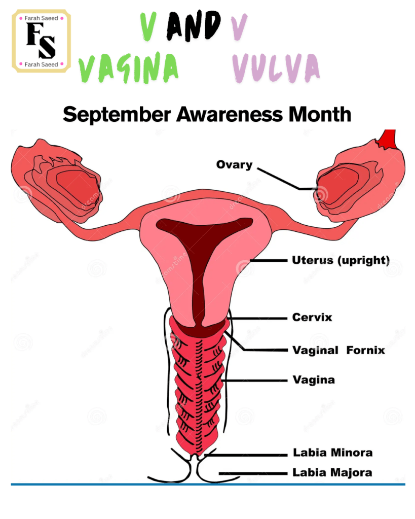
How Does Vaginal Cancer Spread?
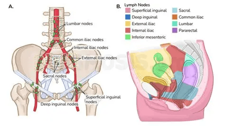
Gentle Reminder:
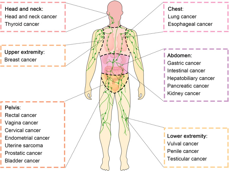
Metastatic vaginal cancer may appear as a result of:
Organs in the female reproductive system: cervix, vulva or the endometrium (womb).
Organs in other parts of the body: Kidney, breas and lung
How does it spread?
There are several main pathways: lateral, iliac node hypogastric, inguinal, anterior, mesorectal and presacral
Spread from the upper thirds of the vagina/vaginal apex:
56% of cases.
Lateral pathway

Internal iliac hypogastric pathway
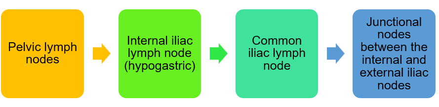
After the common iliac lymph node, it goes to the para-aortic lymph node and then aortic
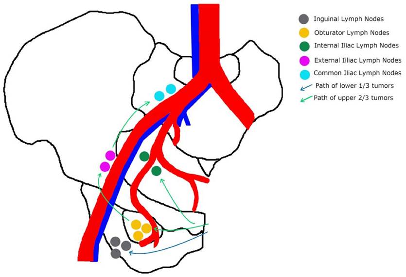
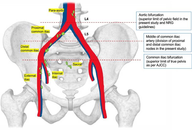
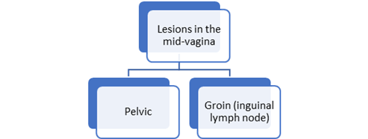
Spread from the middle of the vagina : 13% of cases.
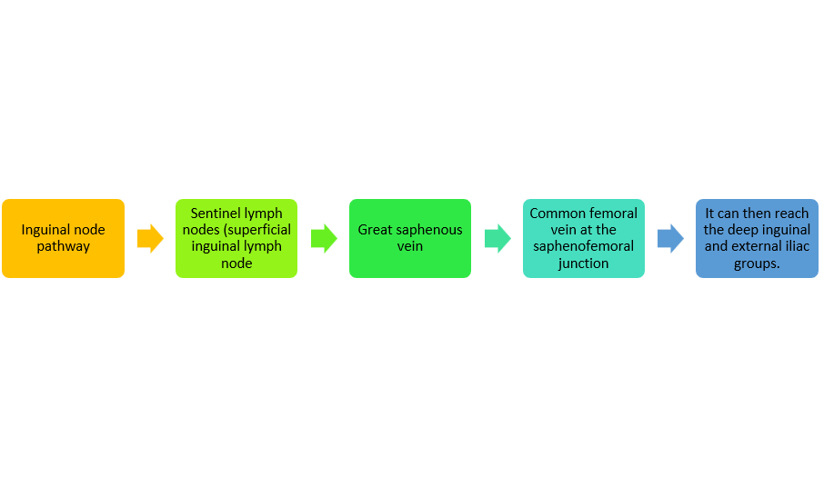
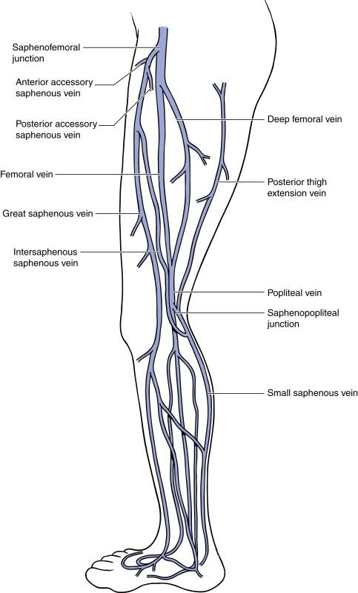
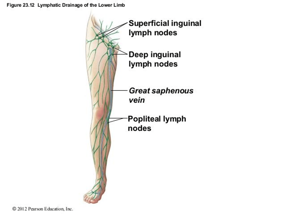
Lesions in the mid-vagina may follow both the pelvic and groin routes
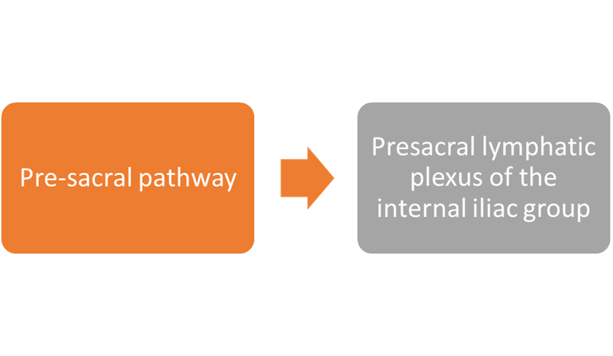
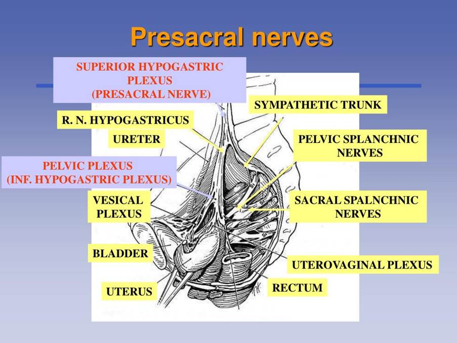
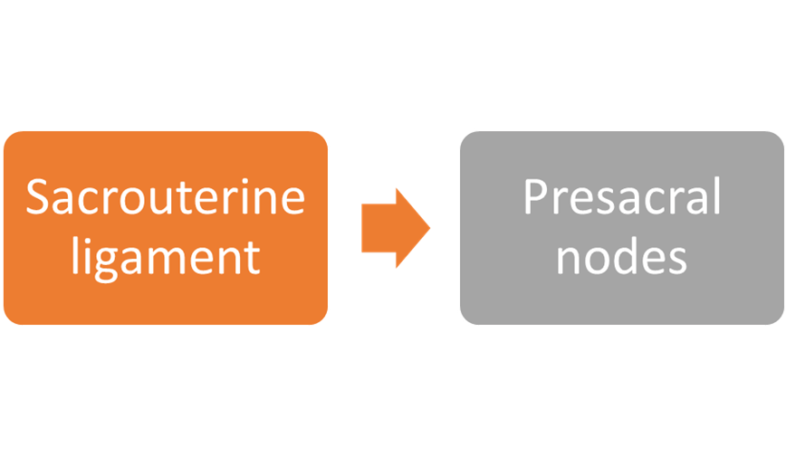
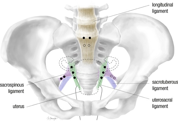
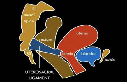
Pre-sacral pathway
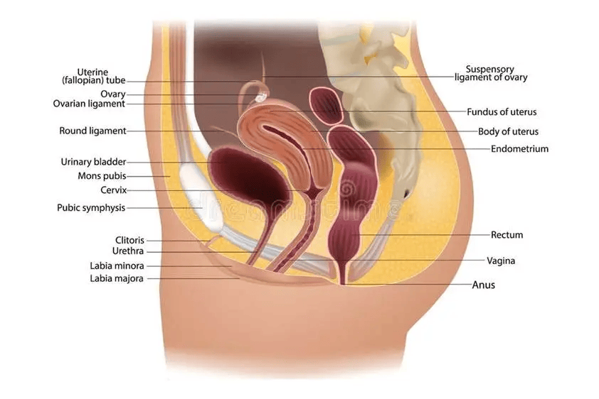
Cancers at the back wall of the vagina (posterior) that may appear in the upper third of the vagina, rectovaginal septum involves the presacral and mesorectal nodes.
Simply to help: cancer may grow over the vagina (paravaginal tissue), parametria (connective tissue to the womb), urethra, bladder and then rectum.
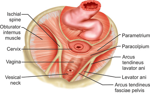
Cervical cancer
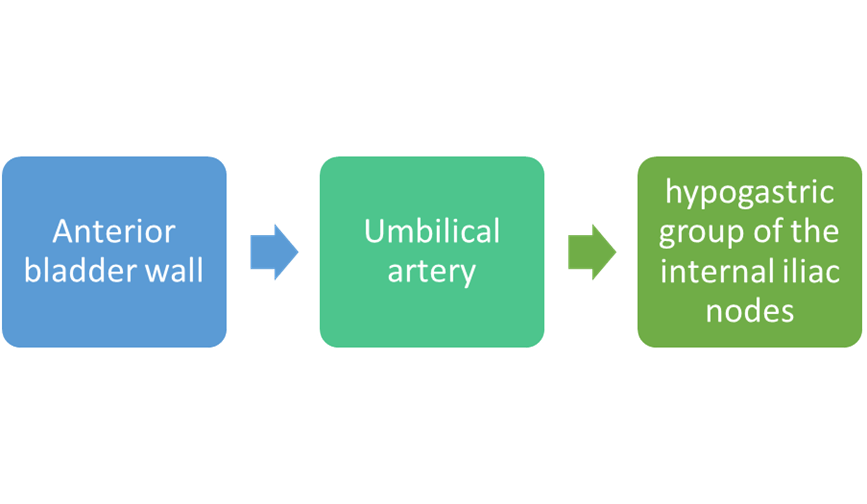
Anterior pathway
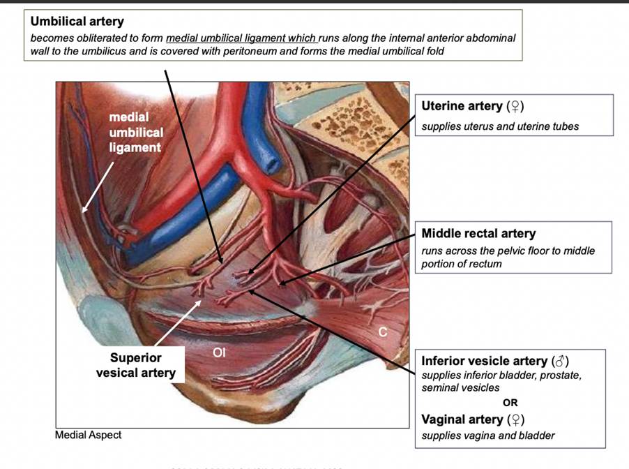
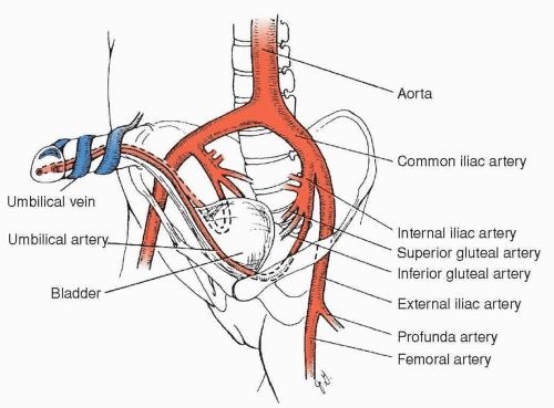
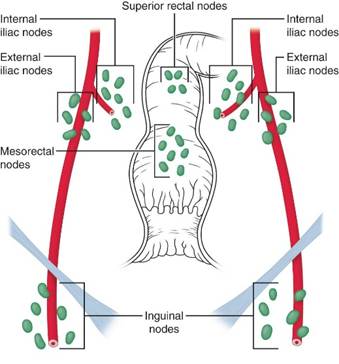
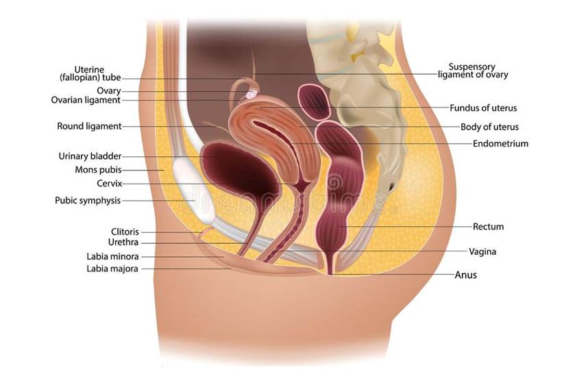
Spread from the lower third of the vagina: 31% of cases.

Hematogenous spread
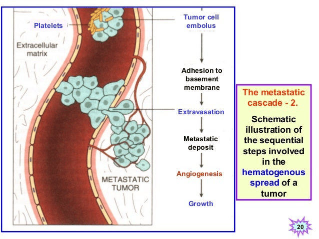
Platelets are small fragments that help clot the blood to prevent blood loss. The structure of the blood vessels contains a thin delicate membrane (basement) that connects to the epithelium (lining). An embolus can cause obstruction to the blood flow. This commonly may appear sudden after a myocardial infarction (heart attack). However, in terms of cancer, this leads to sticking to the basement membrane and results in leaking the contents i.e. tumour cells by a process known as extravasation. This is then metastasised leading to invasion, angiogenesis (production and sprouting of new blood vessels) that facilitate the growth of the cancer in a phase known as metastasis. By this mechanism, it is referred as haematogenous spread of the tumour.
Understanding Why Lymph Nodes Work Is Important.
Why?
Experiencing lymphadenopathy (abnormalities in the lymph node) in a non-local area is classified as distant metastasis even though it is limited to lymph nodes.
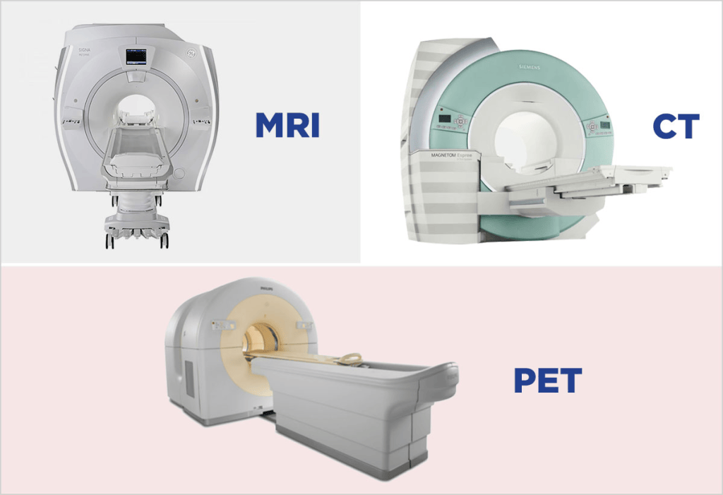
However, there are various imaging techniques used to detect the nodes:
Magnetic Resonance Imaging (MRI) to assess the tumour,
Contrast-enhanced CT (CECT) and FDG PET/CT can evaluate the nodal and distanct metastasis.
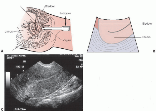
Pelvic ultrasound can help assess the lymph nodes in the groin and facilitate biopsy (removal of a tissue sample from the area for microscopic evaluation).
However,
There are important challenges to detect micrometastasis and comparing the differences between benign and malignant lymph nodes
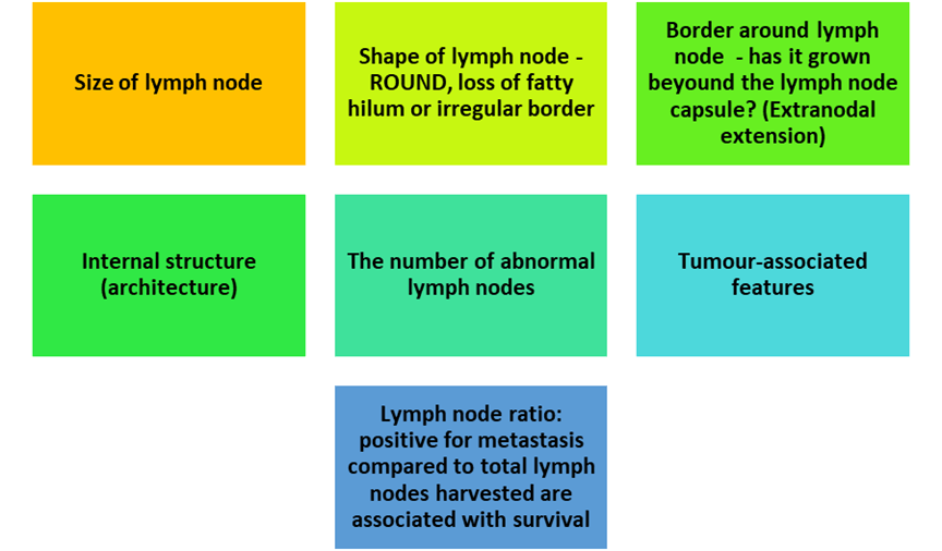
How To Differentiate From Metastatic And Non-Metastatic Lymph Node?
Short-axis diameter of 8mm in the pelvis and 10 mm in the abdomen.
Axial planes recommended
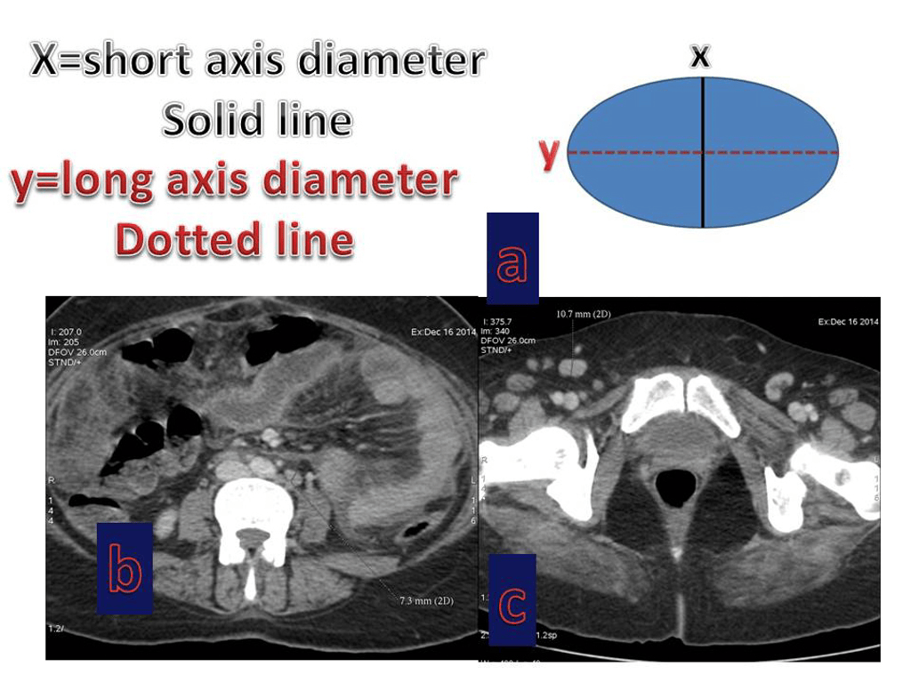
Anyone who is interested further about pelvic dimensions, please visit:
Epomedicine. Pelvic Dimensions : Mnemonics [Internet]. Epomedicine; 2018 Jul 27 [cited 2026 Oct 17]. Available from: https://epomedicine.com/medical-students/female-pelvis/ (Accessed: 20th October 2026)
Other sizes are also common…
…this helps to distinguish between benign (noncancerous) and possible metastasis…
BENIGN
Non-regional inguinal lymph node – smaller than 15 mm in short axis.
Macroscopic fat and calcifications though it may appear in the ovary or even bladder.
POSSIBLE METASTATIC
Perivisceral lymph nodes in rectal cancer in less than 5mm.
However, enlargement of the lymph nodes may be caused by infection e.g. or rective hyperplasia (continuous growth).
METASTATIC
Nodes along a specific tumour pathway.
Central necrosis.
Hetereogenous (different forms).
Homogenous (same form) this may appear in benign and malignant.
Later, in the sections, we will discuss the staging technique of vaginal cancer using the FIGO classification.
Recommended Article by Pederson and Babu (2021)
References
Epomedicine (2018) Pelvic Dimensions : Mnemonics [Internet. Available from: https://epomedicine.com/medical-students/female-pelvis/. (Accessed: 20th October 2026)
Kulkarni, A., Dogra, N. and Zigras, T. (2022). Innovations in the management of vaginal cancer. Current Oncology, 29(5), pp.3082–3092. doi:10.3390/curroncol29050250.
Nout, R.A., Calaminus, G., Planchamp, F., Chargari, C., Lax, S., Martelli, H., McCluggage, W.G., Morice, P., Pakiz, M., Schmid, M.P., Stunt, J., Timmermann, B., Vokuhl, C., Orbach, D. and Fotopoulou, C. (2023). ESTRO/ESGO/SIOPe Guidelines for the management of patients with vaginal cancer. International Journal of Gynecologic Cancer, [online] 33(8). doi:10.1136/ijgc-2023-004695.
Pedersen, C.K. and Babu, A.S. (2021). Understanding the lymphatics: An updated review of the N category of the AJCC 8th edition for urogenital cancers. American Journal of Roentgenology, 217(2), pp.368–377. doi:10.2214/ajr.20.22997.
Scholarly Community Encylopaedia (n.d) Vaginal Cancer. Available at: https://encyclopedia.pub/entry/25552 (Accessed: 20th October 2026)


Leave a Reply