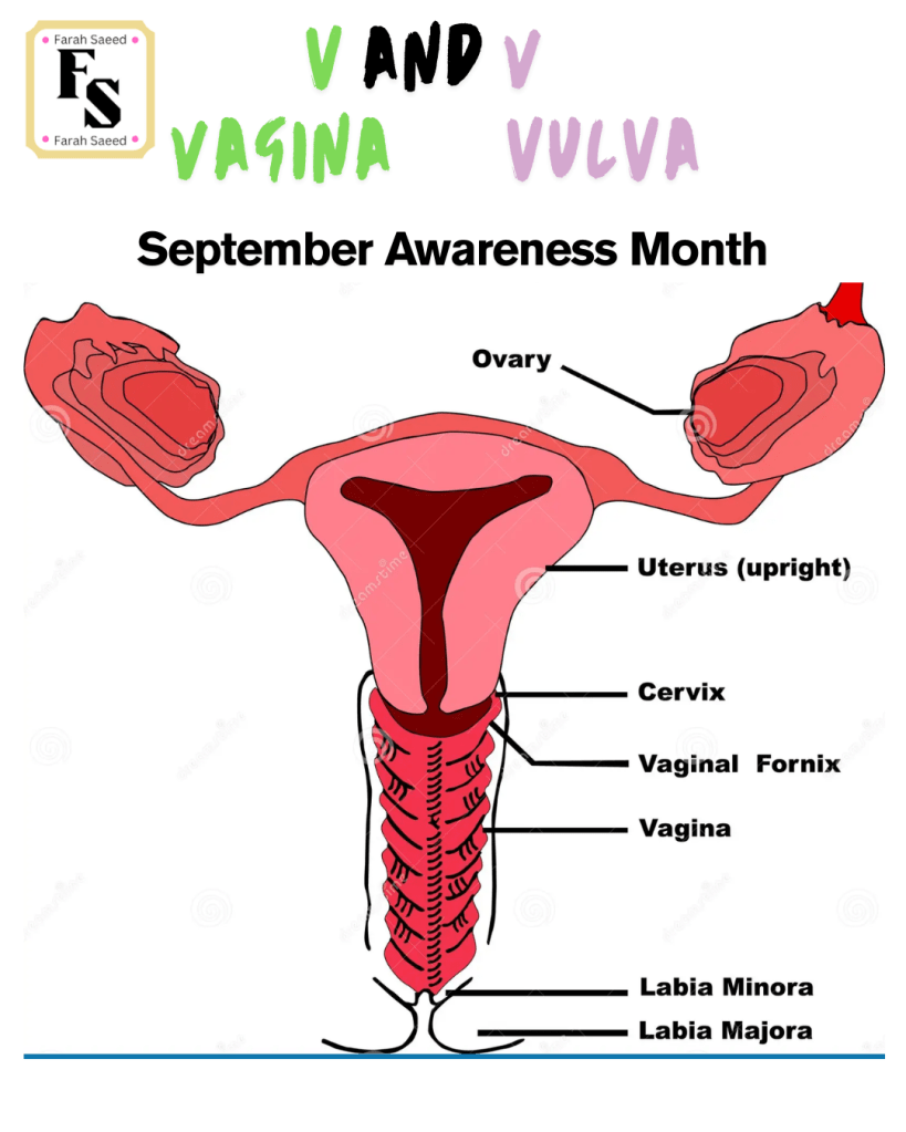
Diagnosis Of Vaginal Cancer
Below summarizes the main points for the histological classification, signs, symptoms, diagnosis and treatments in accordance to the European Society of Gynaecological Oncology (ESGO), European Society for Radiotherapy and Oncology (ESTRO) and the European Society of Paediatric Oncology (SIOPe) 2023 guidelines. A 2025 update of Vaginal cancer has been provided by experts from the following reference:
Adams, T.S., Maluleke, J.C. and Cuello, M.A. (2025). Cancer of the vagina: 2025 Update. International Journal of Gynecology & Obstetrics, 171(S1), pp.48–59. doi:10.1002/ijgo.70325.
Please read the guidelines attached for further details.
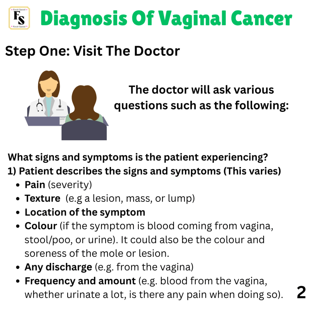
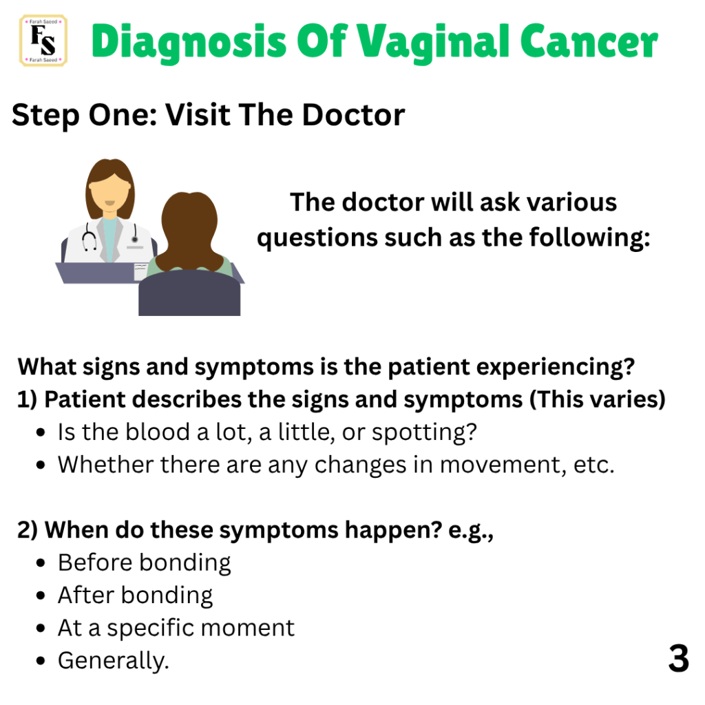
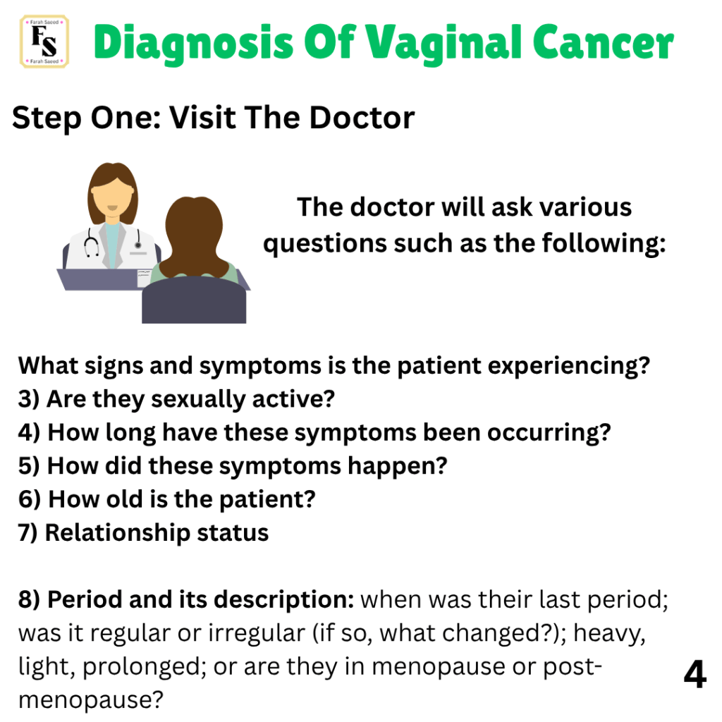
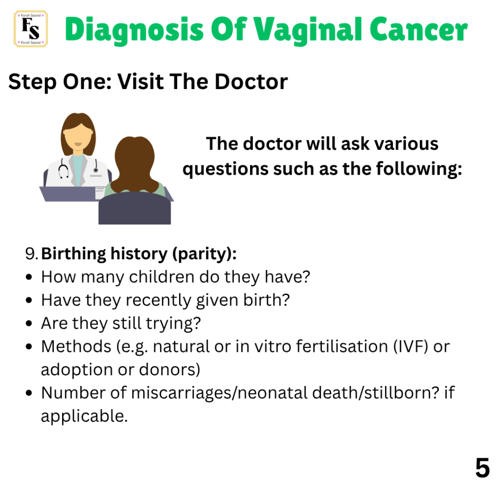
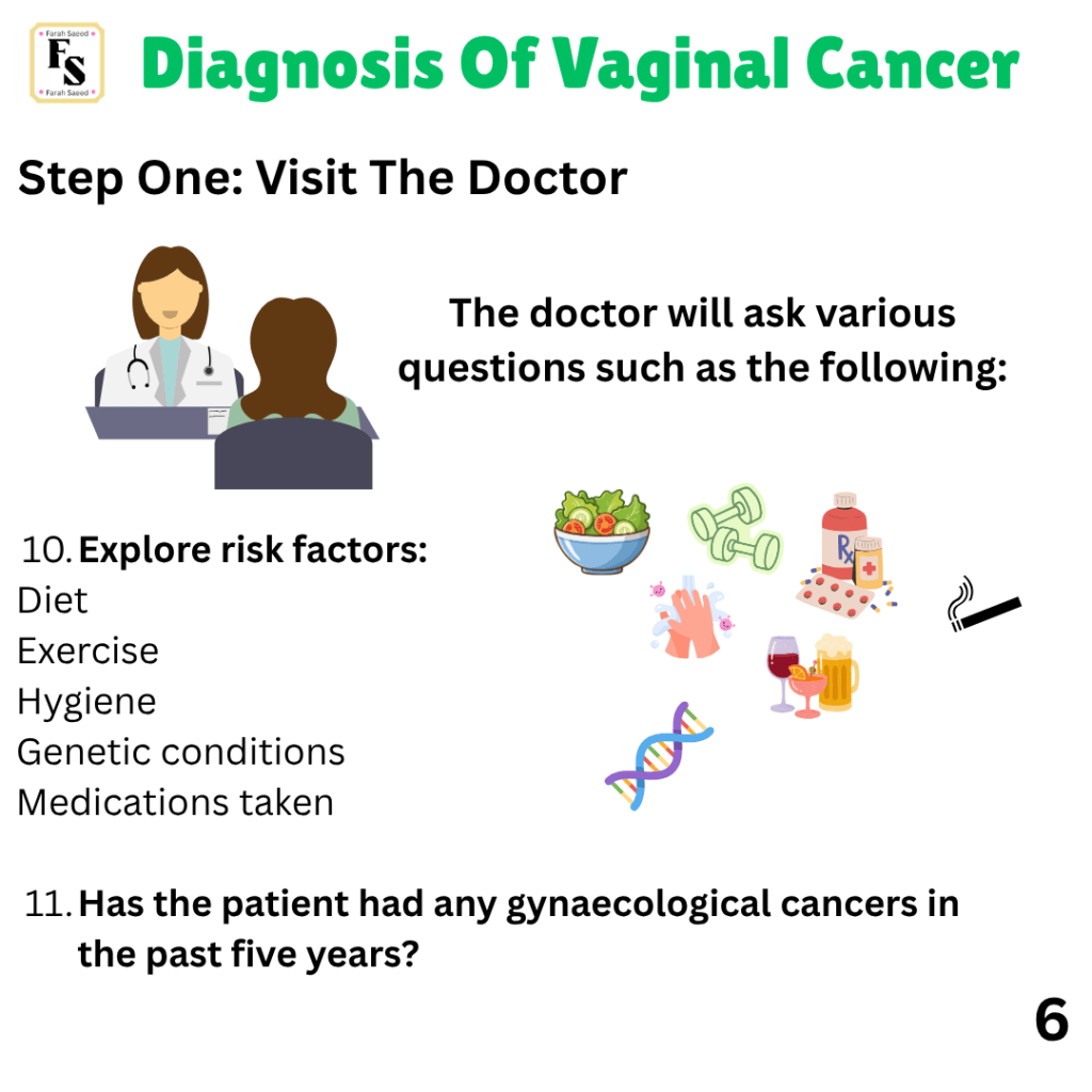
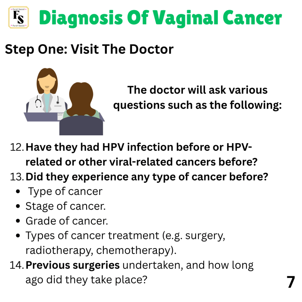
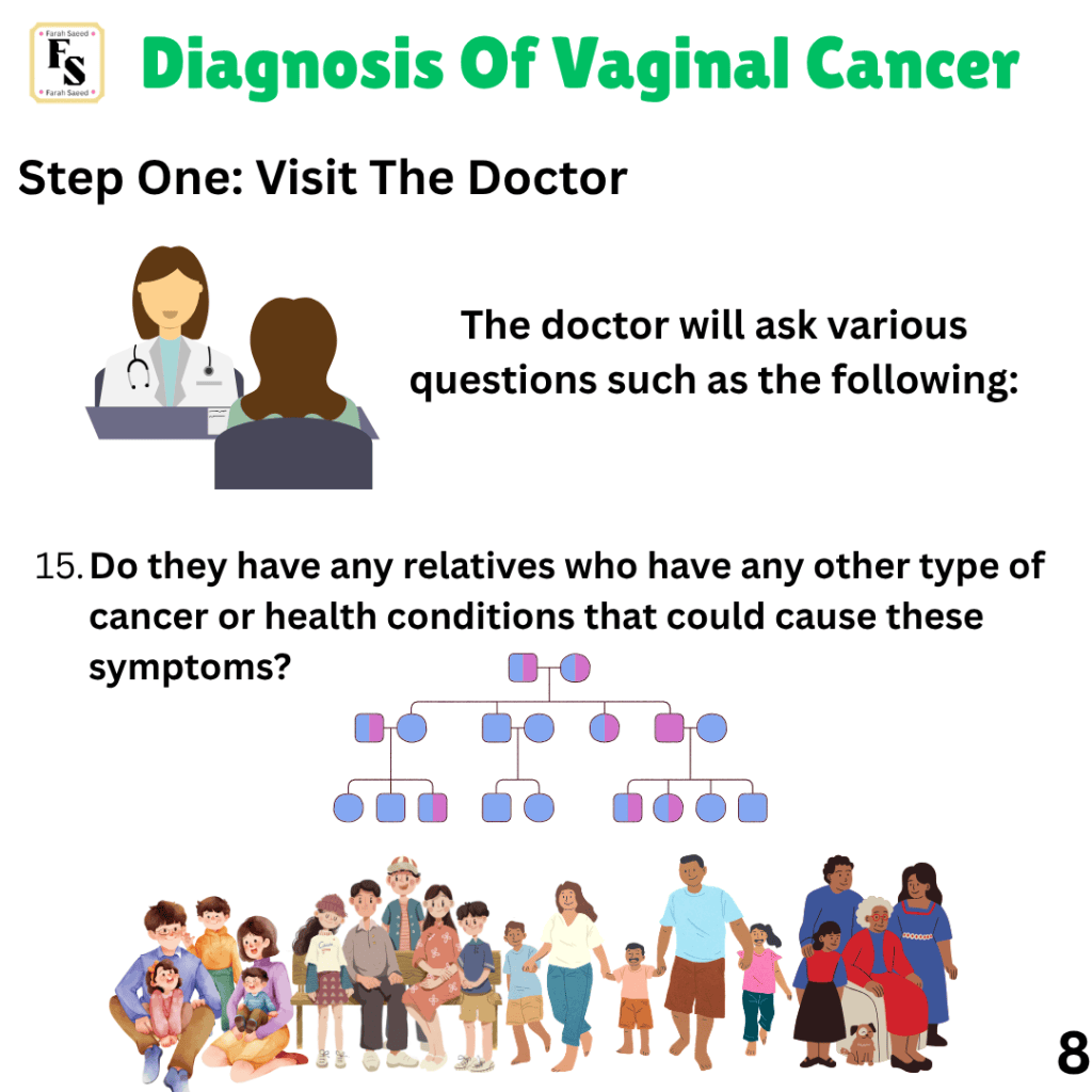
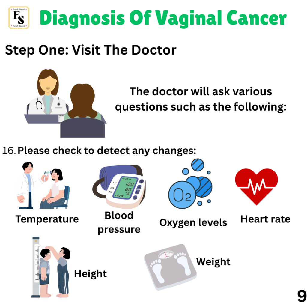
Step One: Visiting The Doctor, Communication and Understanding

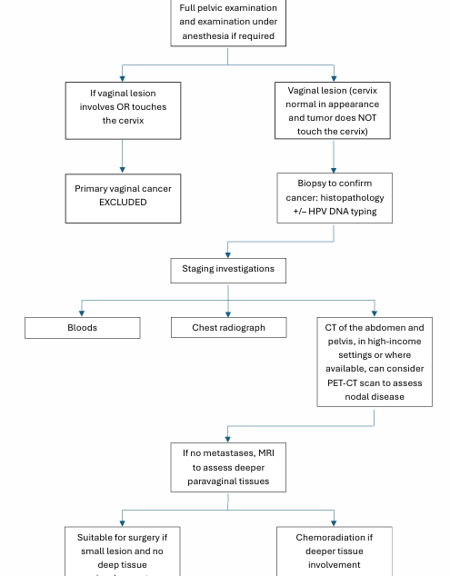
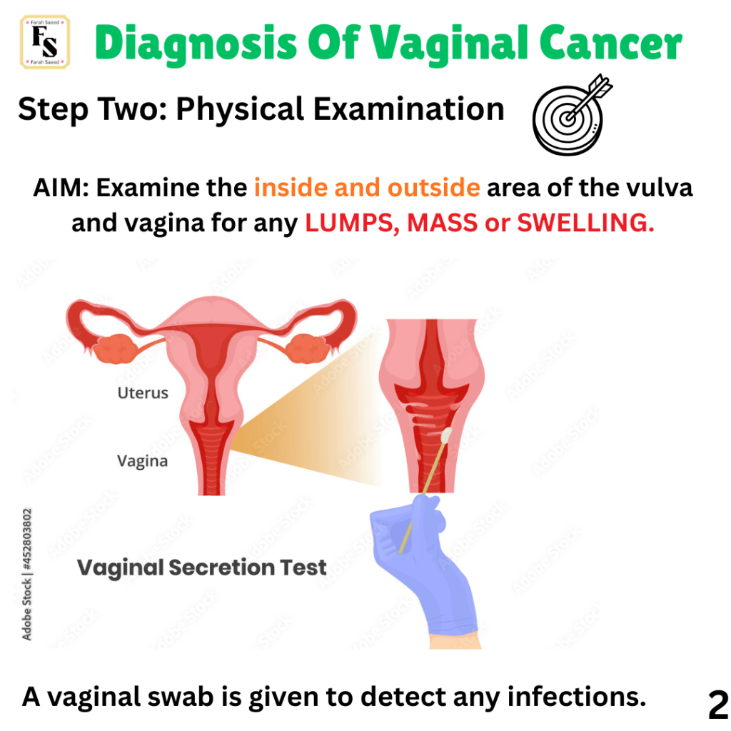
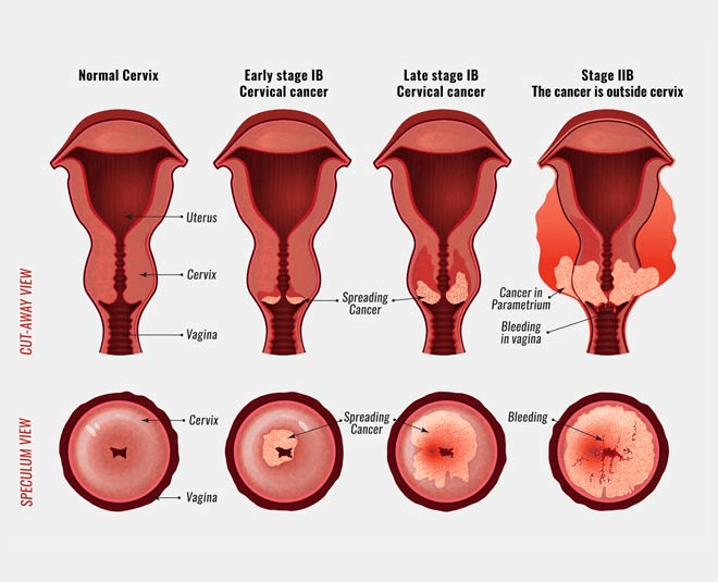
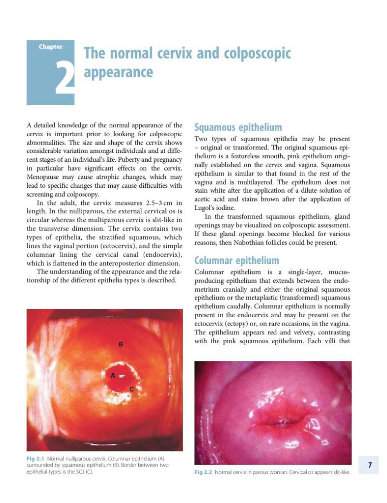
Step Two: Physical Examination
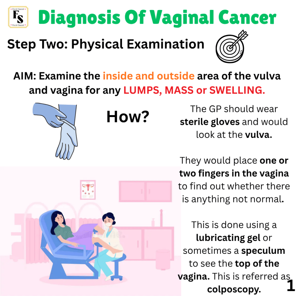
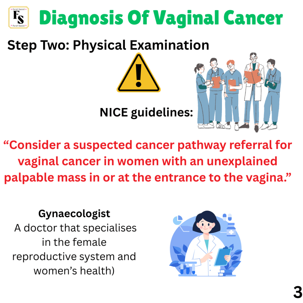
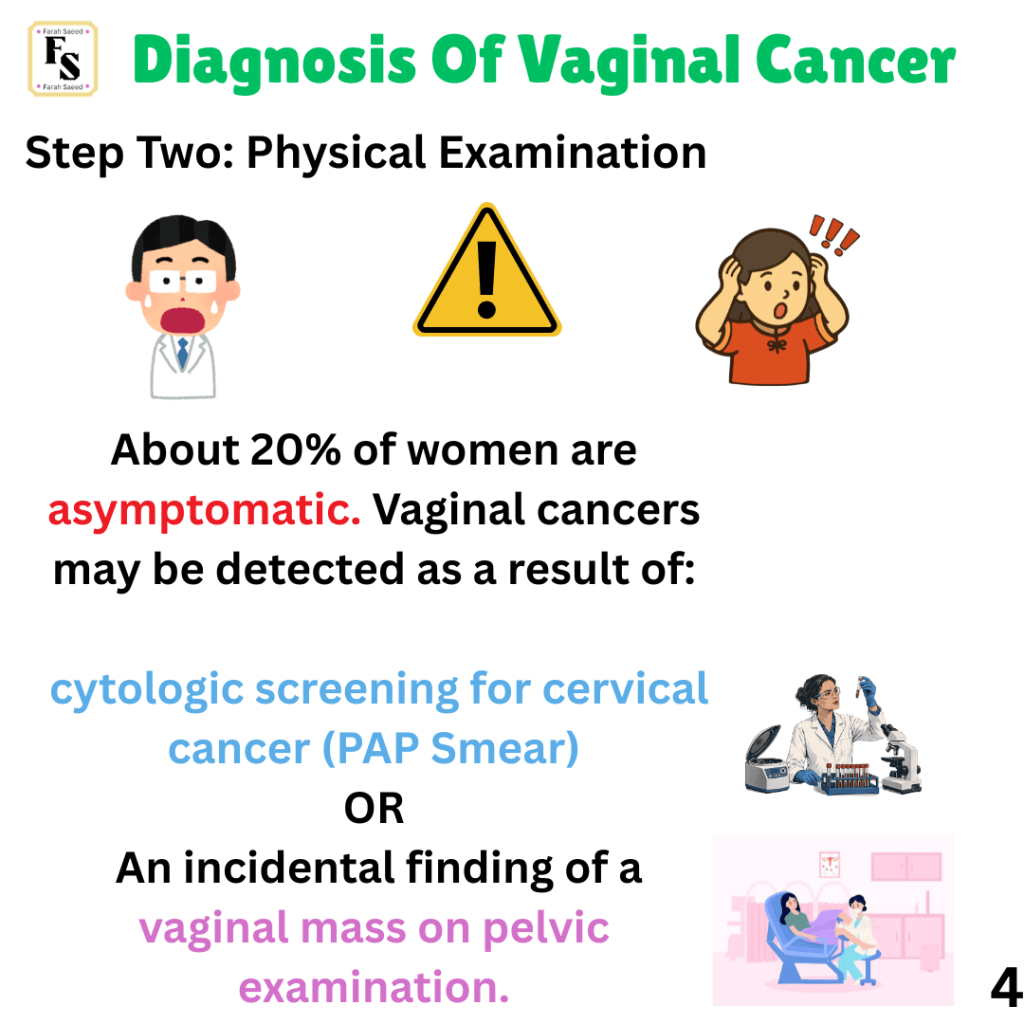
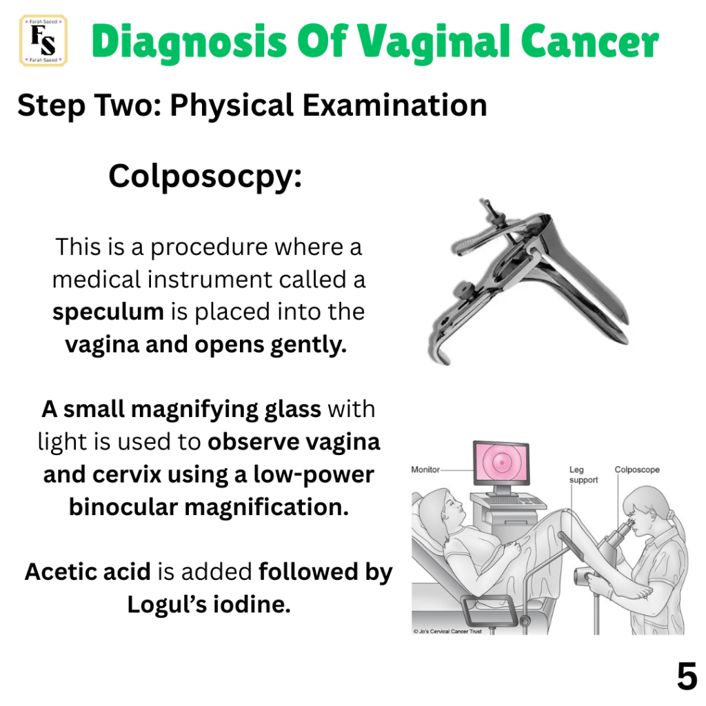
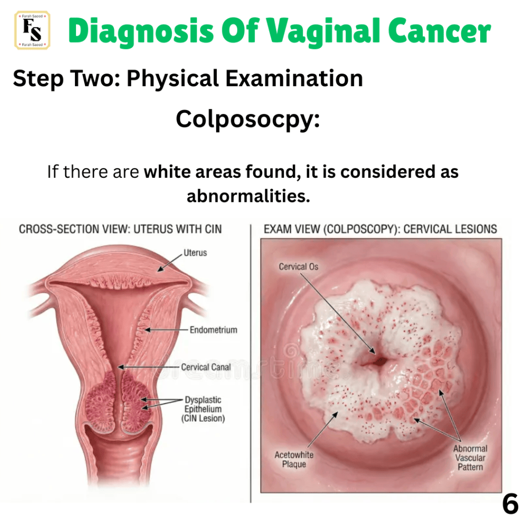
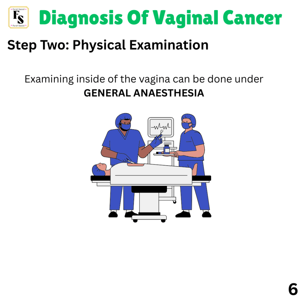
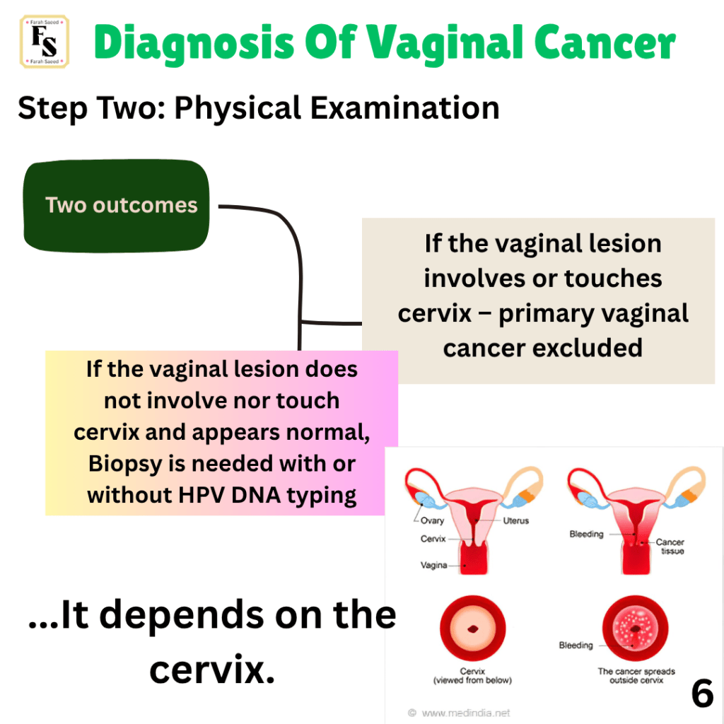
Things to consider when performing a pelvic examination for specific types of vaginal cancer:
ADENOCARCINOMA
Patients with DES in utero – Their first gynaecological examination at menarche (first period) with assessment of the cervix and vagina every year using two tools:
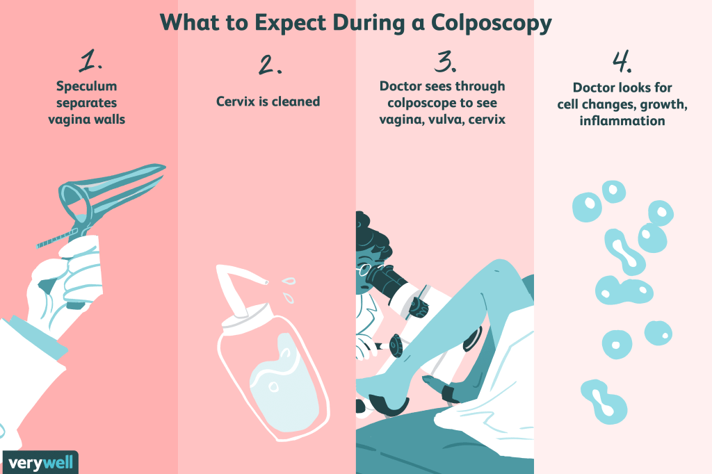
Colposcopy
Examine the cells in both areas (cytology)
MELANOMA
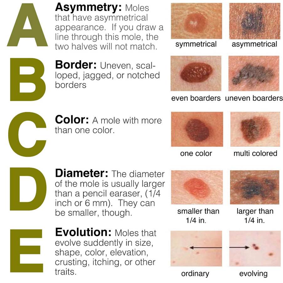
Assess the mole, papule, or nodule using the ABCDE examination.
How to Collect a Vaginal Swab Sample | Step-by-Step Guide
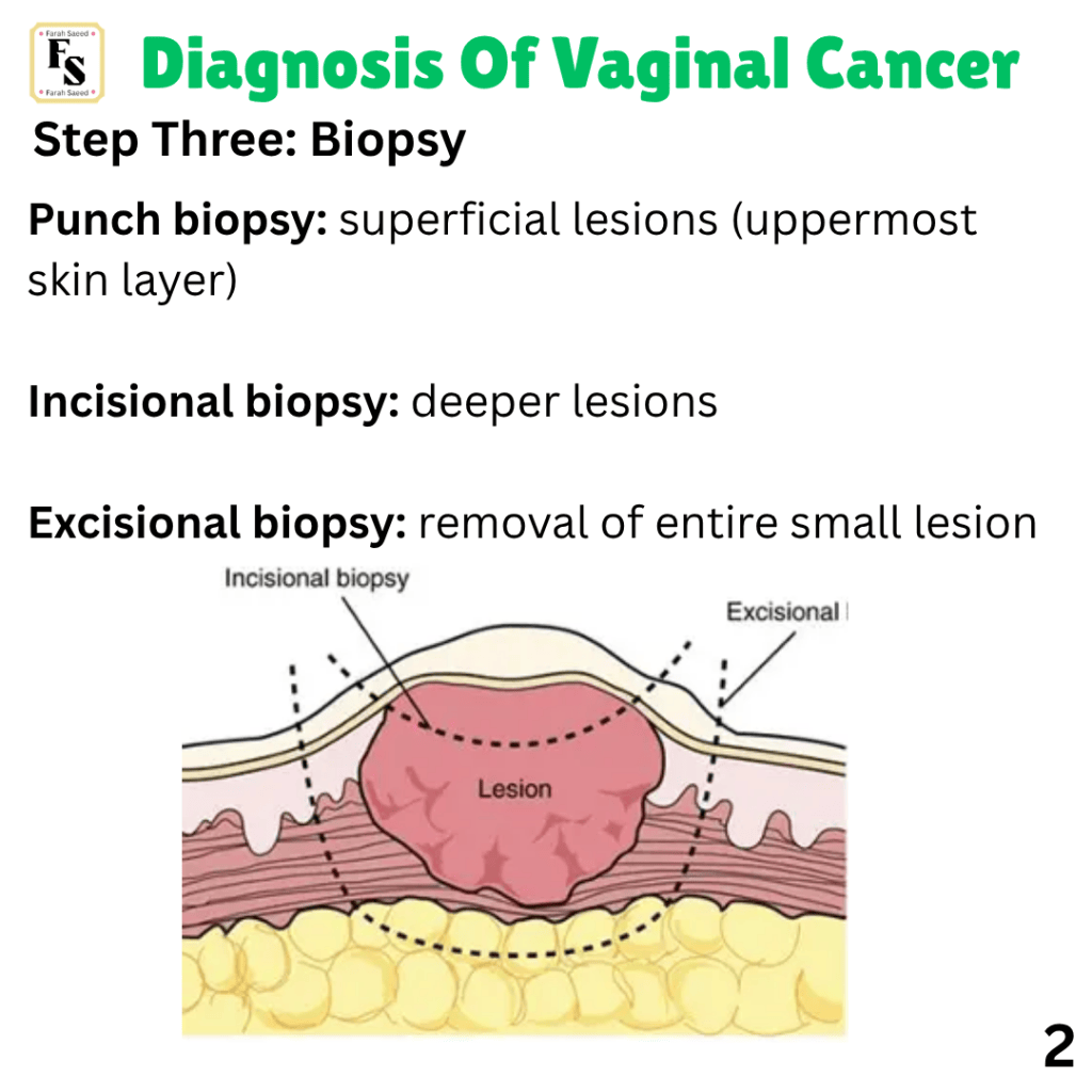
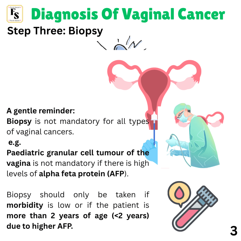
Step Three: Biopsy
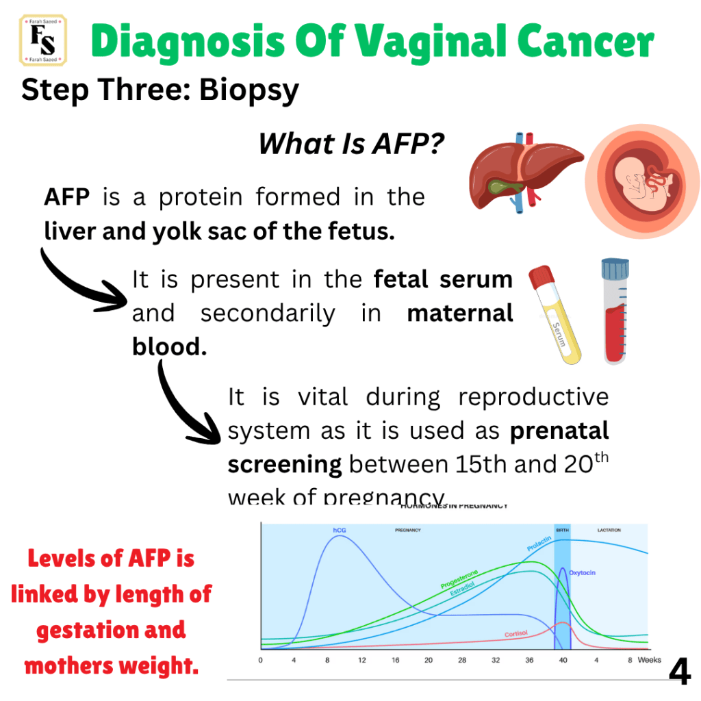
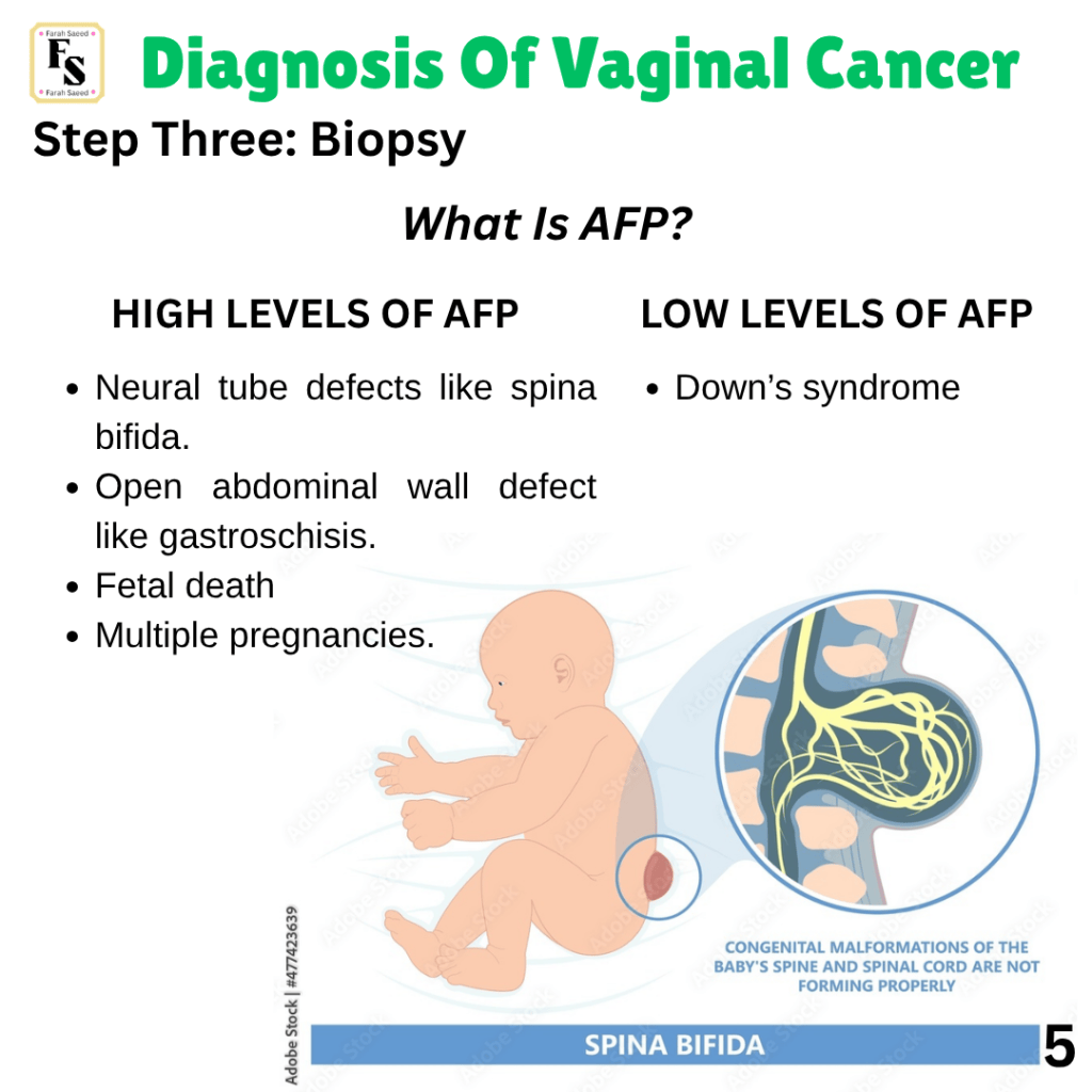
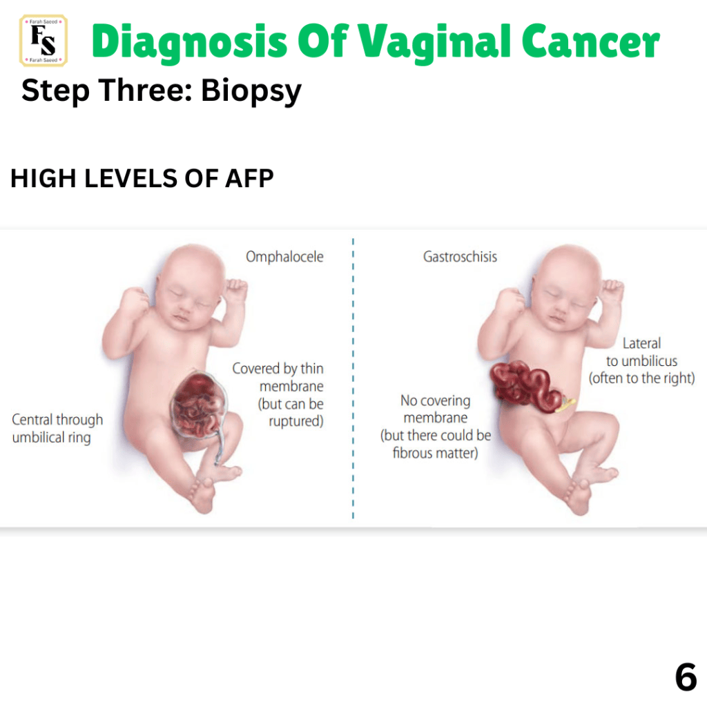
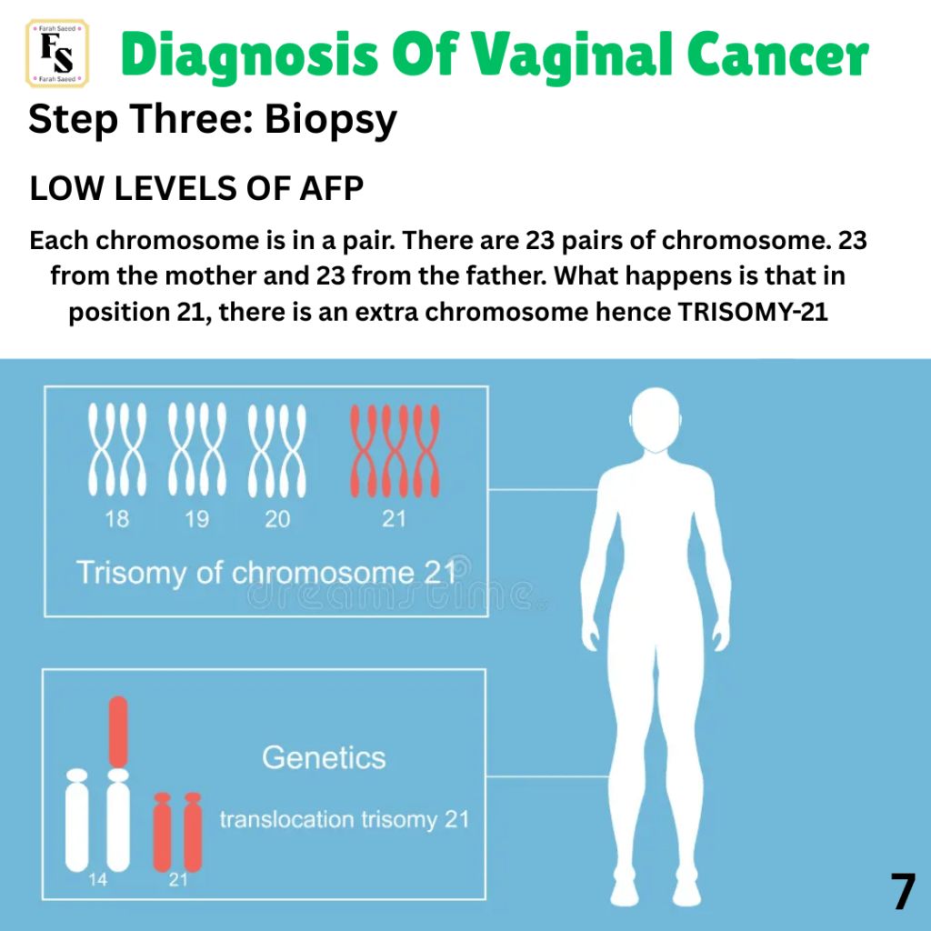
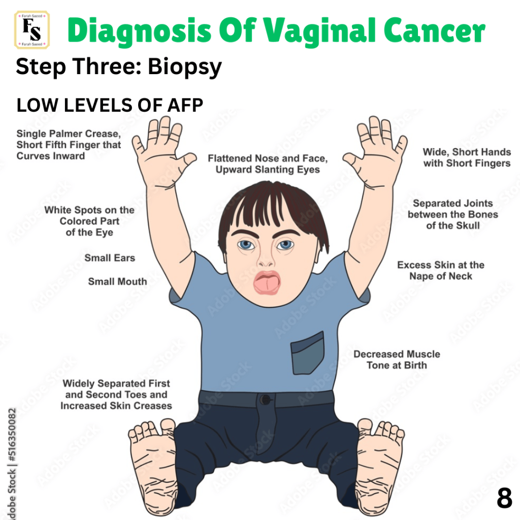
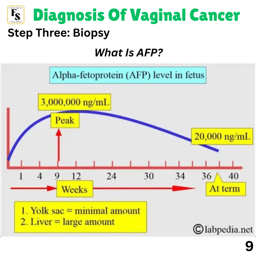
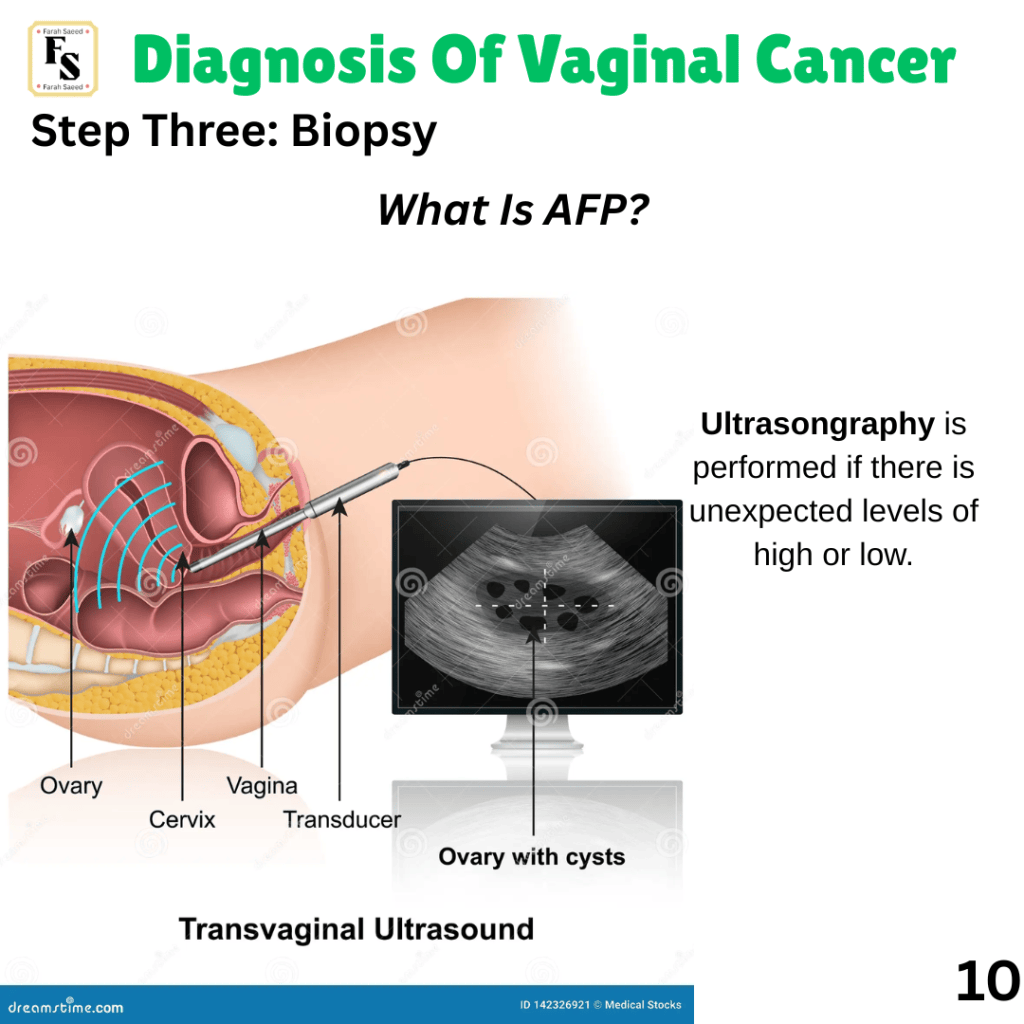
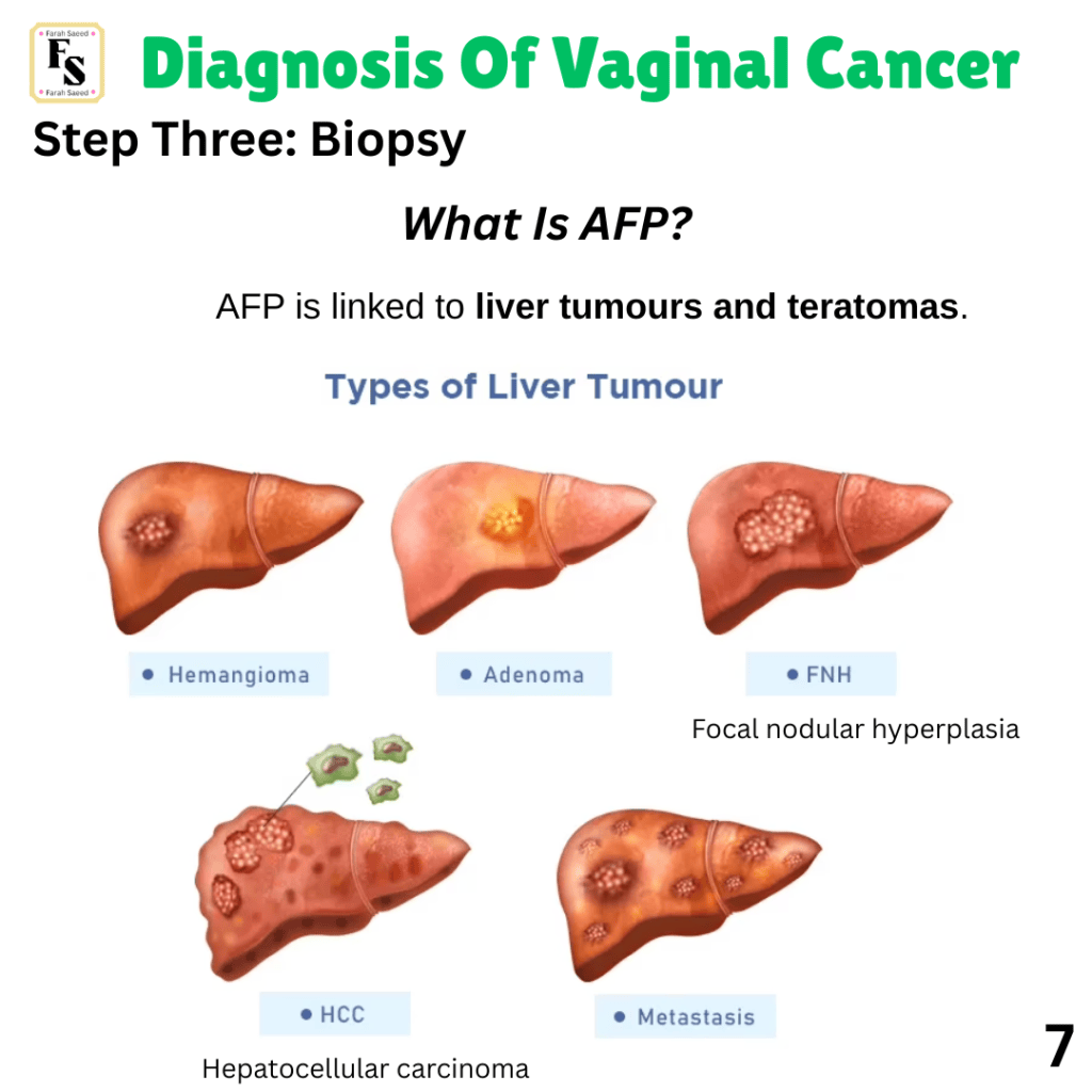
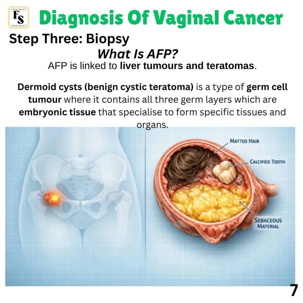
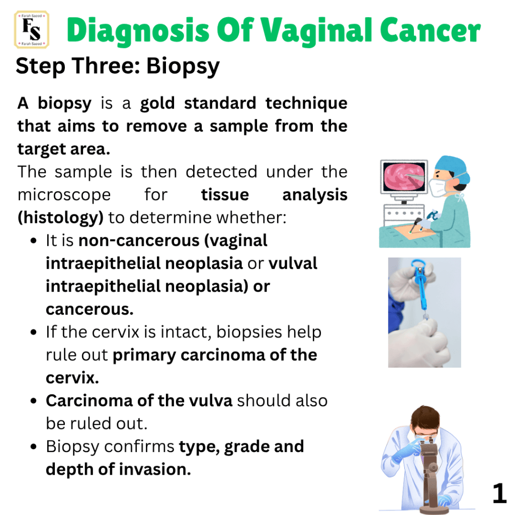
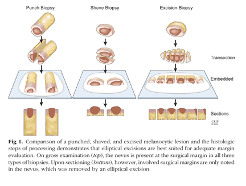
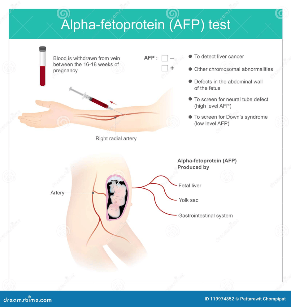
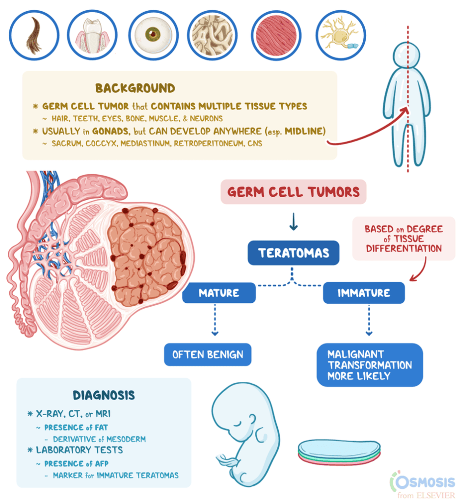
Recommended reading: Dermoid cystic teratoma of the vagina
Step Four: Histological Examination
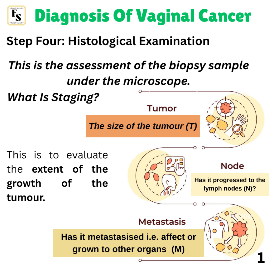
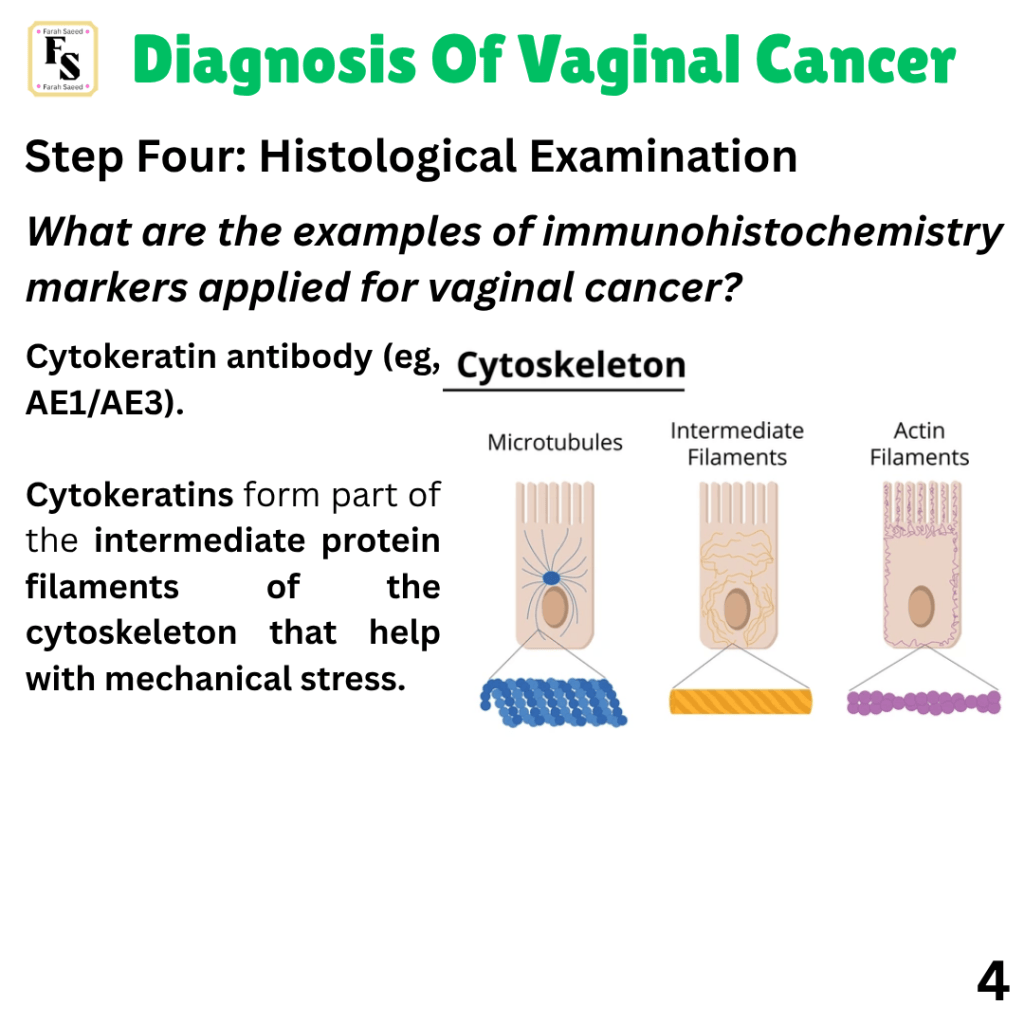
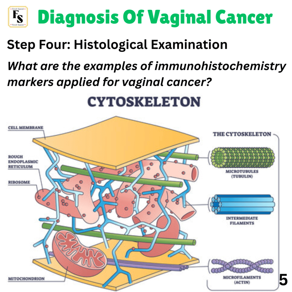
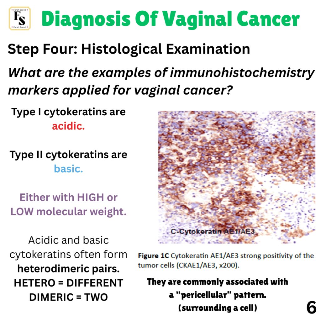
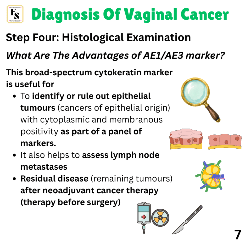
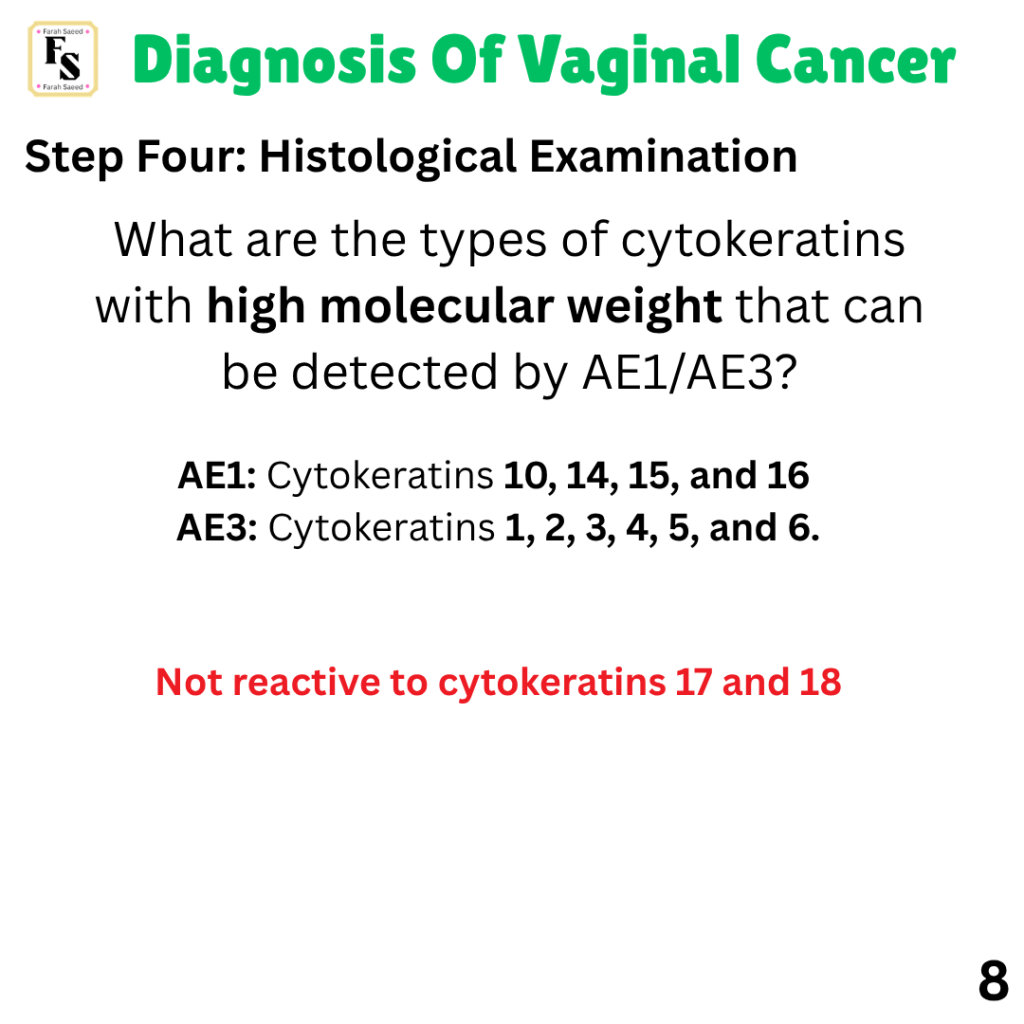
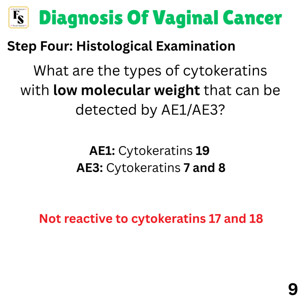
Sentinel Lymphadenectomy And The Histology Journey
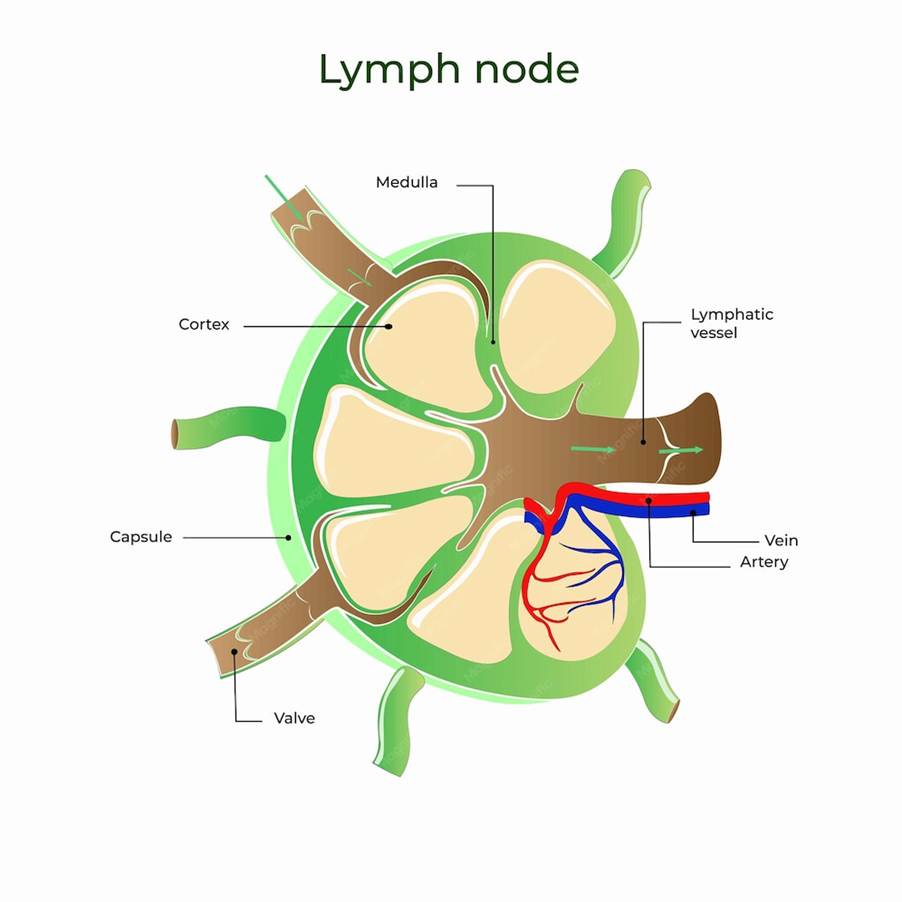
The first lymph node that is affected after a cancer grows from the primary site is the SENTINEL NODE. It is a useful technique that helps with intraoperative assessment.
HOWEVER…
It may miss micrometastases and isolated tumour cells.
Histology helps to detect infections, degenerative diseases, inflammatory conditions, and cancers
How does the histology journey start?
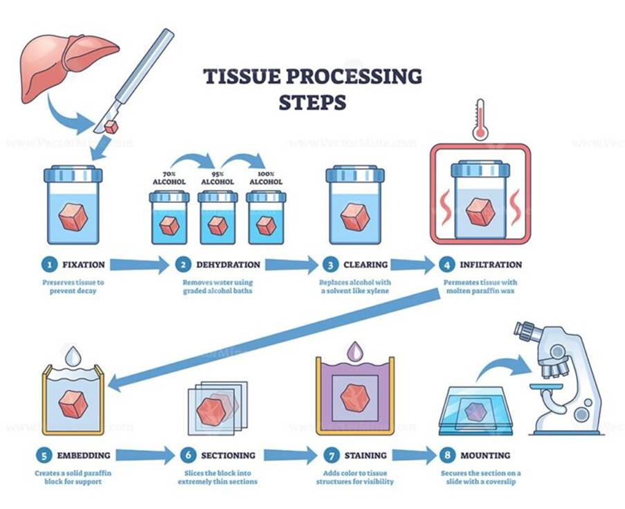
1) The tissue sample is fixed with 4% buffered formalin (normally 10% buffered formalin) – they are then assigned a unique digital barcode for tracking.
Purpose of Fixation: This helps to stabilise the cellular structure for a period of 6 to 24 hours and maintain the architecture.
2) Gross examination – The pathologist measures the size of the tissue and sections it (cuts).
3) The samples are then placed in cassettes for further processing.
How?
4) The samples are then dehydrated using alcohol.
Purpose: Dehydration because the later step that involves paraffin wax does not mix with water.
5) The samples are then cleared using xylene and embedded in paraffin wax.
Purpose: To provide support.
6) The paraffin blocks are then ultrastaged and sectioned. This is achieved using a microtome. The estimated thickness is 3 to 5 micrometres.
Purpose: The sample of tissue needs to be thin to be able to be detected easily under the microscope.
7) The samples are placed on glass slides and then stained.
Purpose: The different parts of the cells are stained to provide a clear overview of cell architecture.
What is the most common stain used in histology?
The most common stain is Haematoxylin and Eosin (H and E) – nuclei stained blue-purple.
Eosin stains cytoplasm and extracellular matrix (e.g., connective tissue) pink.
Other examples of stains are:
PAS: Glycogen (type of sugar storage) and basement membrane
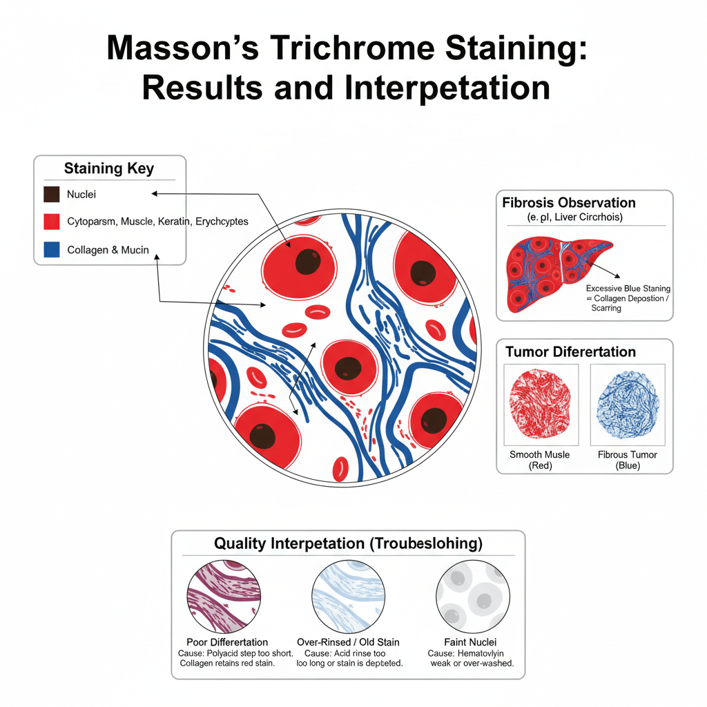
Masson’s trichrome – red keratin and muscle fibers. Green/Blue for collagen fibers and bone.
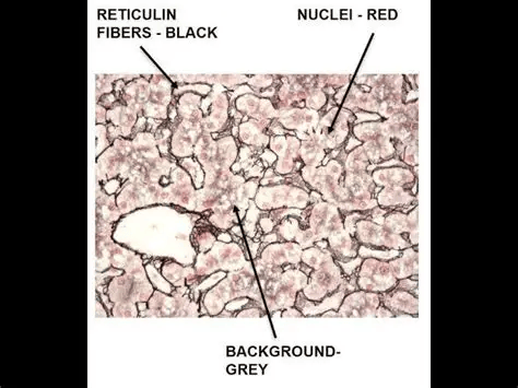
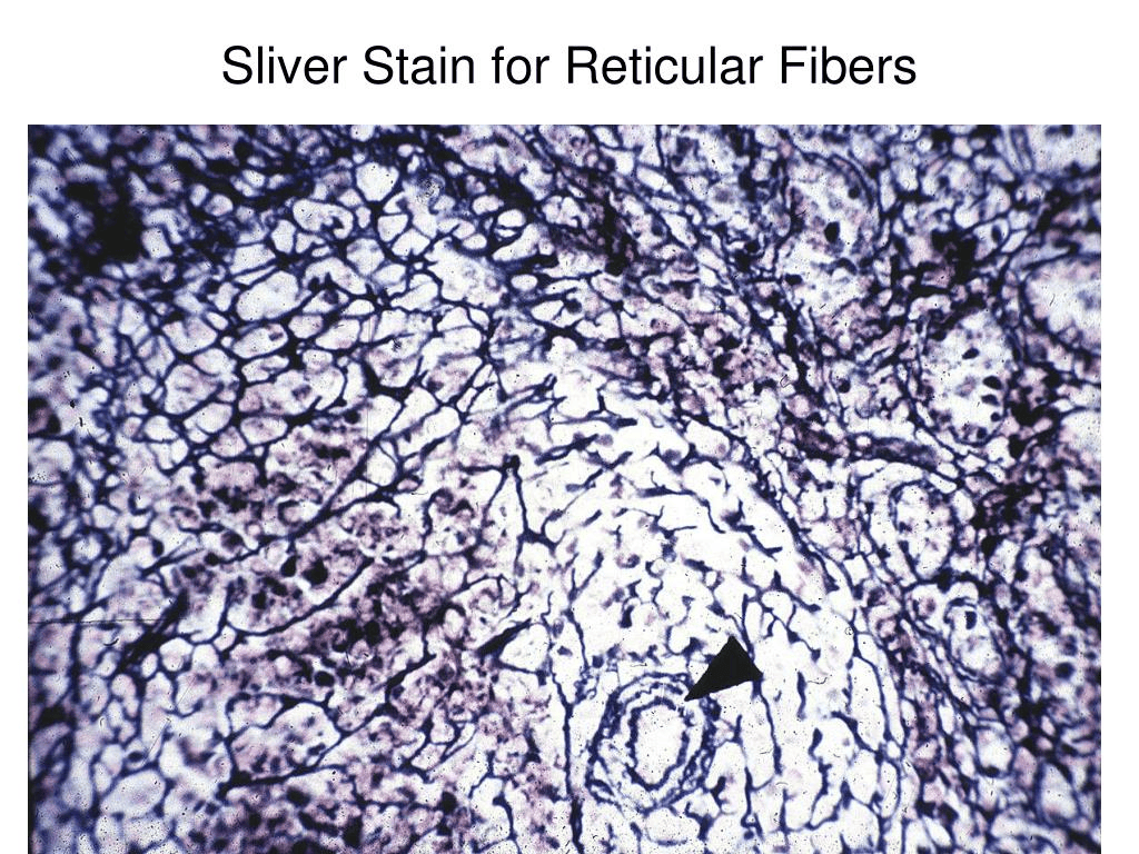
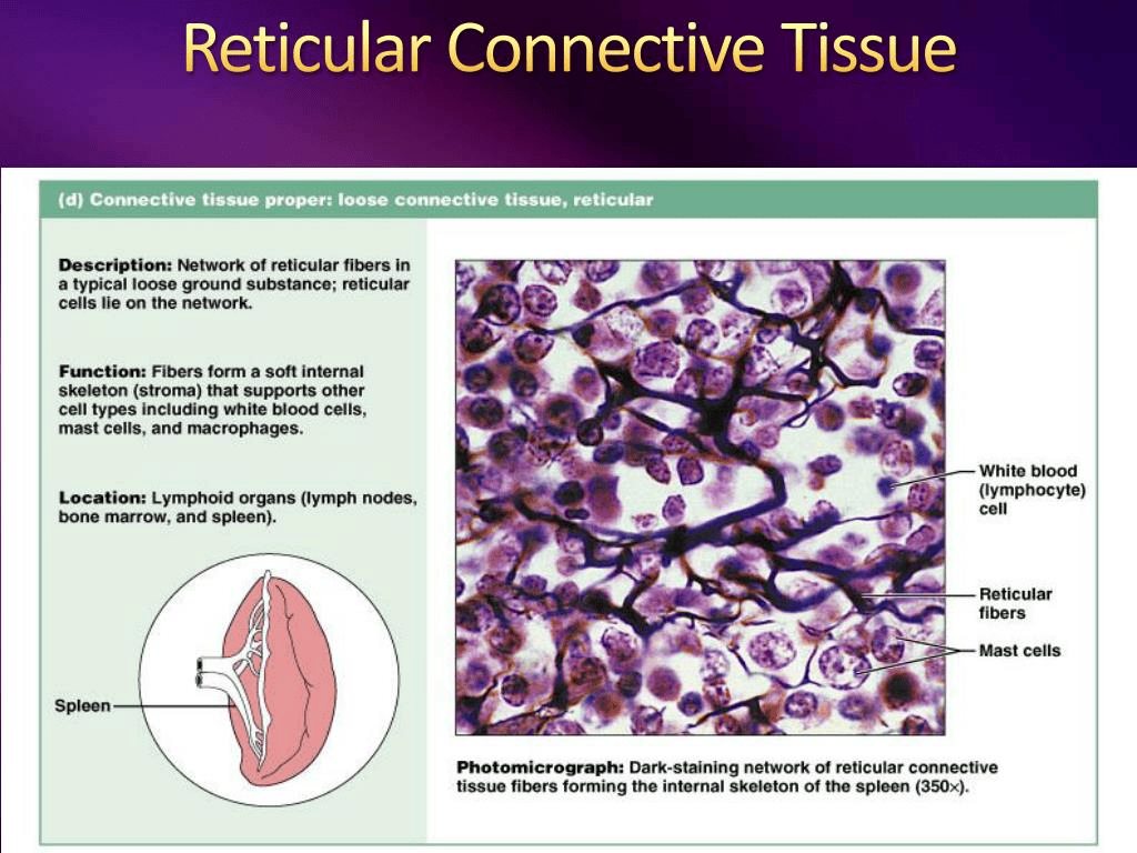
Silver stains – reticular and nerve fibers
9) Immunohistochemistry markers:
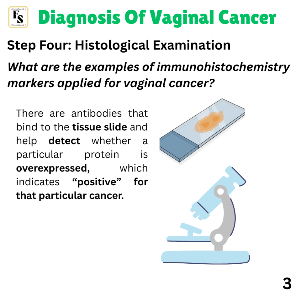
Several immunohistochemistry markers are used to highlight whether a protein is overexpressed by antibodies binding to the slide and are specific for gynaecological cancers.
Immunohistochemistry markers for vaginal cancer is similar to cervical carcinoma if there is no metastasis present in the first section.
Examples of immunohistochemistry markers for specific types of vaginal cancers…
Further information about Histology…
Histology – Lab Workflow By Med Lab Technology 3D
HISTOLOGY: Tissue Preparation & Staining By MBBS Clinical Lectures
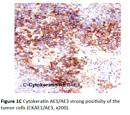
Source: Mouhamed, H.A. (2017). The diagnostic utility of immunohistochemistry in undifferentiated ovarian carcinoma. Archives in Cancer Research, 05(02). doi:10.21767/2254-6081.1000143.
Immunohistochemistry markers for specific vaginal cancers
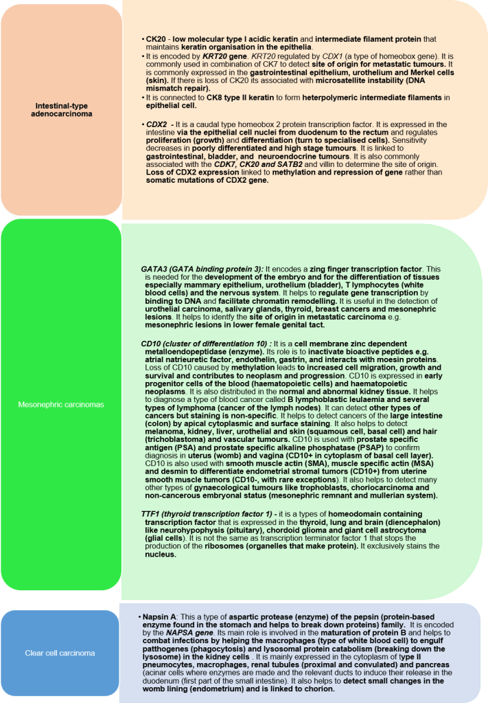
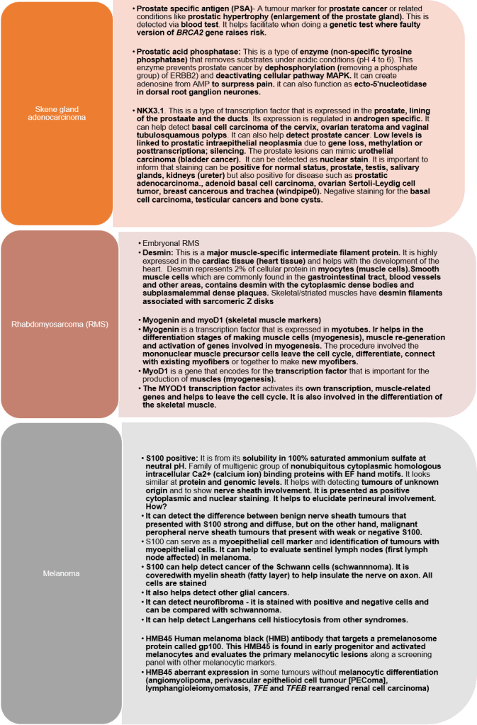
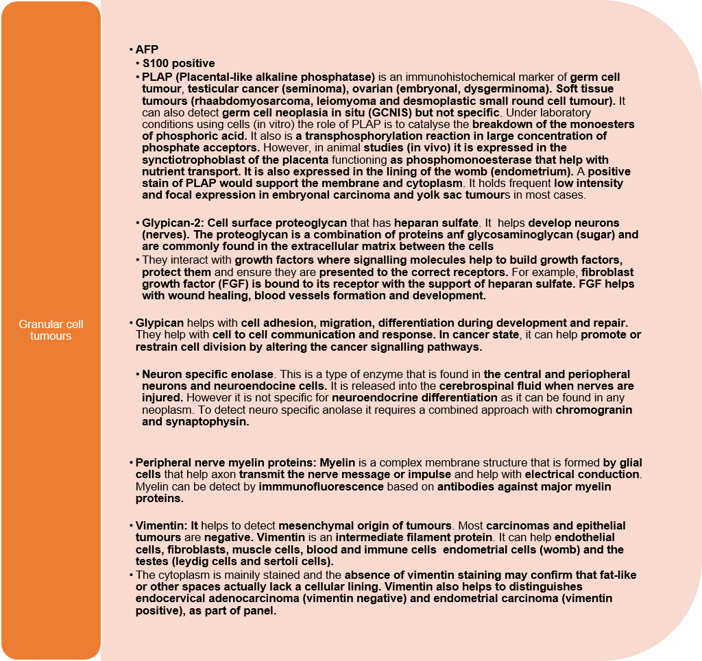
A Summary File Is Downloadable…

Are You Interesting In Finding Out What A Pathologist Adds To Surgical Report?
The International Collaboration on Cancer Reporting (ICCR) has developed standardized dataset-reporting proformas for several cancers, based on the ESTRO/ESGO/SIOPe Guidelines for managing patients with vaginal cancer.
The datasets has designed by clinicians including pathologists that present what to include in the pathology report.
It is important to note that most of the vaginal carcinoma dataset contains all types of vaginal carcinoma and sarcomas. However, it excludes melanomas, blood (haematopoietic) neoplasms, metastatic tumours, and mesenchymal tumours.
It is important to note that most of the vaginal carcinoma dataset contains all types of vaginal carcinoma and sarcomas. However, it excludes melanomas, blood (haematopoietic) neoplasms, metastatic tumours, and mesenchymal tumours.
Clinical history
Previous treatment for cervical or vulval to exclude primary vaginal carcinomas.
Has the patient been exposed to diethylstilbestestrol (DES) though condition is rare as withdrawn in 1970s.
What operation has the patient had or is having? Was it partial or full removal?
About The Tumour
What organ was submitted?
Block Identification Key
This is for internal or external review about the origin of each block to give a specialist opinion. This needs to be presented either in the pathology report or computer system for the pathologist to see. It also helps when further tests are needed for: immunohistochemical or molecular analysis, research studies or clinical trials.
What is the stage and grade of the cancer?
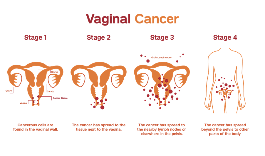
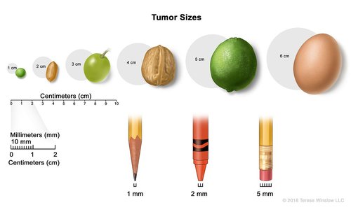
What is the size of the tumour (I.e. dimension – the horizontal extent (length) and the depth of invasion? This helps to know how radical a resection has been undertaken.
Please note measurement should be in millimetre (mm) from the base of the epithelium to the deepest point of invasion.
To provide the final tumour dimensions, a previous specimen done (excisional biopsy) should be measured. Maximum horizontal extent need to be added together. Maximal depth of invasion is the maximum depth in either specimen.
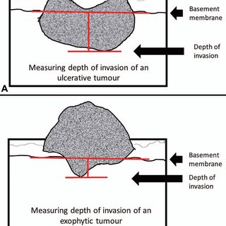
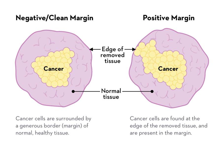
What is the margin status?
“Resection margins should be microscopically clear of disease, but no minimum cut- off is defined and no large negative margins are required. Tumour involvement of a margin or tumour distance from the margins (nearest peripheral mucosal margin and the deep margin) should be included in the pathology report.
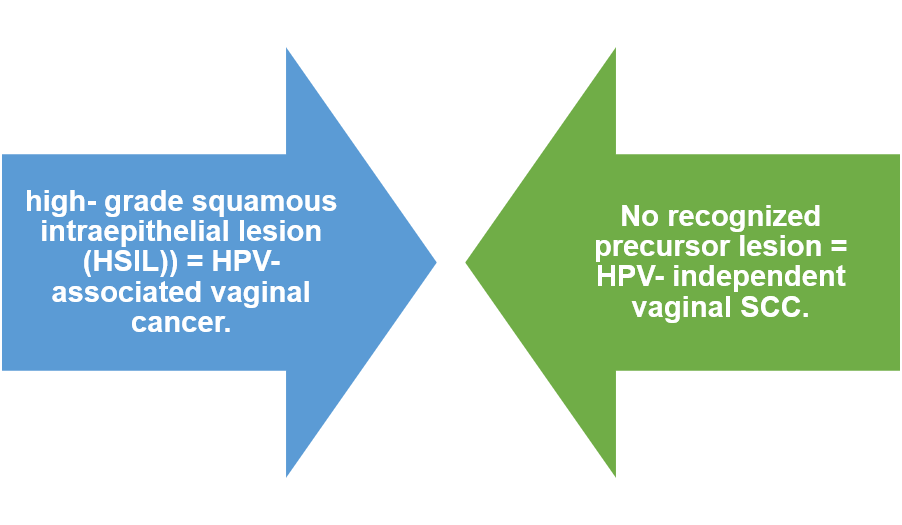
The location of the involved or nearest peripheral margin should be specified if possible. Involvement of a peripheral margin by a high- grade precursor lesion (in particular HPV- associated high- grade squamous intraepithelial lesion (HSIL) should also be recorded and the margin specified if possible.”
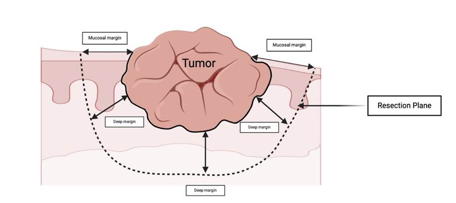
What Is the histological tumour type i.e. what type of tumour is it based on the WHO classification?
Please take care:
“Some primary vaginal cancers, for example those of gastric, HPV- associated and clear cell type may be associated with and arise from adenosis which is usually sporadic but which may be secondary to in utero exposure to DES.
Primary vaginal endometrioid and clear cell carcinomas may arise in endometriosis.
Intestinal- type adenocarcinomas may arise in tubular and tubulovillous adenomas.
Mesonephric adenocarcinomas arise from benign mesonephric remnants.”
Is there presence or absence of squamous intraepithelial lesions? These are precursor lesions before the cancer occurs. This is important in cases of vaginal squamous cell carcinoma to indicate whether it is associated with HPV-associated or not?
Has it spread to the vessels that surrounds tumour i.e lymphovascular invasions?
The guidelines state
“Lymphovascular Space Involvement There is a lack of evidence regarding the prognostic significance of LVSI in primary vaginal carcinomas because of the rarity of these neoplasms. Two studies have evaluated Lymphovascular Space InvolvementI as a prognostic factor in primary vaginal carcinomas; this was not statistically significant in one study and no conclusion was documented in the other.However, the presence or absence of LVSI should be included in the pathology report since it may alter clinical management in some cases and also by extrapolation from cervical and vulval carci nomas, many of which are biologically similar to vaginal carcinomas”
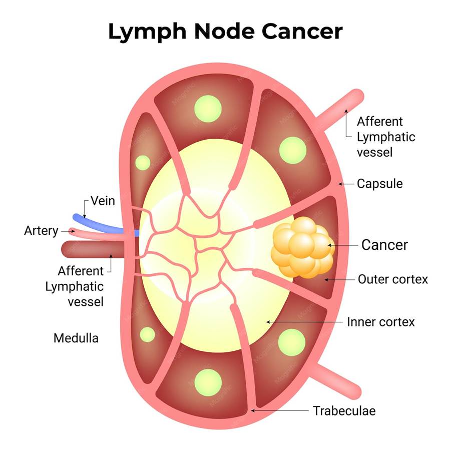
What is the Lymph node status?
How many positive lymph nodes affected?
Size of largest tumour deposit?
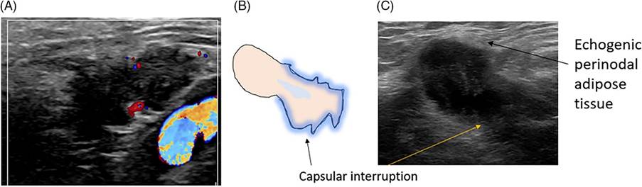
Presence of extracapsular spread? This is identified by the presence of perinodal fat
Know metastatic disease.
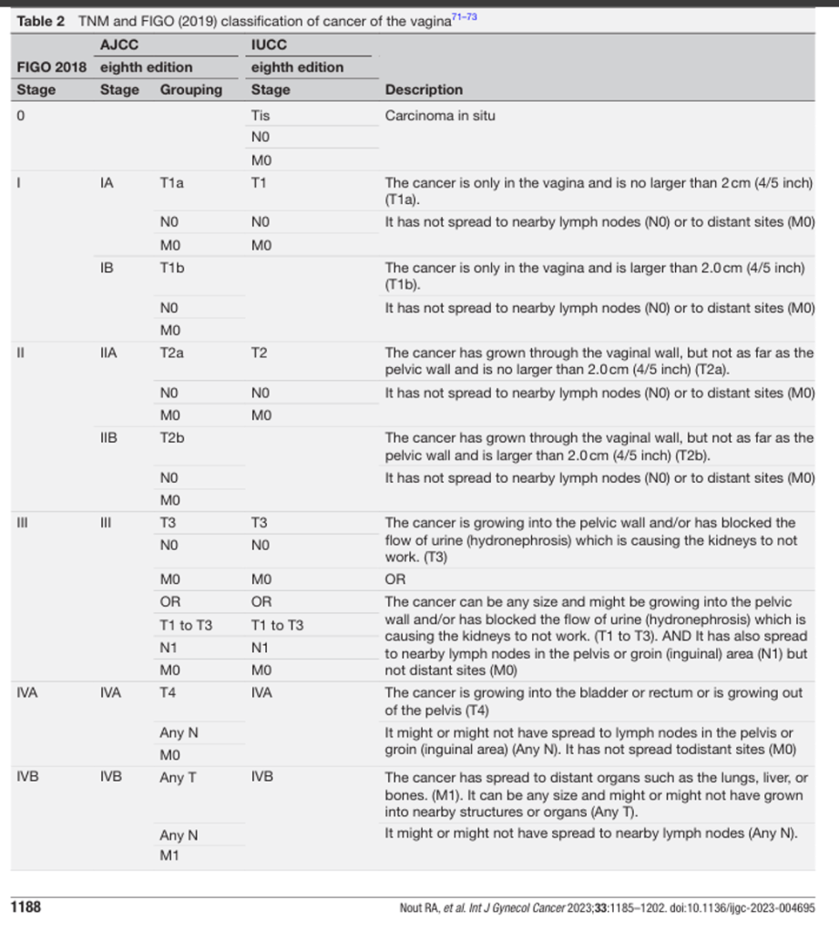
The staging of epithelial and non-epithelial malignancies should be performed when specific staging systems are not available.
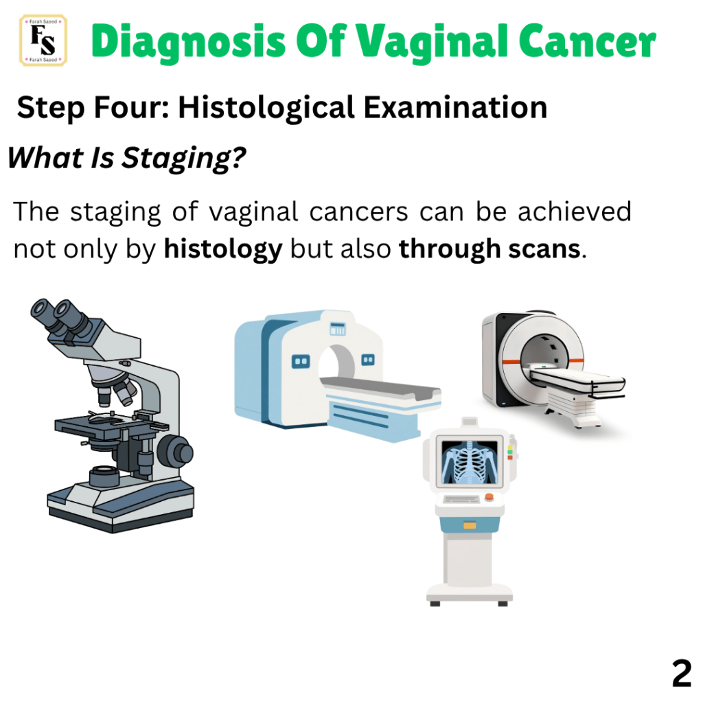
Gentle reminder: Staging can be done clinically, using radiology and also pathology.
Original tumour stage = Does Not Change Following Treatment.
TNM tumour staging system = This Allows Staging To Be Performed On Resection Specimen Following Non-surgical Treatment.
What is the “y” symbol? This presents that it is post-therapy stage.
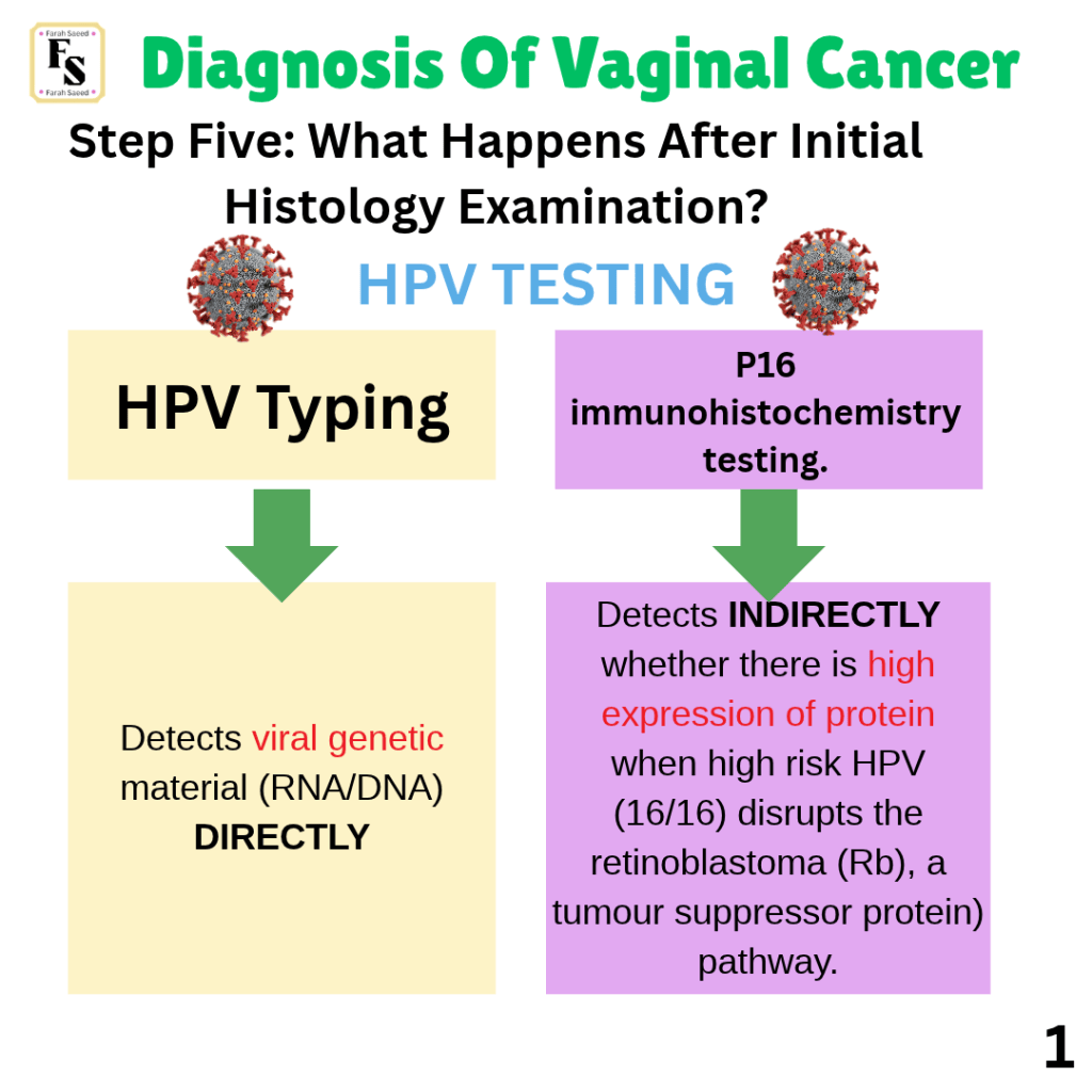
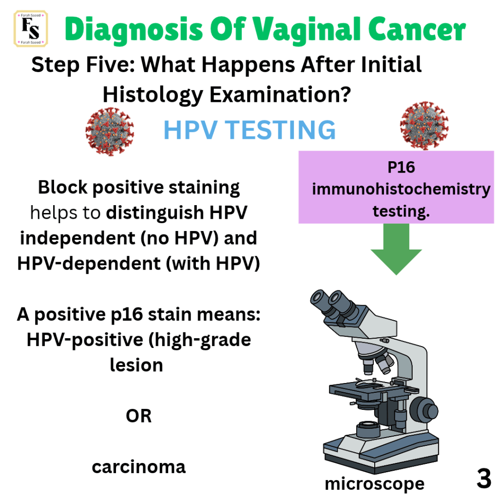
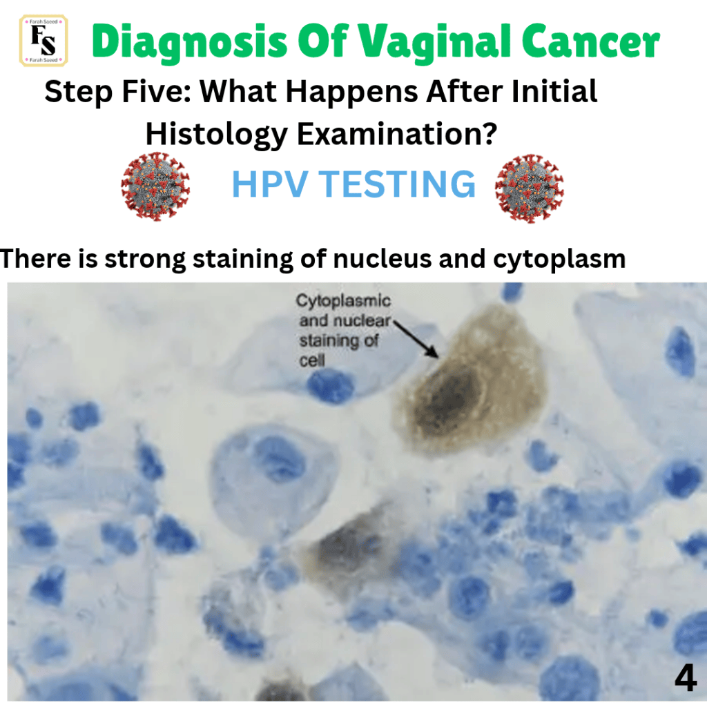
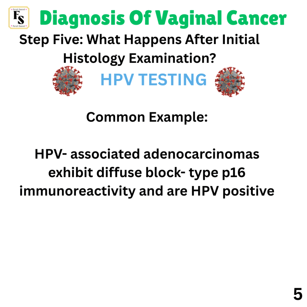
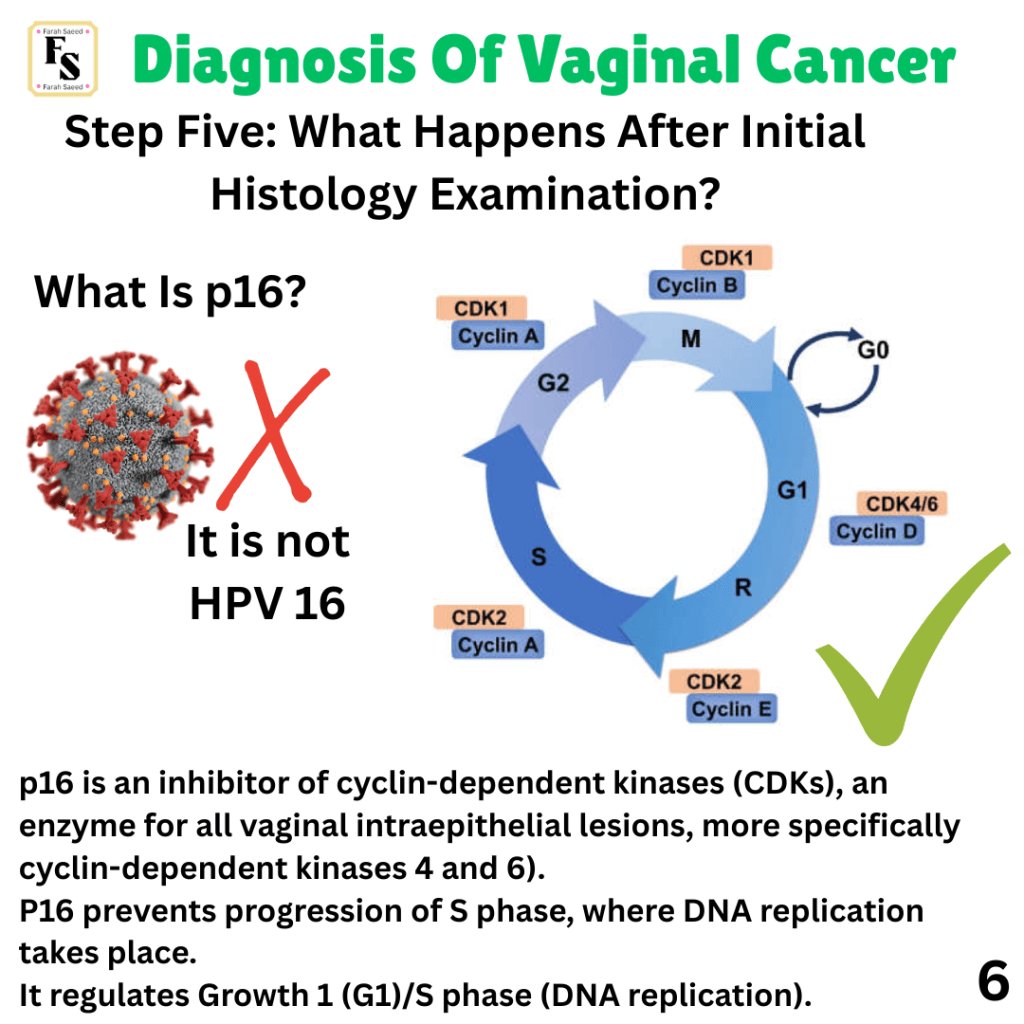
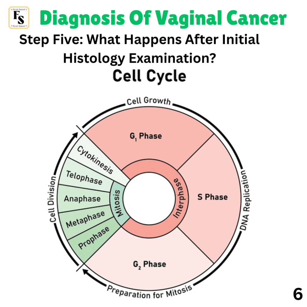
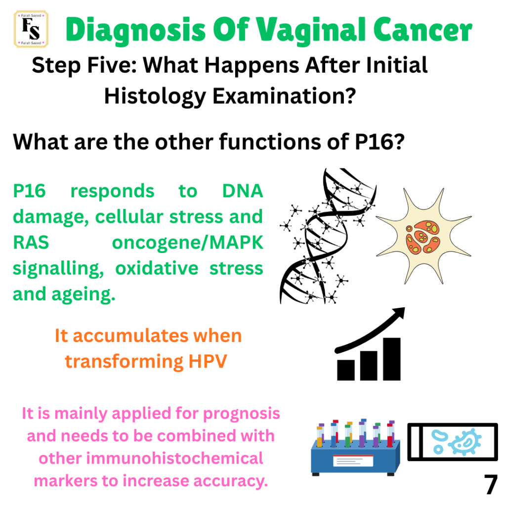
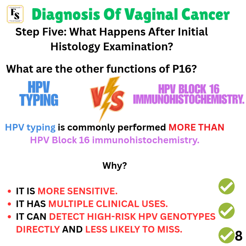
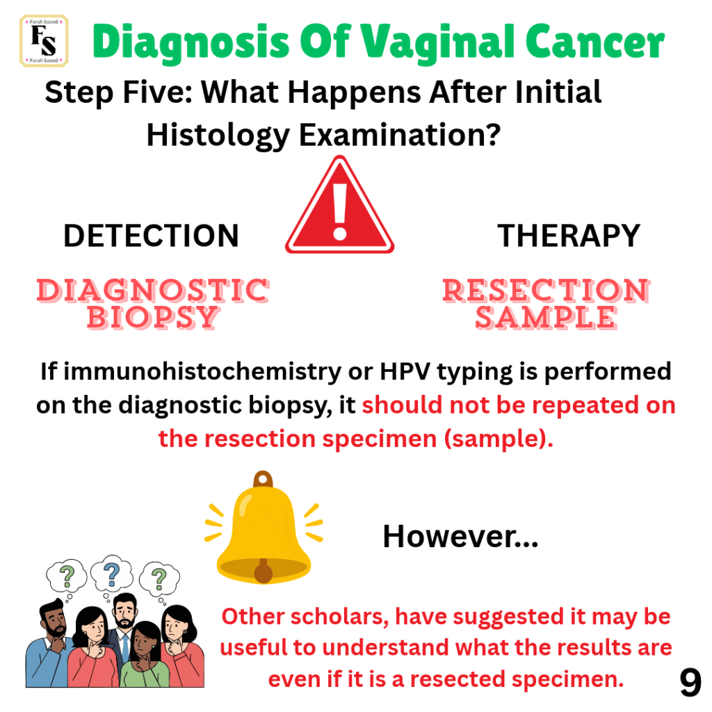
Step Five: What Happens After Initial Histology Examination?
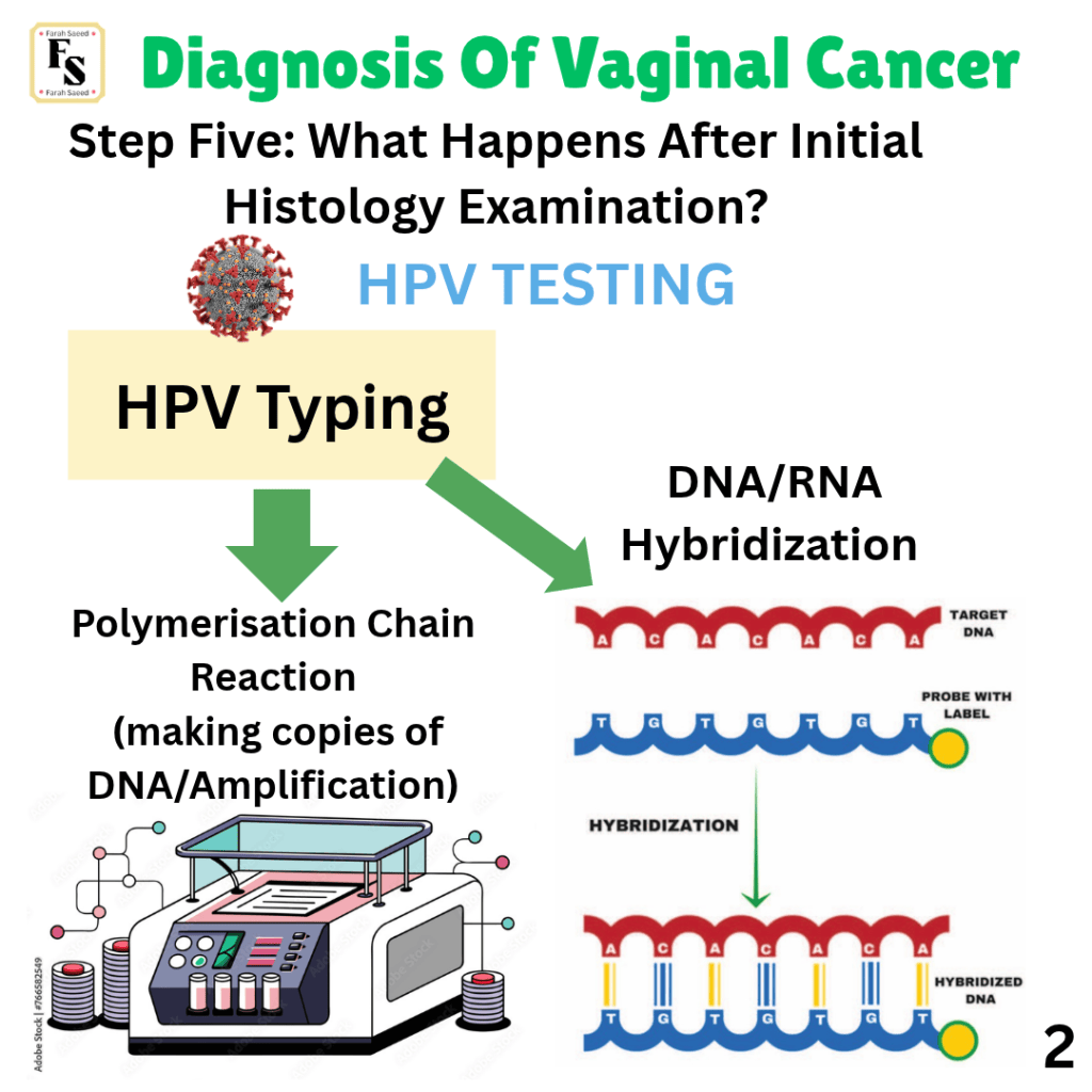
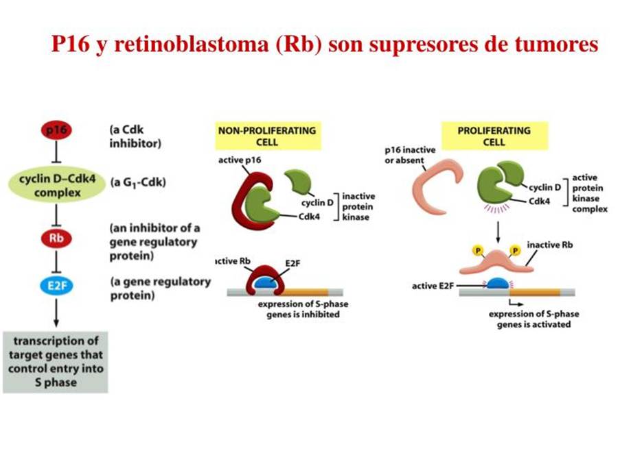
Polymerase Chain Reaction (PCR): DNA Amplification By AMBOSS: Medical Knowledge Distilled
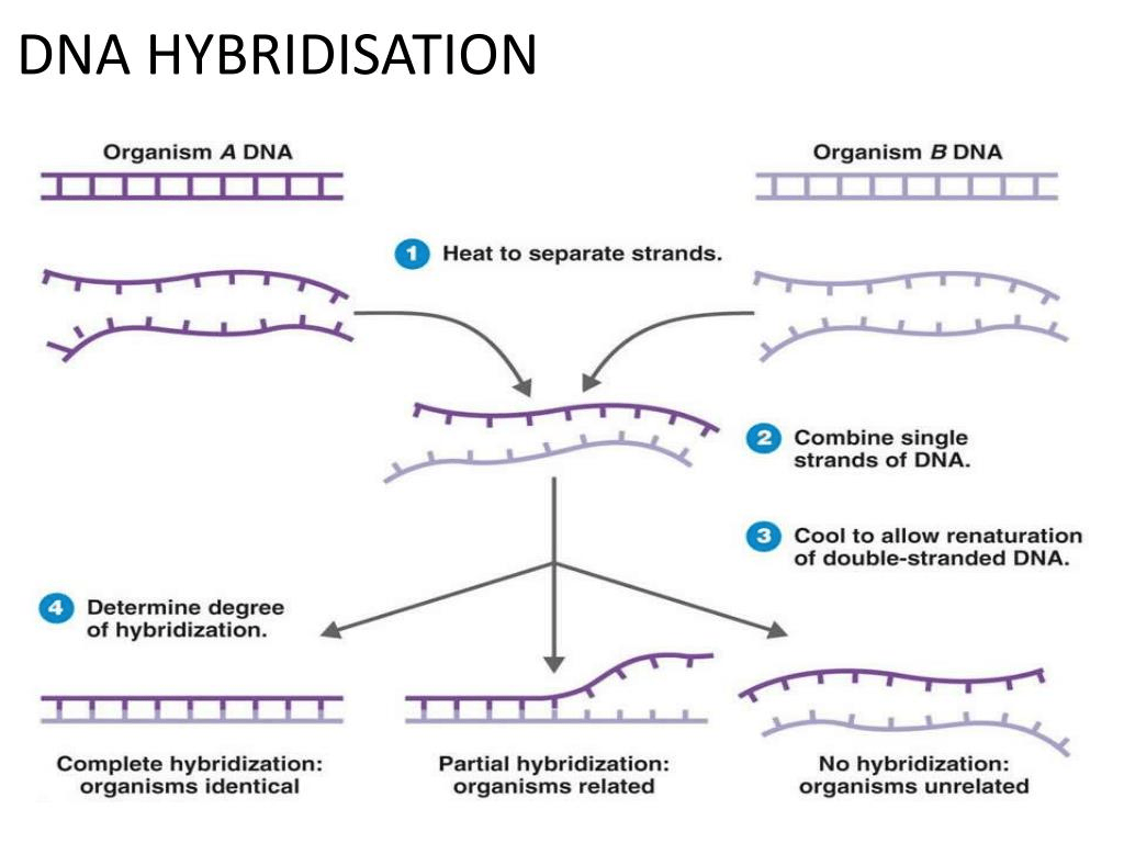
Nucleic acid hybridization | DNA hybridization Shomu’s Biology
The Journey Continues…
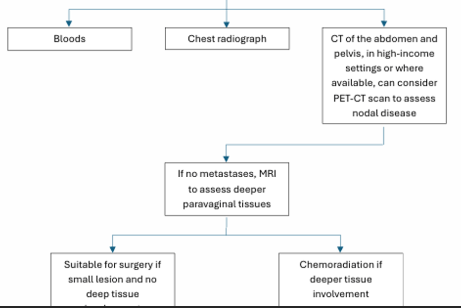
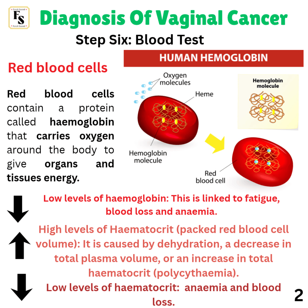
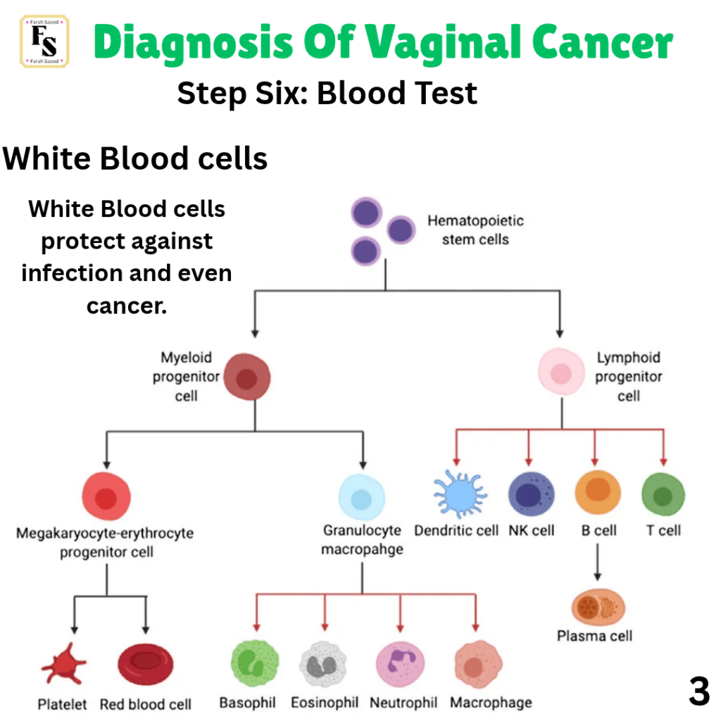
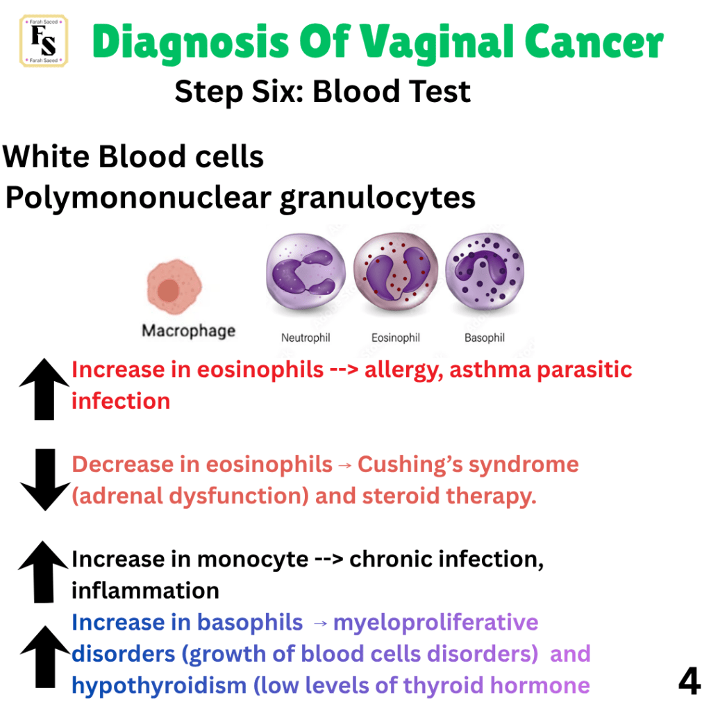
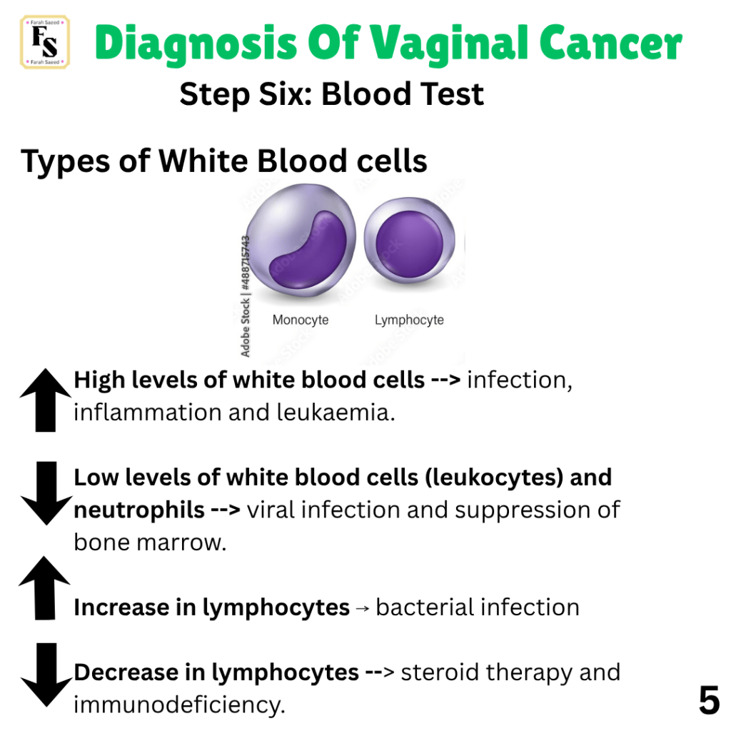
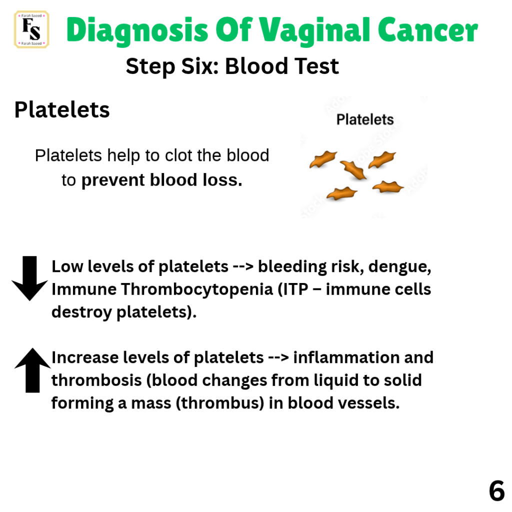
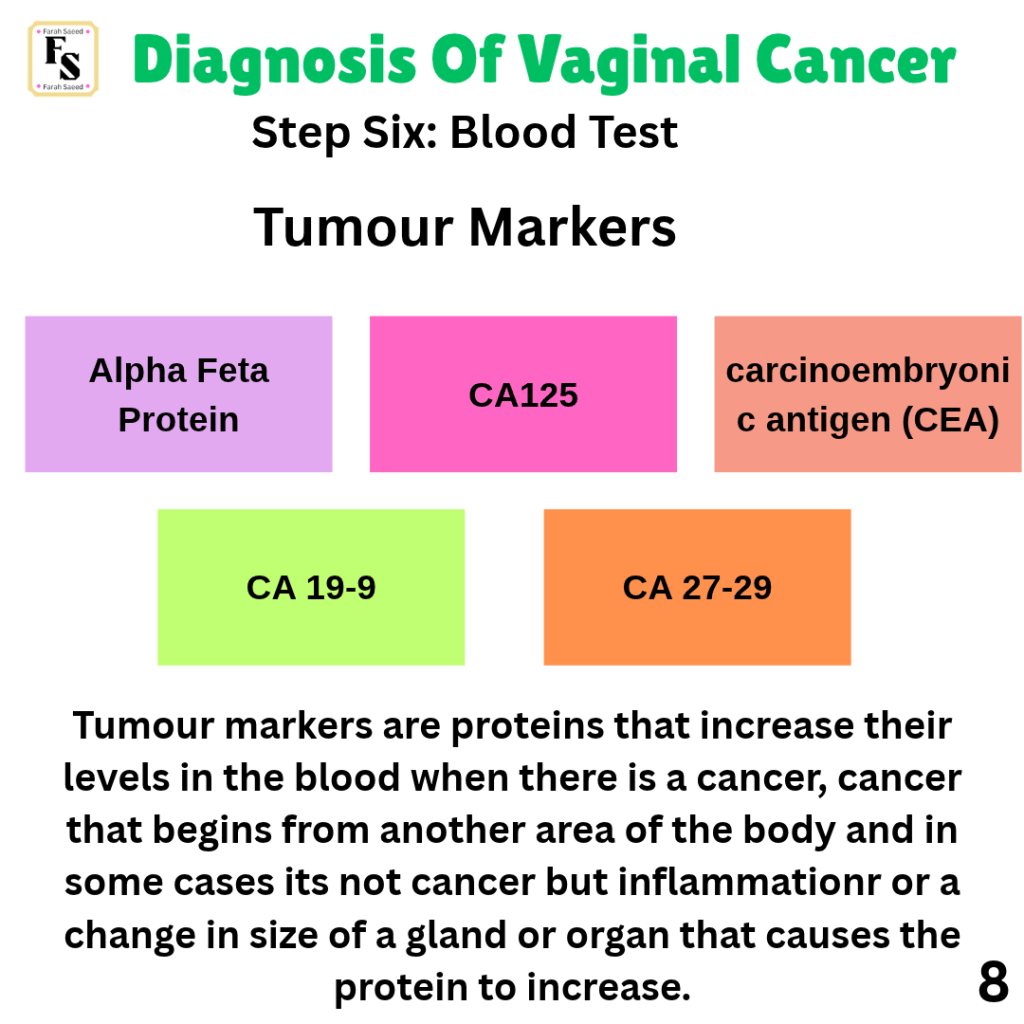
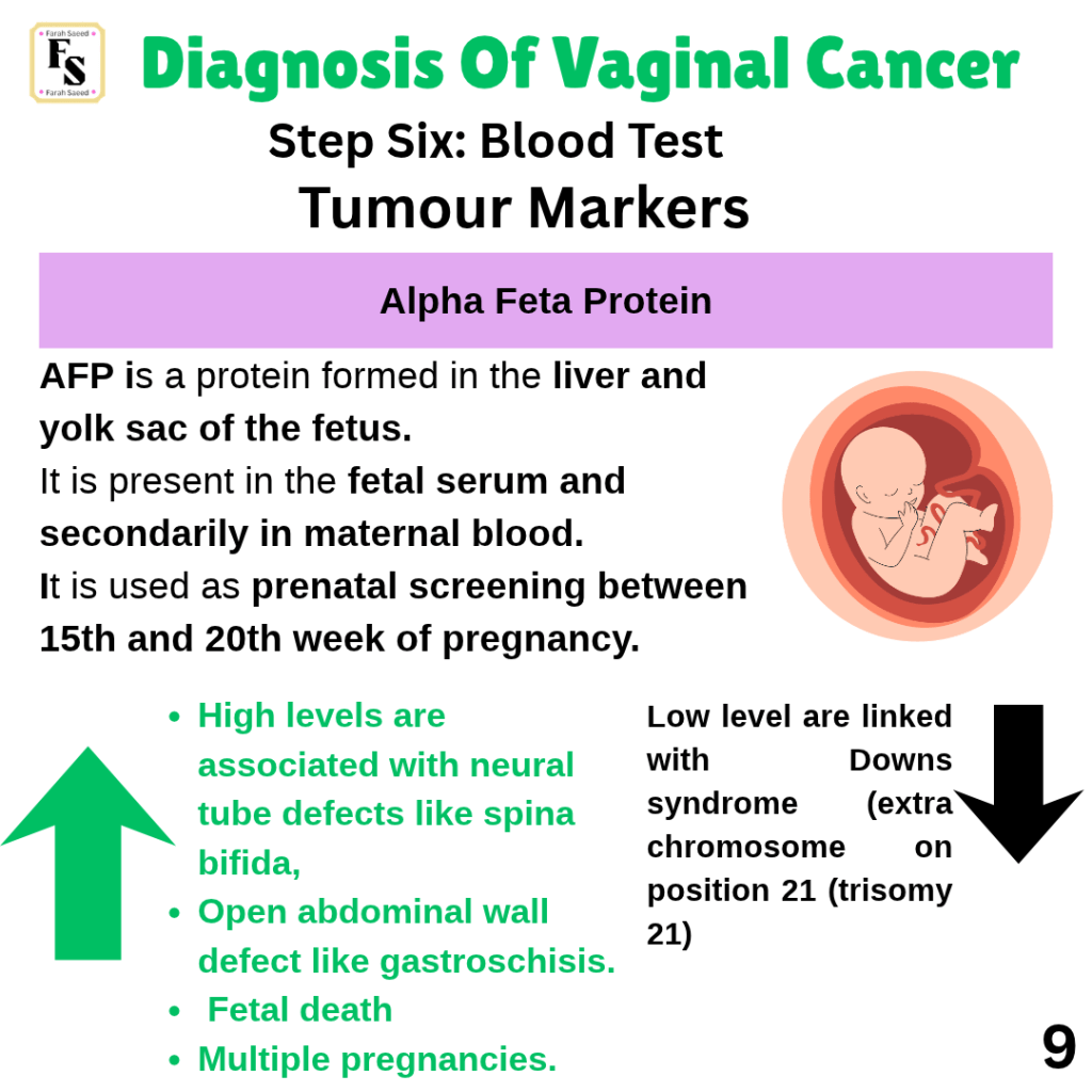
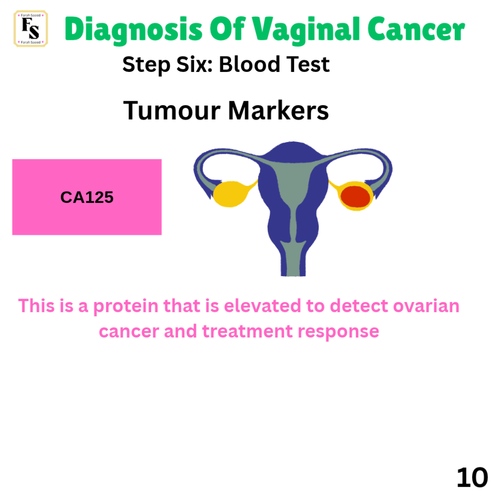
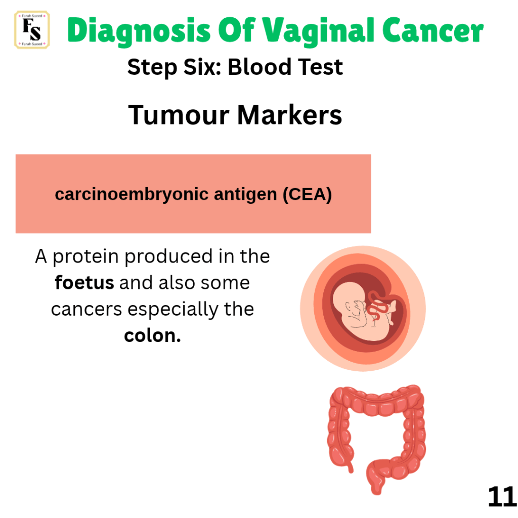
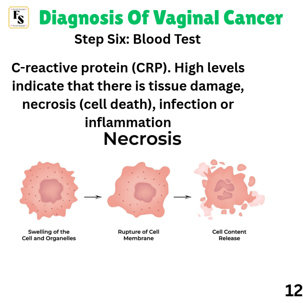
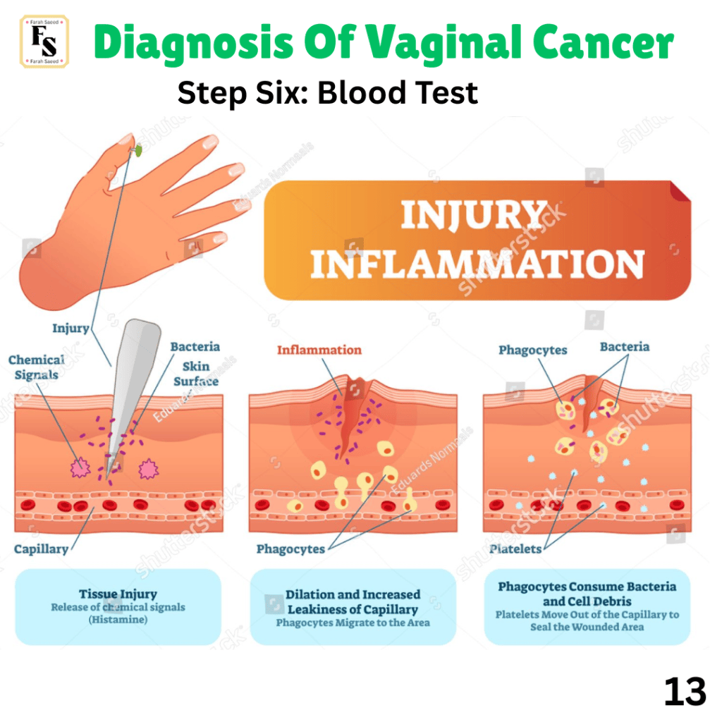
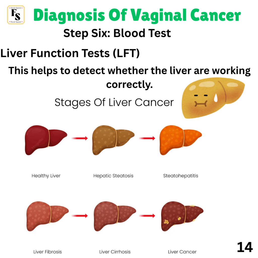
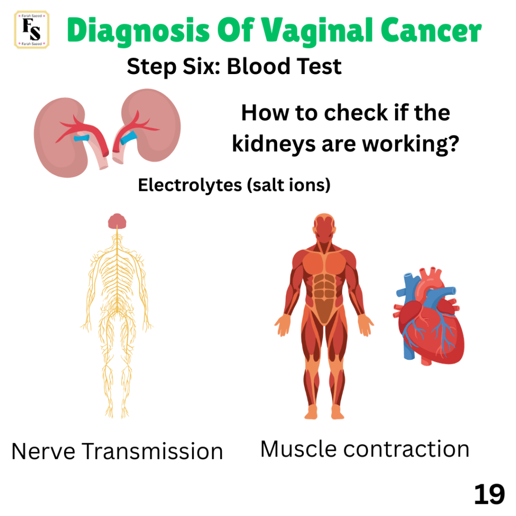
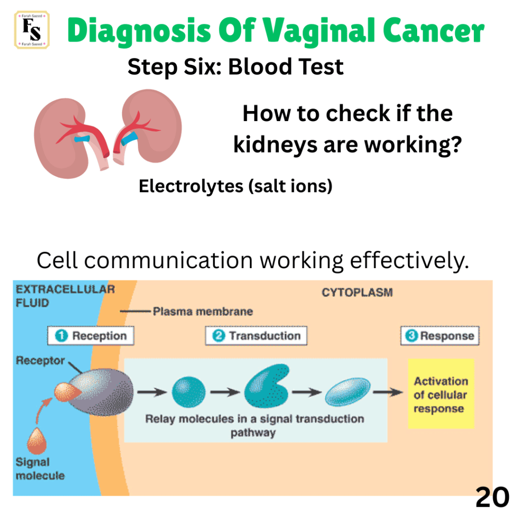
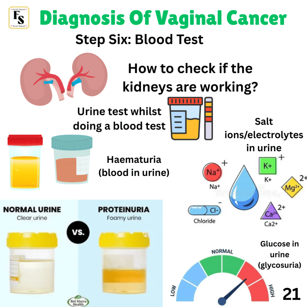
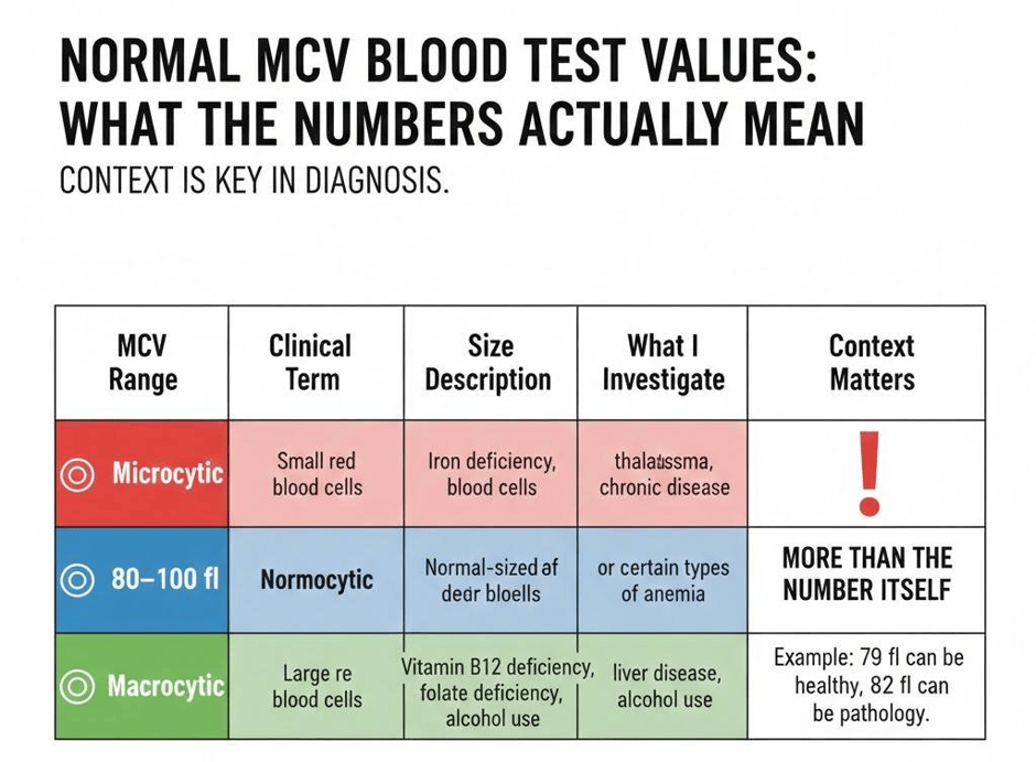
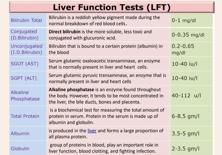
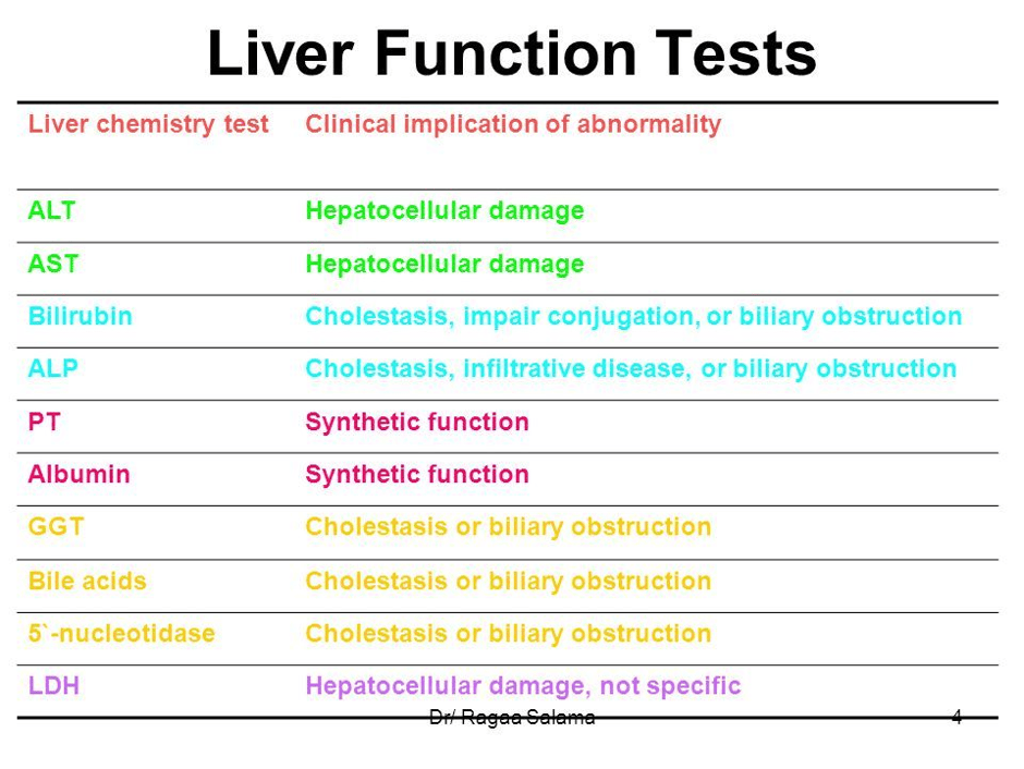
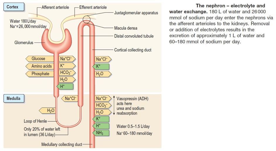
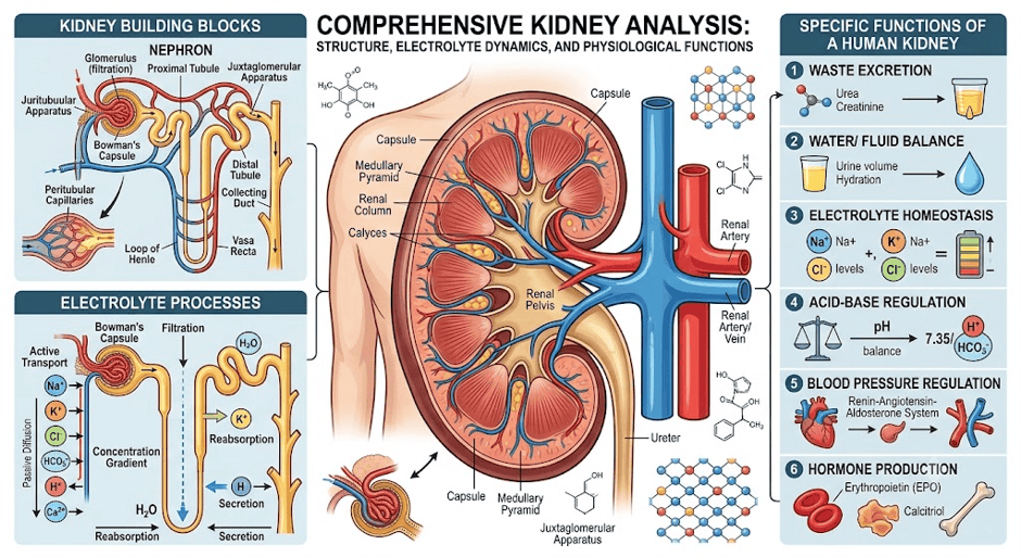
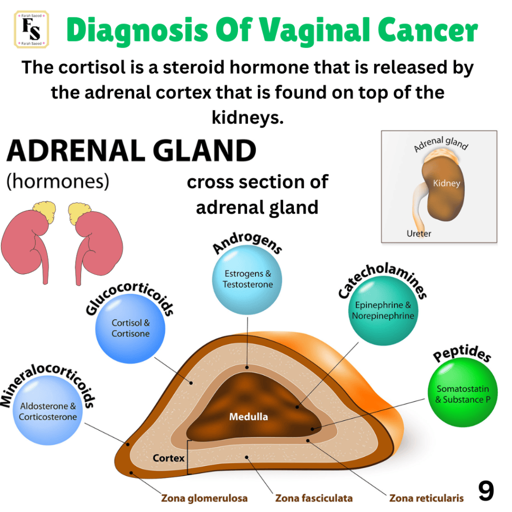
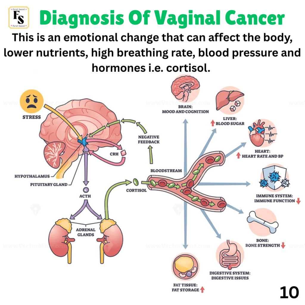
Step Six: Blood And Urine Test
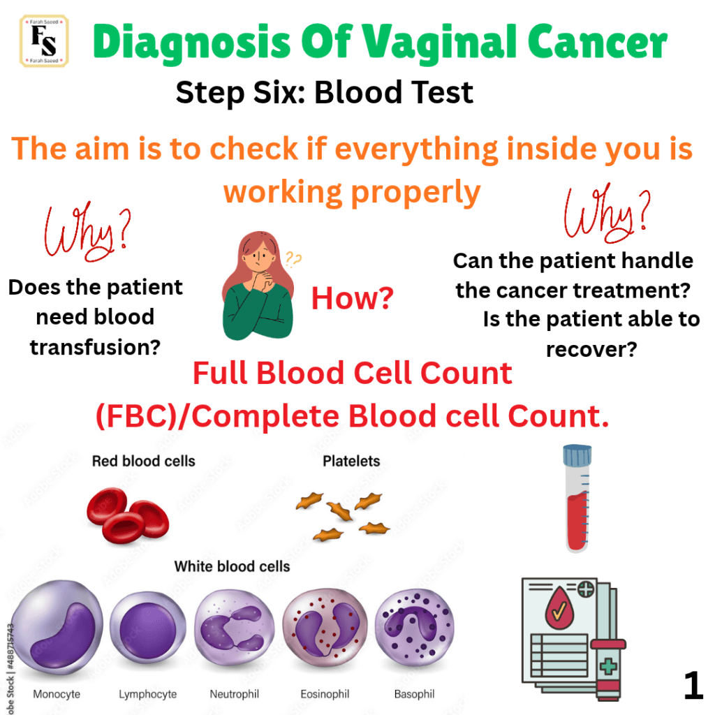

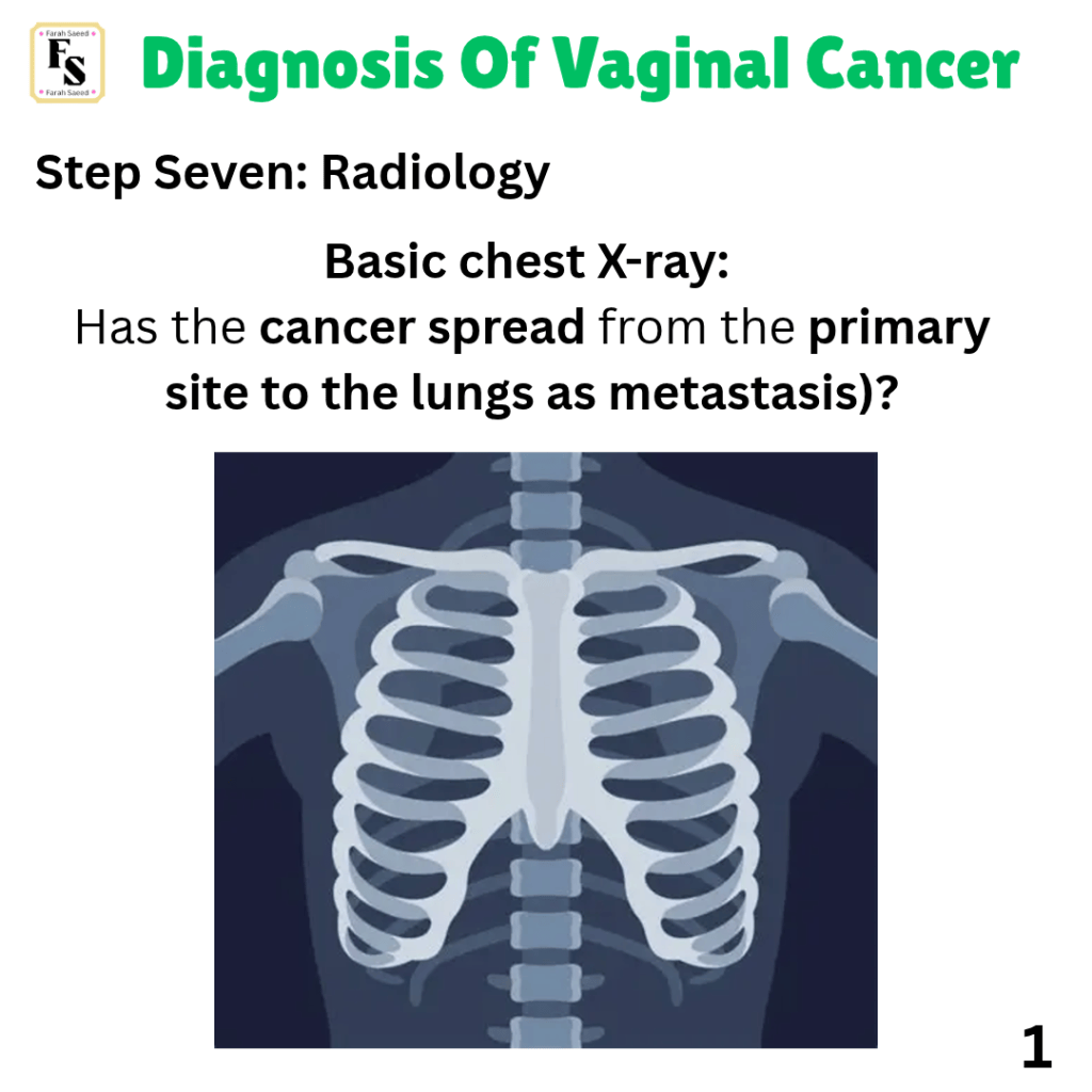
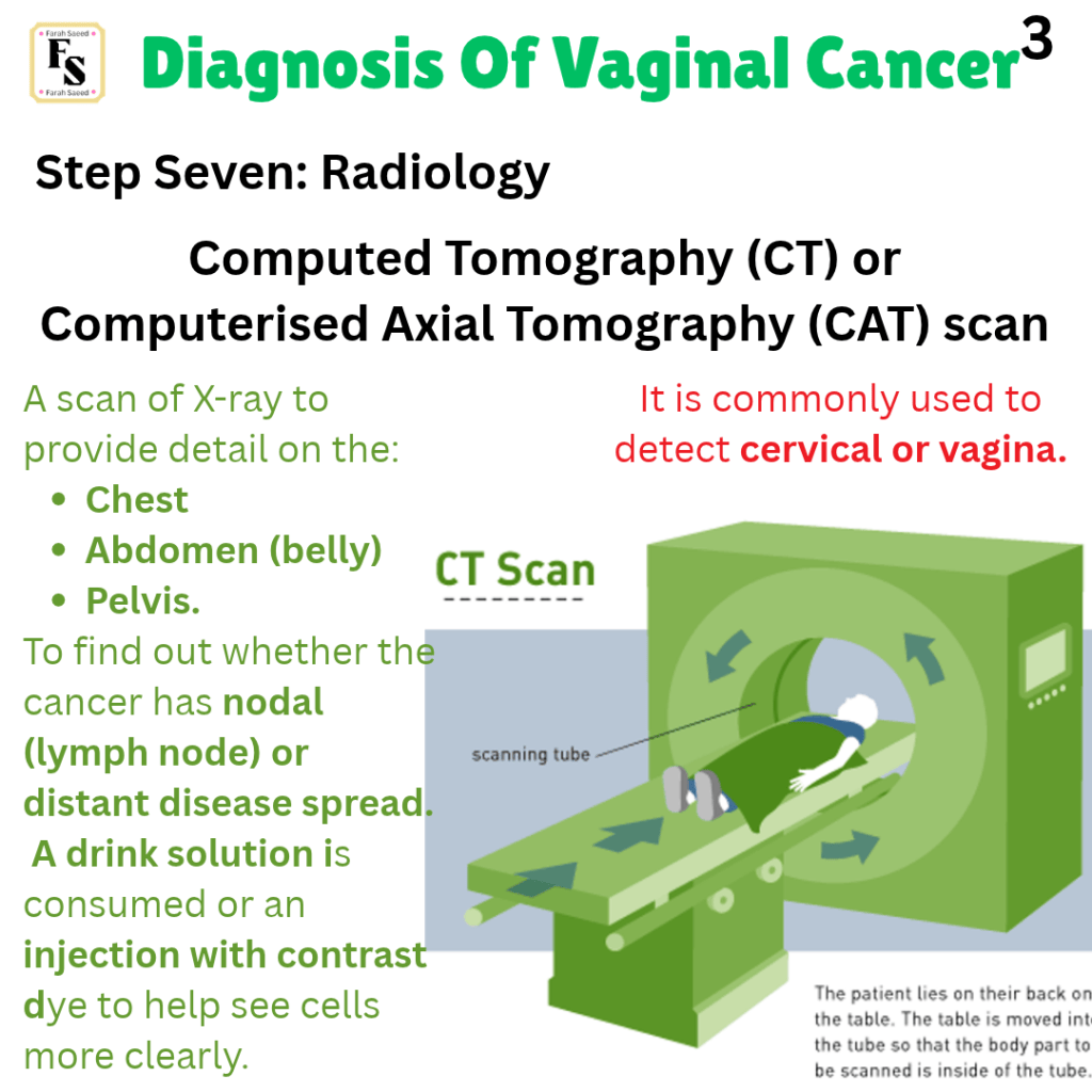
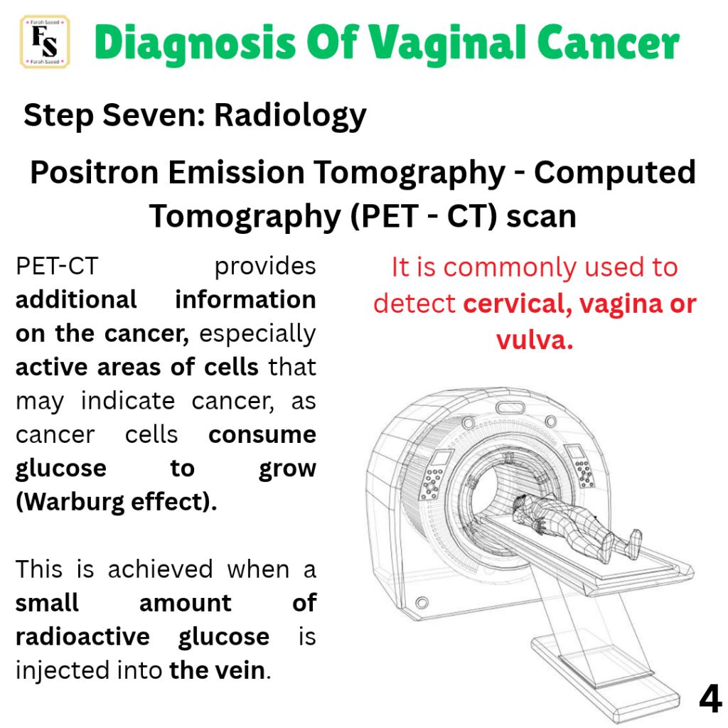
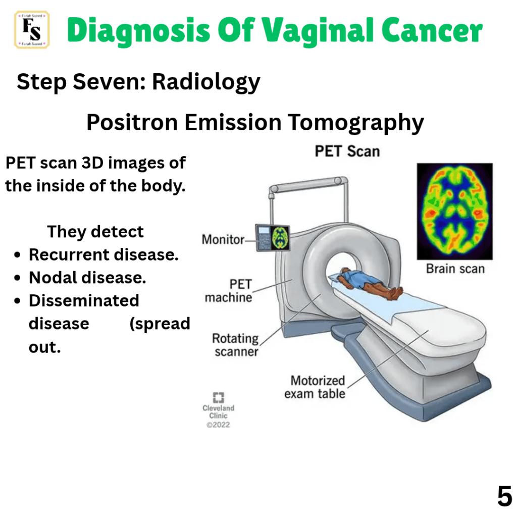
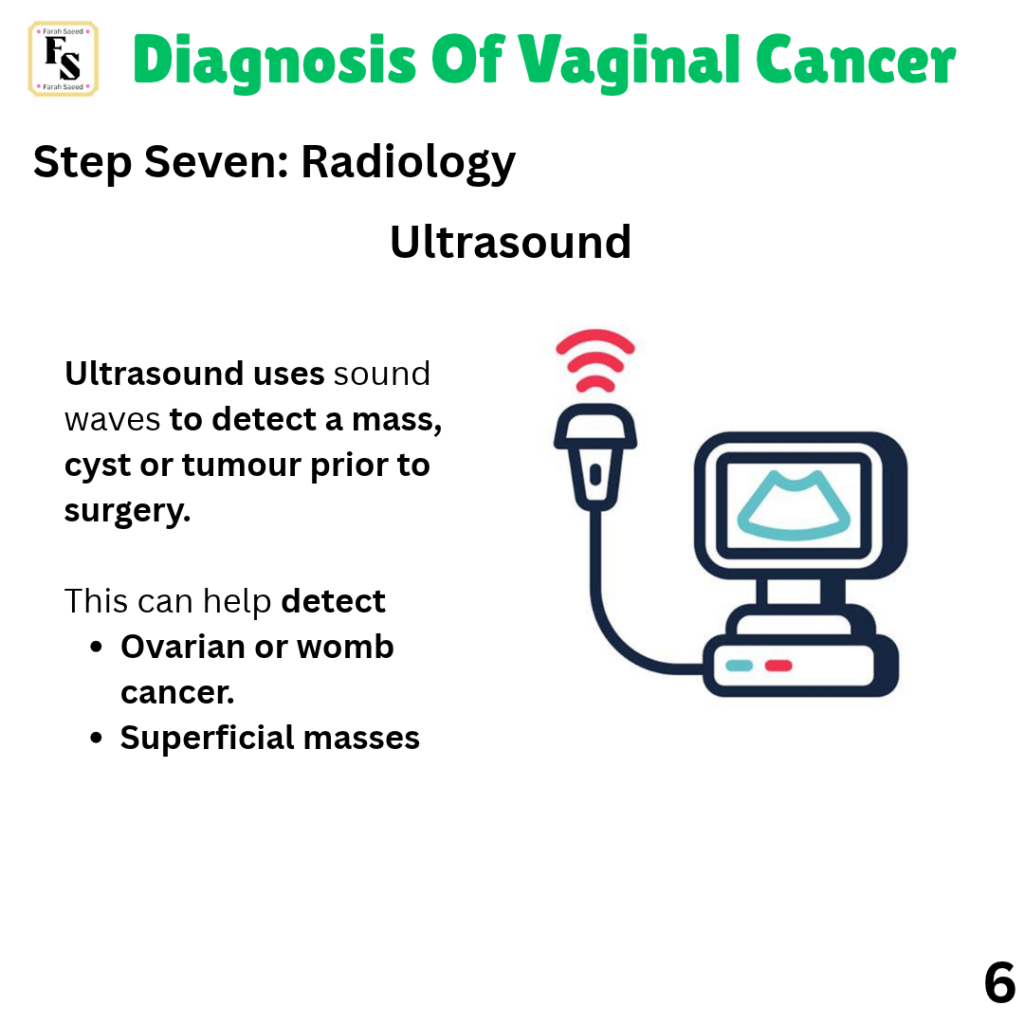
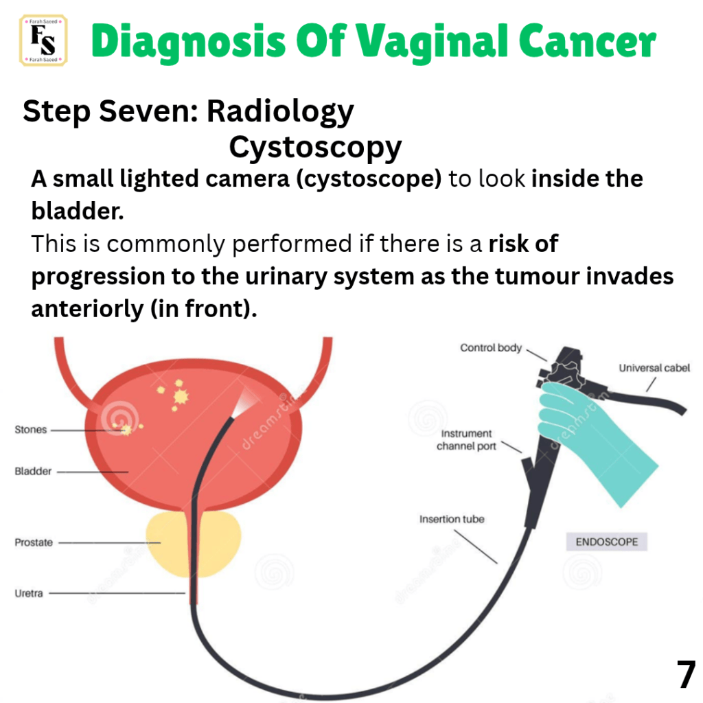
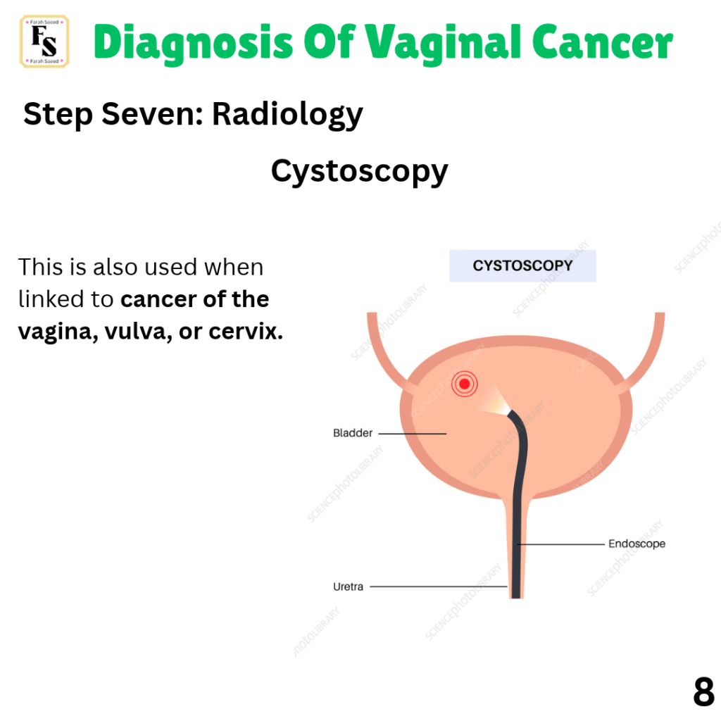
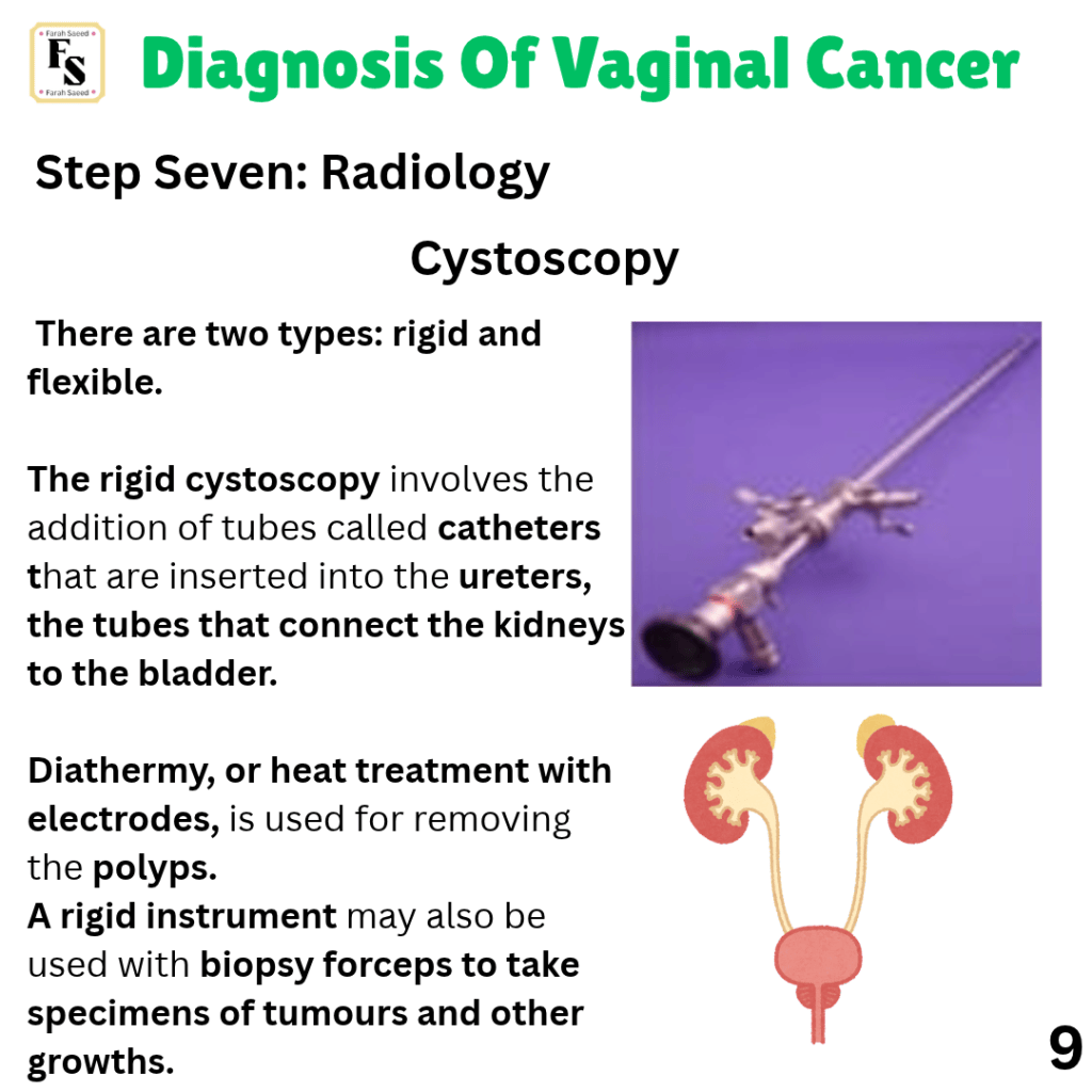
Step Seven – Radiology


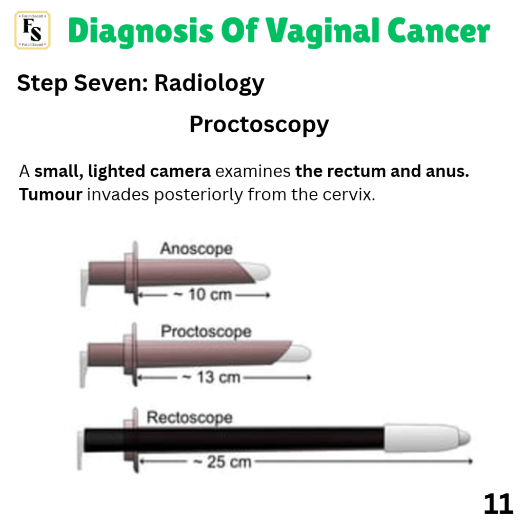
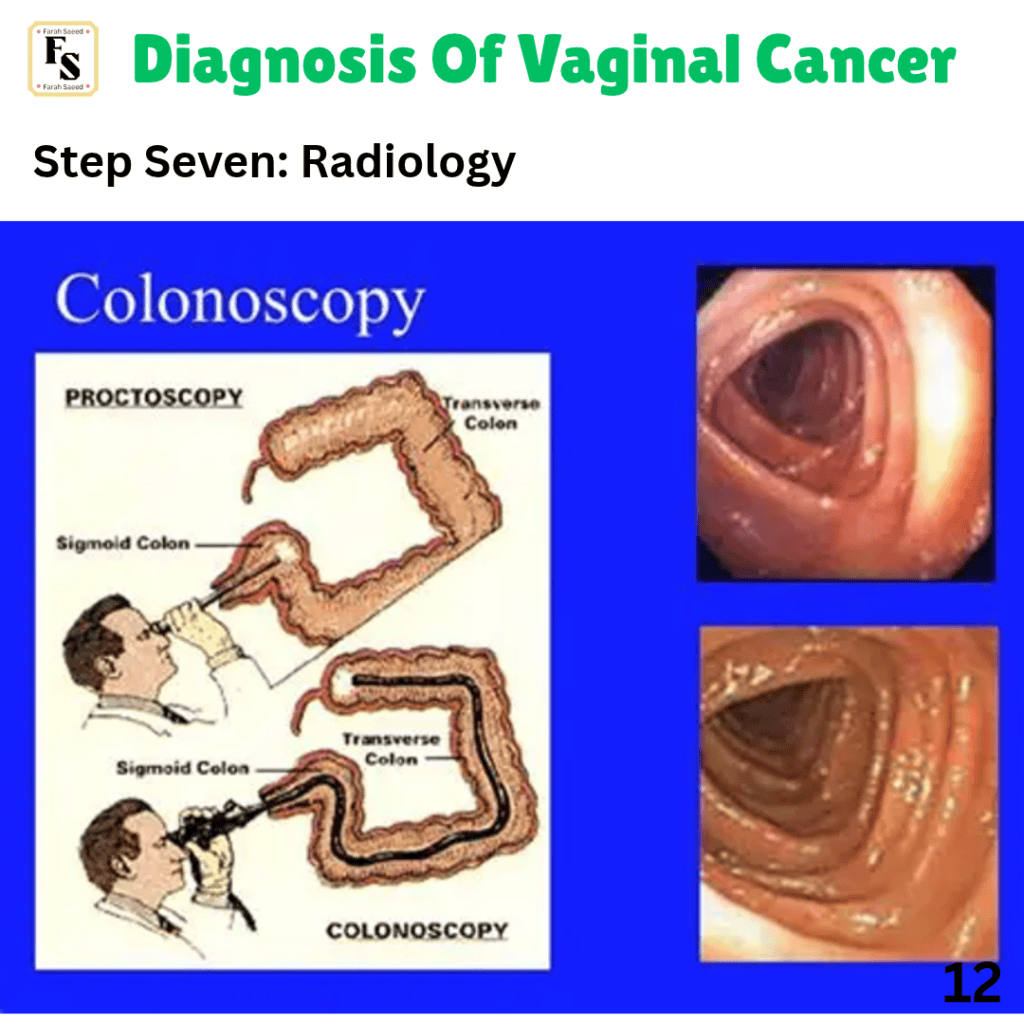
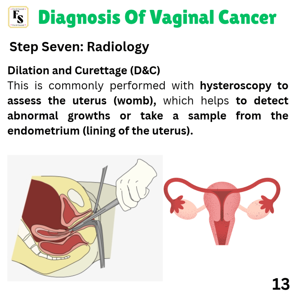
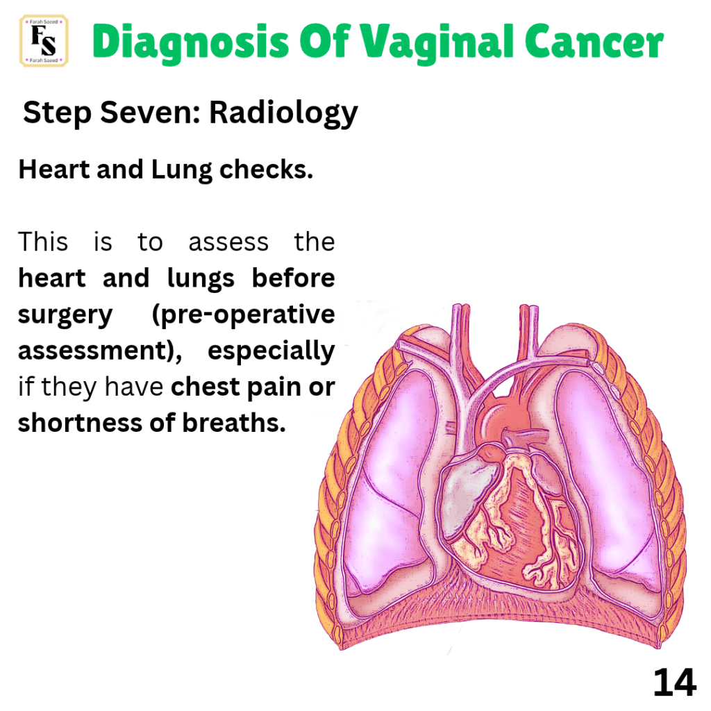



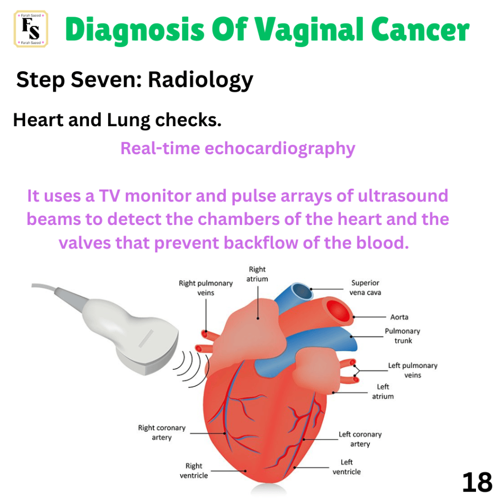
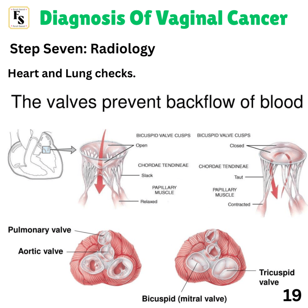
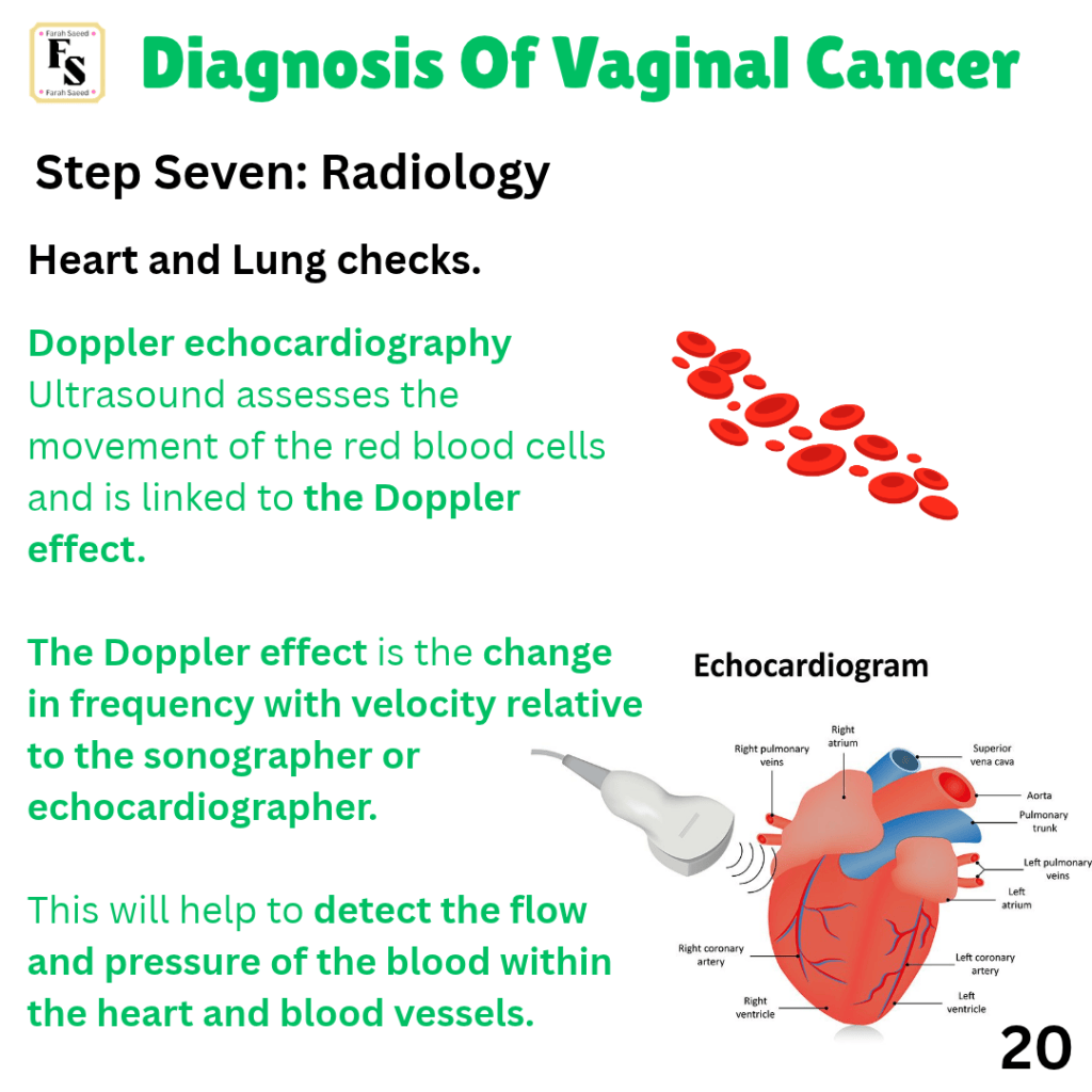

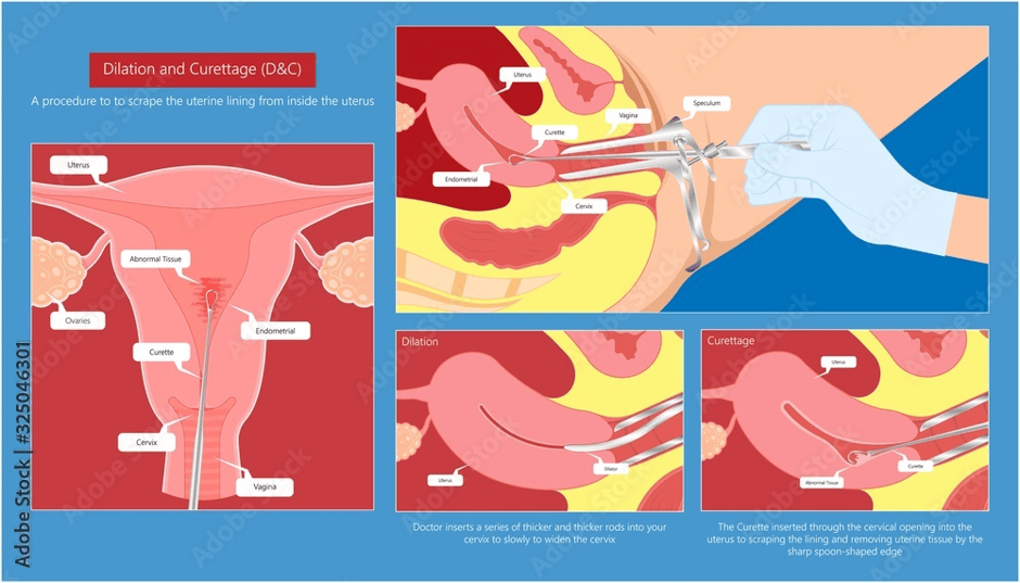
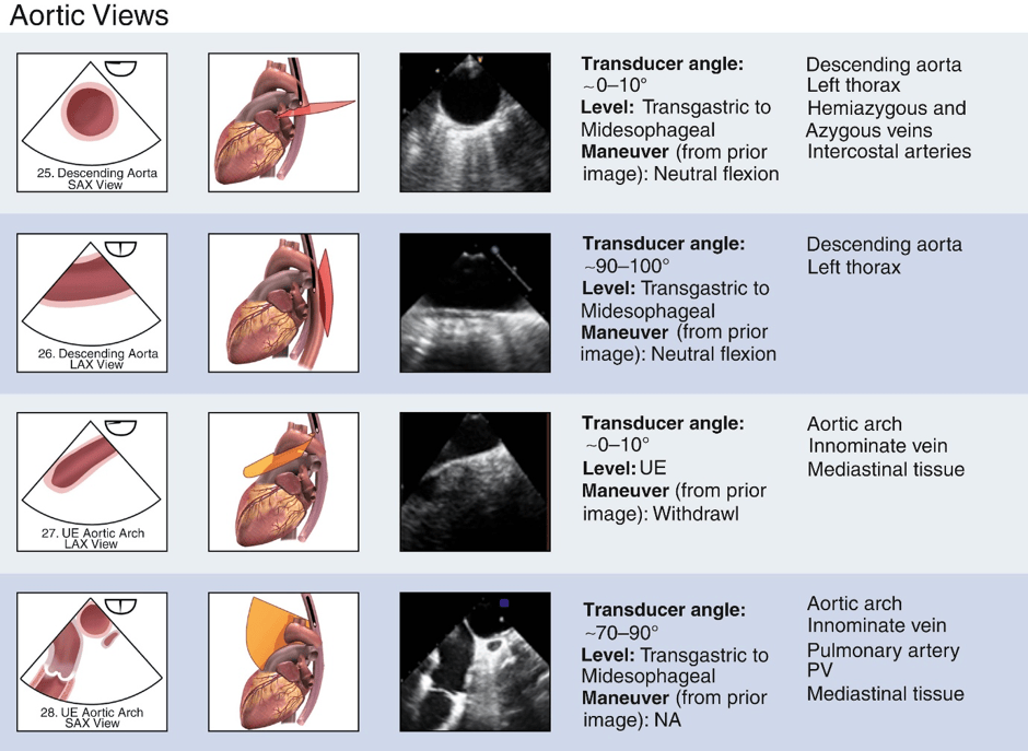
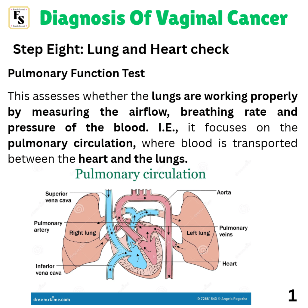
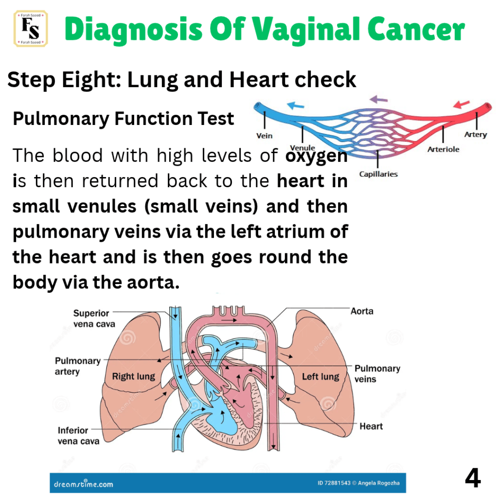
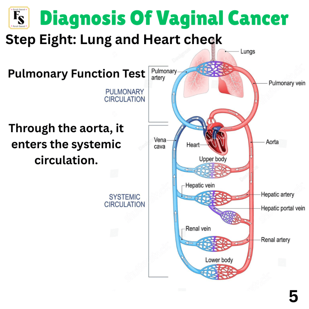
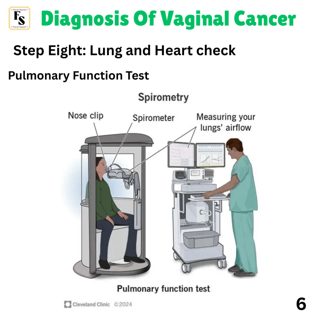
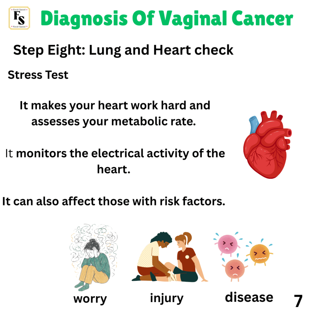
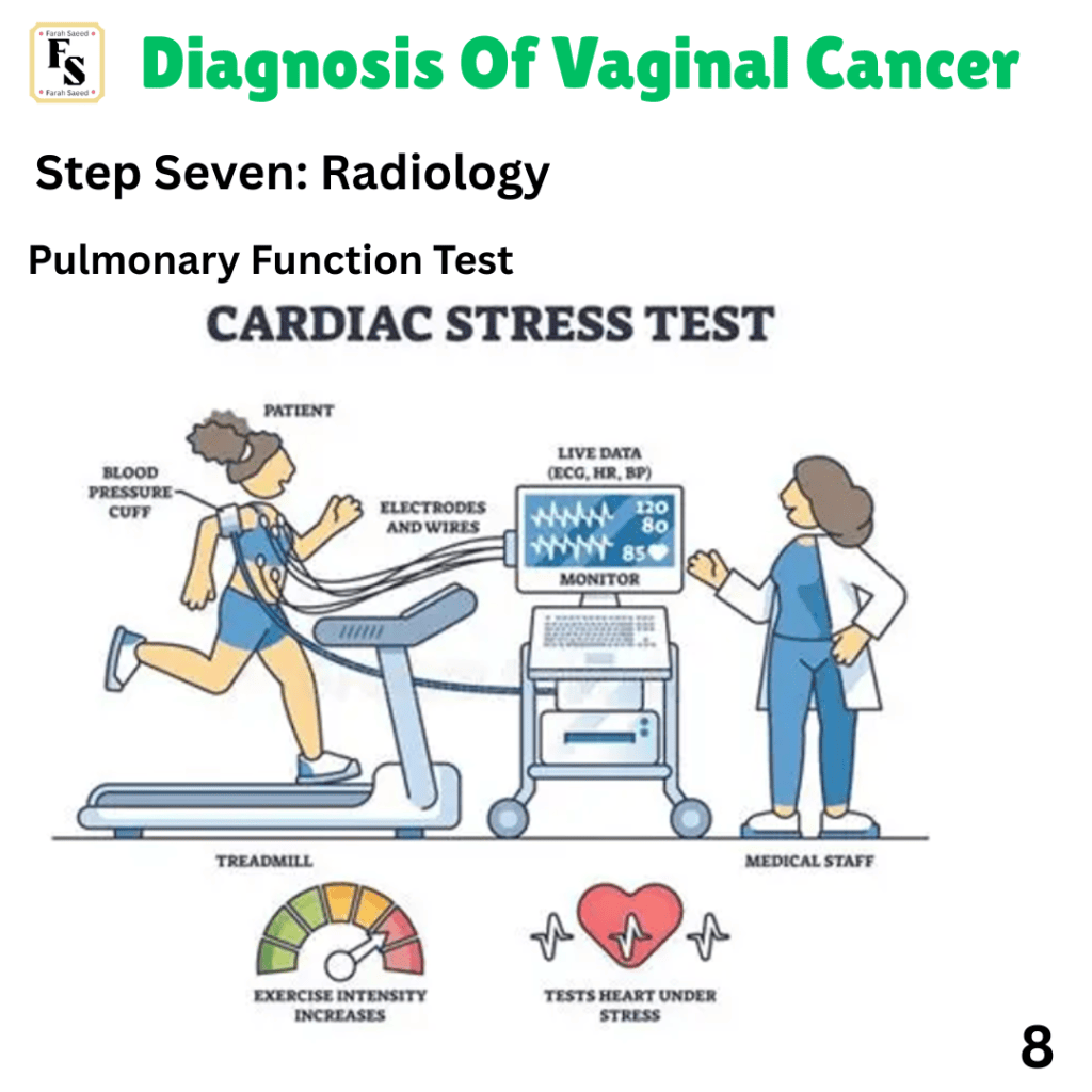
Step Eight – Heart and Lung Check
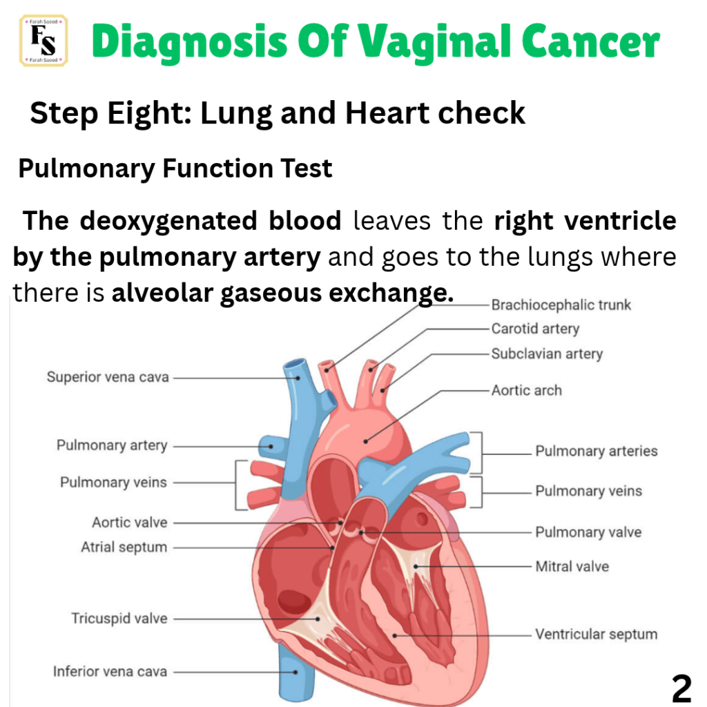
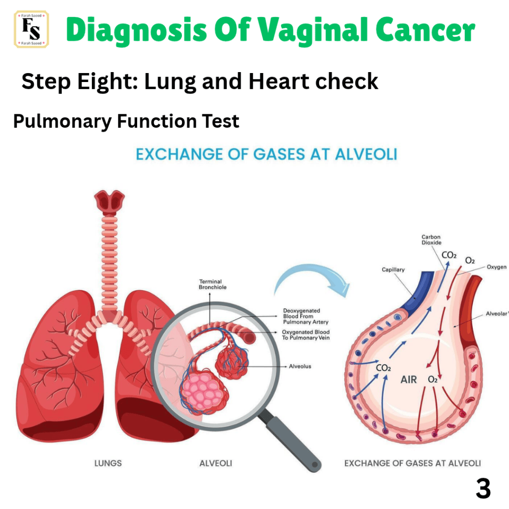
References
References
Baniak, N. (2026) Stains (IHC and special) Napsin A. Available at: https://www.pathologyoutlines.com/topic/stainsnapsina.html (Accessed: 29th October 2026)
Cerilli, L.A. and Wick, M.R. (2006). Immunohistology of Soft Tissue and Osseous Neoplasms. Elsevier eBooks, [online] pp.65–120. doi:10.1016/b978-0-443-06652-8.50009-0.
Chen, Y-T. (2026) Stains (IHC and special) MelanA/MART1. Available at: https://www.pathologyoutlines.com/topic/stainsmart1.html (Accessed: 29th October 2026)
Chowdhury, K. (2026) What Are Proteoglycans and What Is Their Function? Available at: https://biologyinsights.com/what-are-proteoglycans-and-what-is-their-function/ (Accessed: 29th October 2026)
Clay, M. (2026) Soft tissue. Available at: https://www.pathologyoutlines.com/topic/softtissueembryonalrhabdo.html (Accessed: 11th October 2026)
Fenech, M. (2023). Sonographic localisation and description of inguinofemoral lymph nodes in patients with vulvar squamous cell carcinoma. Sonography, 10(2), pp.66–75. doi:10.1002/sono.12349.
Foundation For Women’s cancer (n.d.) Diagnostic tests. Available at: https://foundationforwomenscancer.org/gynecologic-cancers/gynecologic-cancer-basics/diagnostic-tests/ (Accessed: 29th October 2026)
Gene Cards (2026) MYOD1 gene. Available at: https://www.genecards.org/card/MYOD1 (Accessed: 19th October 2026)
Khan, A., Obeidin, F. and Topilow, A. (2026). Stains (IHC and special) S100 Available at: https://www.pathologyoutlines.com/topic/stainss100.html (Accessed: 29th October 2026)
Mouhamed, H.A. (2017). The diagnostic utility of immunohistochemistry in undifferentiated ovarian carcinoma. Archives in Cancer Research, 05(02). doi:10.21767/2254-6081.1000143.
National Health Service (n.d.) PSA test. Available at: https://www.nhs.uk/tests-and-treatments/psa-test/ (Accessed: 29th October 2026)
Ohashi, R. (2026). Stains (IHC and special) Vimentin. Available at: https://www.pathologyoutlines.com/topic/stainsvimentin.html/ (Accessed: 29th October 2026)
Orsatti, A. and Lobo, J. (2026). Stains (IHC and special) PLAP. Available at: https://www.pathologyoutlines.com/topic/stainsplap.html (Accessed: 29th October 2026)
Pernick, N. (2026a) Stains (IHC and special) CK20. Available at: https://www.pathologyoutlines.com/topic/stainsck20.html (Accessed: 29th October 2026)
Pernick, N. (2026b) Stains (IHC and special) CDX2. Available at: https://www.pathologyoutlines.com/topic/stainsCDX2.html (Accessed: 29th October 2026)
Pernick, N. (2026c) Stains (IHC and special) GATA3. Available at: https://www.pathologyoutlines.com/topic/stainsgata3.html (Accessed: 29th October 2026)
Science Direct (n.d.) Neuron-Specific Enolase. Available at: https://www.sciencedirect.com/topics/neuroscience/neuron-specific-enolase (Accessed: 29th October 2026)
Siegele, B. (2026) Stains (IHC and special) CD10. Available at: https://www.pathologyoutlines.com/topic/cdmarkerscd10.html (Accessed: 29th October 2026)
Thakur, S. and Enwere, E. (2026) Stains (IHC and special) NKX3.1. Available at: https://www.pathologyoutlines.com/topic/stainsnkx3.html (Accessed: 29th October 2026)
Thetiot, M., Freeman, S.A., Dezmaxieres, A. (2018) Immunohistochemical Analysis of Myelin Structures. Available at: https://experiments.springernature.com/articles/10.1007/978-1-4939-7862-5_2 (Accessed: 29th October 2026)
Thompson, L.D.R. (2018) Data Set for the Reporting of Oral Cavity Carcinomas. Archives of Pathology and Laboratory Medicine. Available at: https://www.researchgate.net/publication/329488624_Data_Set_for_the_Reporting_of_Oral_Cavity_Carcinomas (Accessed: 29th October 2026)
Ulici, V. and Wang, L.J. (2026) Stains (IHC and special TTF1). Available at: https://www.pathologyoutlines.com/topic/stainsttf1.html (Accessed: 29th October 2026)
UniProt (n.d.) P15309 PPAP_HUMAN. (Prostatic acid phosphatase) Available at: https://www.uniprot.org/uniprotkb/P15309/entry (Accessed: 29th October 2026)
UniProt (n.d.) Q8N158.GPC2_Human (Glypican-2). Available at: https://www.uniprot.org/uniprotkb/Q8N158/entry (Accessed: 29th October 2026)
Van Bockstal, M. (2026) Stains (IHC & special) Cytokeratin AE1 / AE3. Available at: https://www.pathologyoutlines.com/topic/stainsae1ae3.Html (Accessed: 26th October 2026)
Wirth, P. (2026) Stains (IHC and special) SOX10. Available at: https://www.pathologyoutlines.com/topic/stainssox10.html (Accessed: 29th October 2026)


Leave a Reply