Risk Factors of Vaginal Cancer
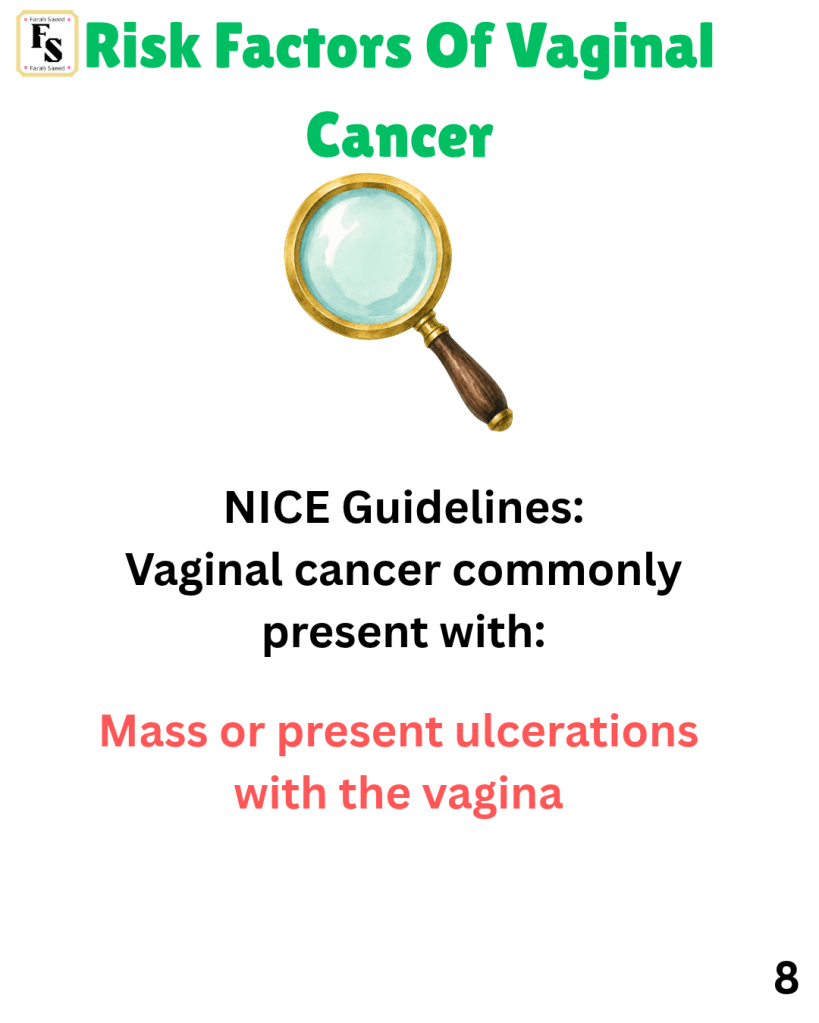
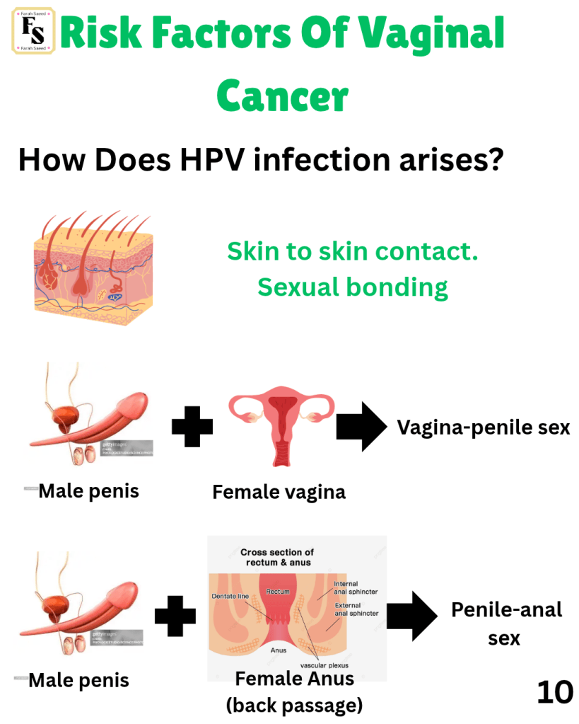
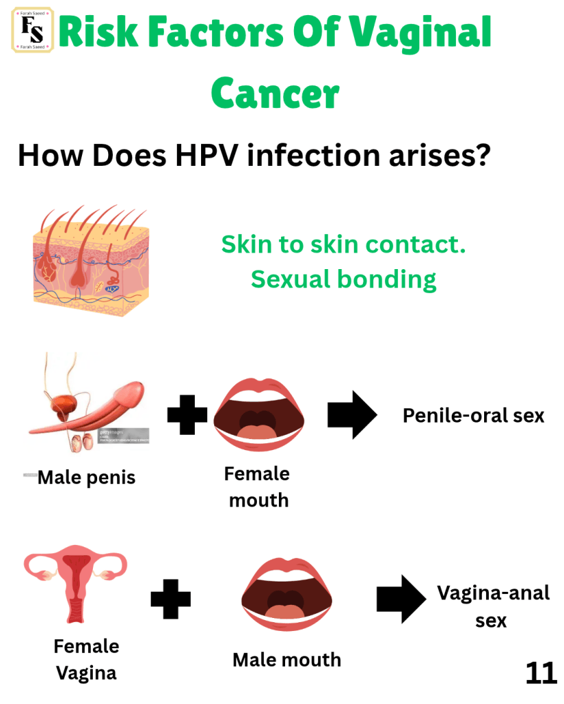
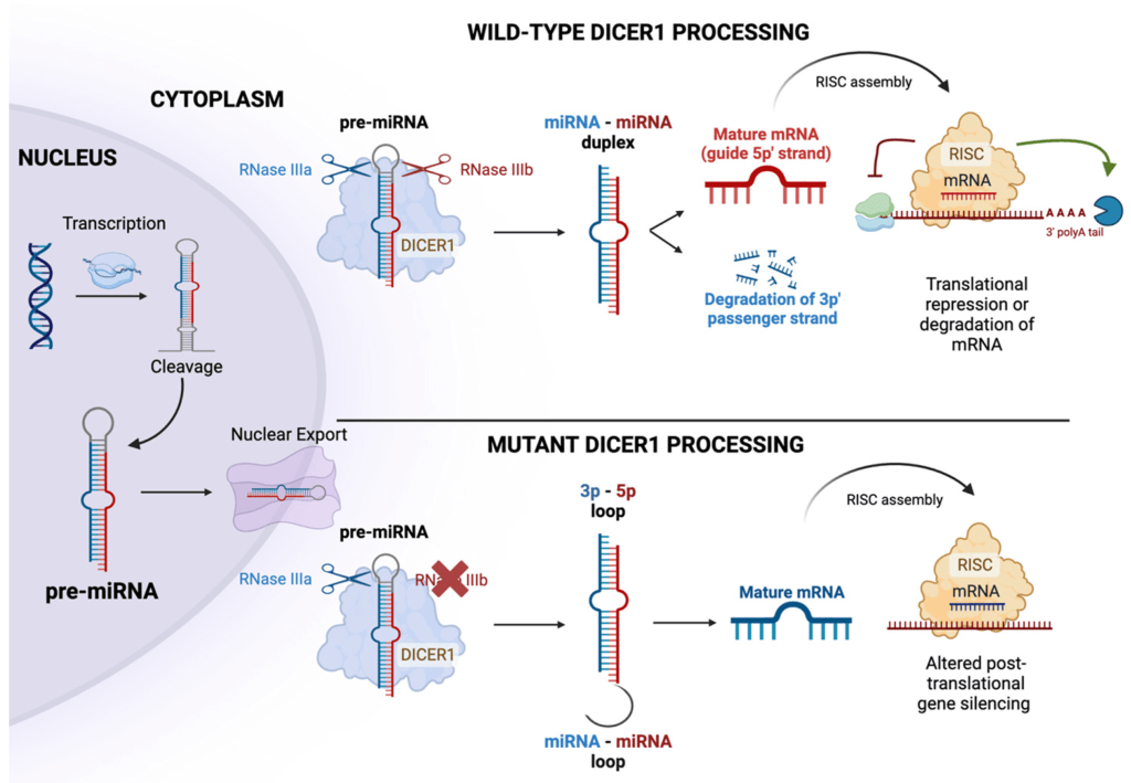
There are various risk factors for vaginal cancer. Some are generic; for instance, lifestyle factors (diet, exercise, smoking, hygiene, stress), environment (poverty, exposure to chemicals). Mutations (changes in the gene) in specific genes involved in suppressing the tumours and regulating how cells divide and grow. Others are more specific to gynaecological cancers (viral infections, the formation of non-cancerous growths and bonding). Bonding refers to the number of sexual partners, early age at first interaction and type of bonding.
Viral Infections
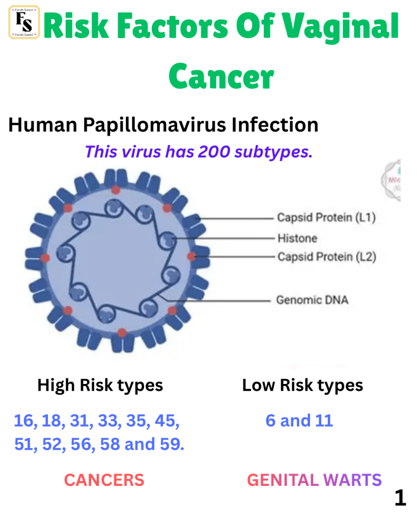
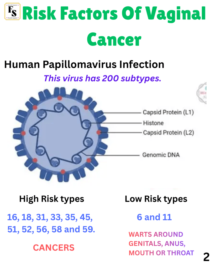
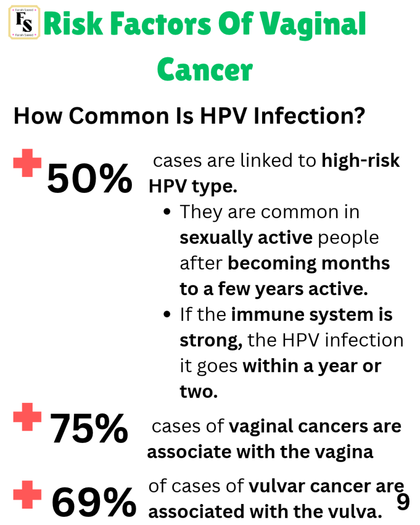
Human Papillomavirus Infection
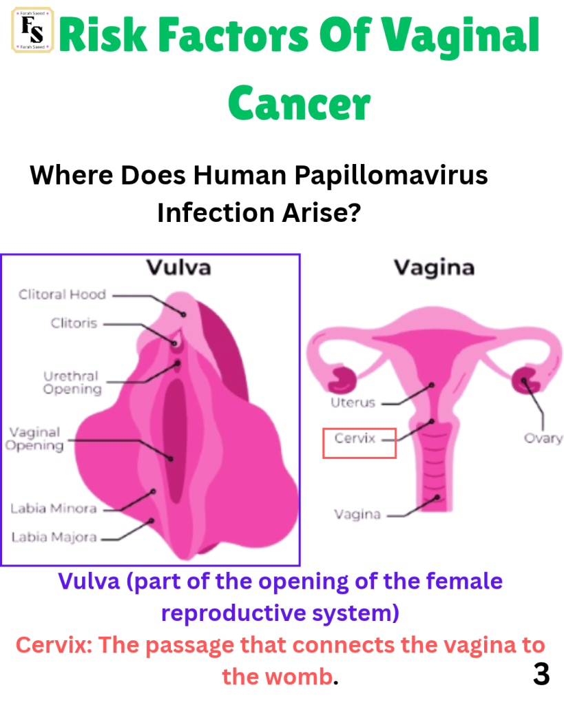
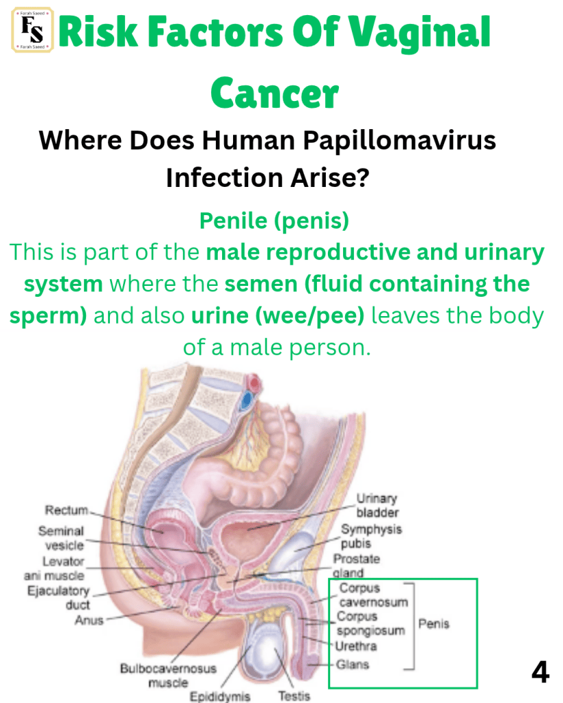
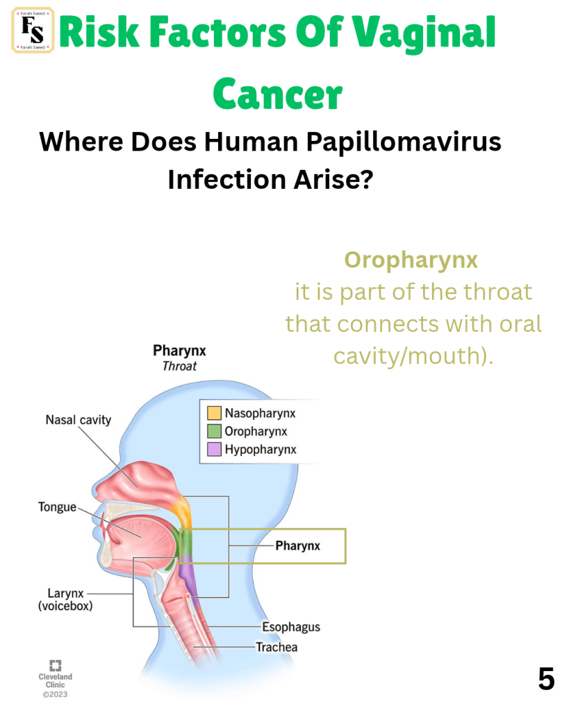
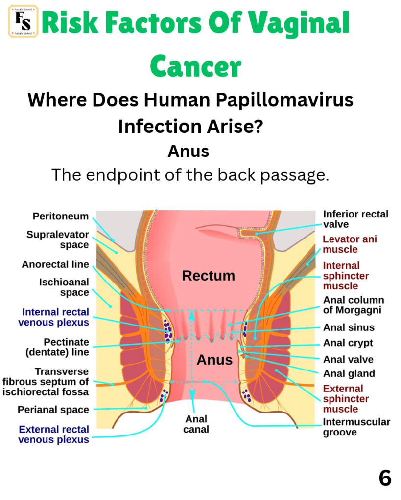
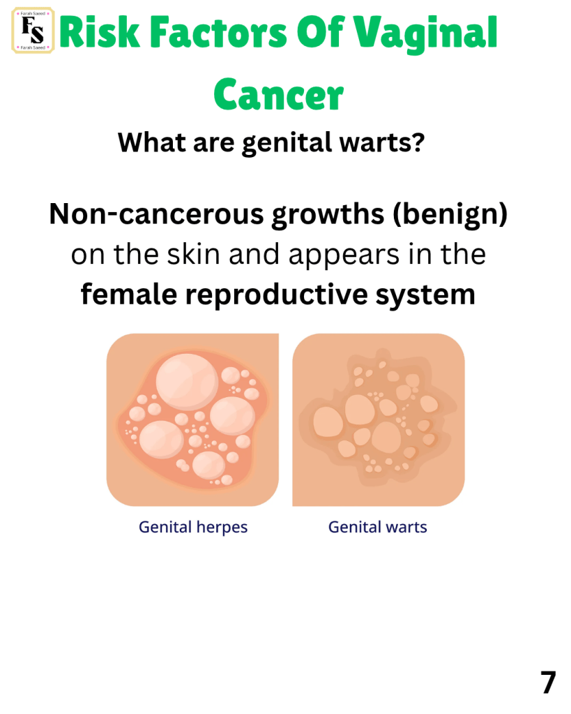

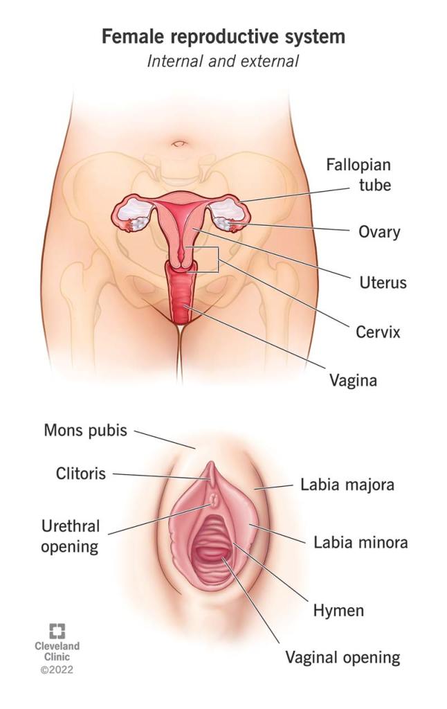
How does HPV affect the structure of the female reproductive system?
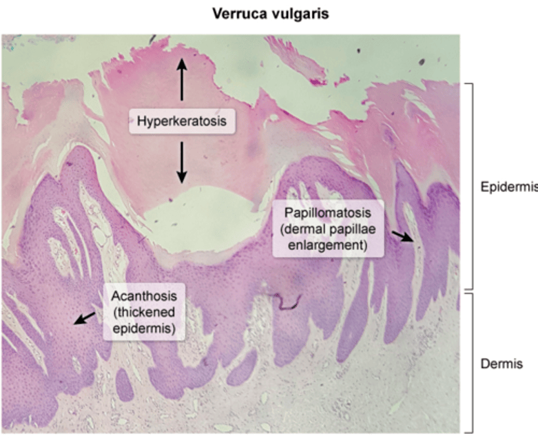
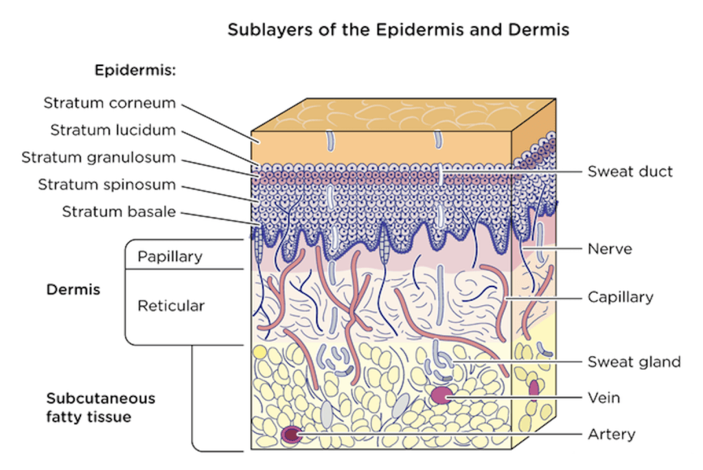
This is when high-risk HPV (16, 18) infects the lining of the reproductive system, predominantly the squamous cell lining. Abnormal growth of this lining is called dysplasia.
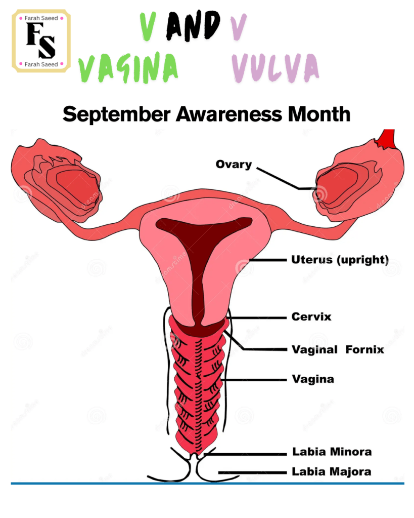
Dysplasia may arise in the cervix, vagina and vulva.
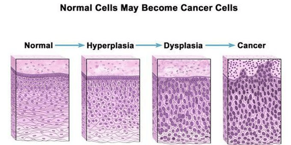
This process is called intraepithelial neoplasia. It can transition from hyperplasia (continuous growth) to low-grade dysplasia (non-cancerous) to high-grade dysplasia (non-cancerous) and then become cancerous if the viral HPV is not cleared.
The most common type of cancer arising from the lining is squamous cell carcinoma.
Abnormal cells in the cervix – cervical intraepithelial neoplasia
Abnormal cells in the vagina – vaginal intraepithelial neoplasia
Abnormal cells in the vulva – vulval intraepithelial neoplasia
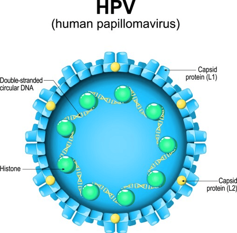
How does HPV infect cells to become cancerous if not treated?
At first, the protein coat (capsid) of the virus binds to the cell and attaches. Then it enters the cell, targets the DNA, replicates (copies DNA), and divides to make little viruses (virions) and releases the new viruses to affect other cells in the lining uncontrollably.
This is recognised by the immune cells. However, some remain and continue to grow and become precancerous and, if not treated, cancer.
Researchers estimated that 5 to 10 years after HPV infection, cervical cells can become cancerous, and 20 years to become cancerous.
Immunosuppressants
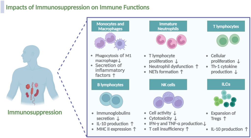
What Are Common Examples Of Autoimmune diseases?
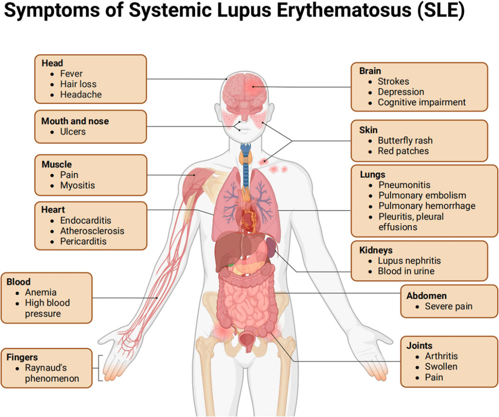
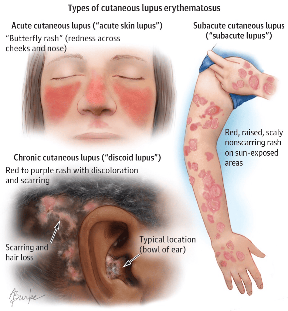
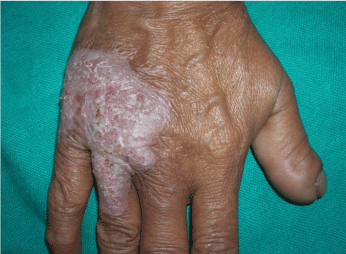
Verrucous Lupus
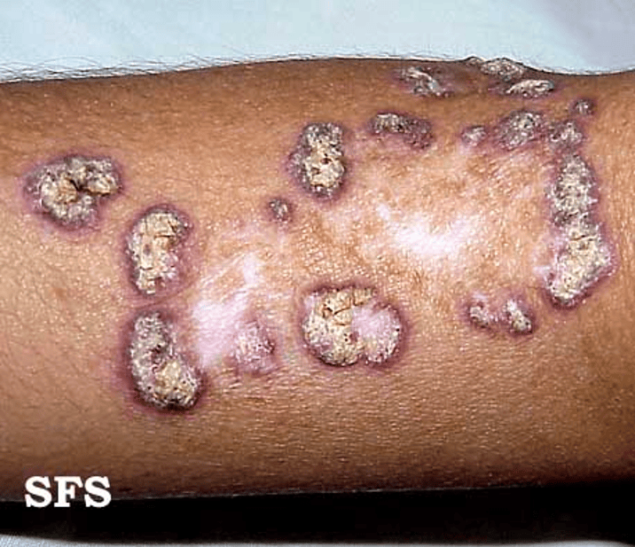
Lupus Vulgaris.
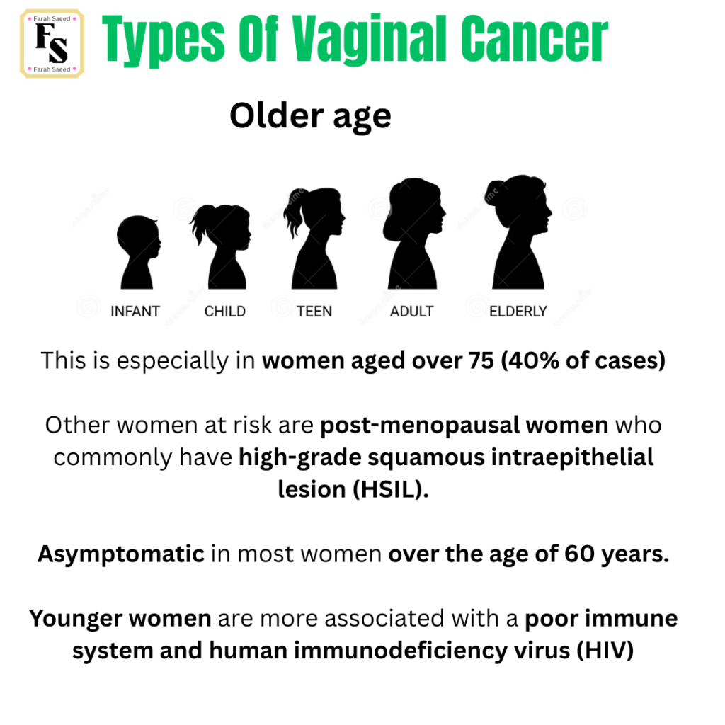
Younger women are more prone to HIV….
What Is HIV?
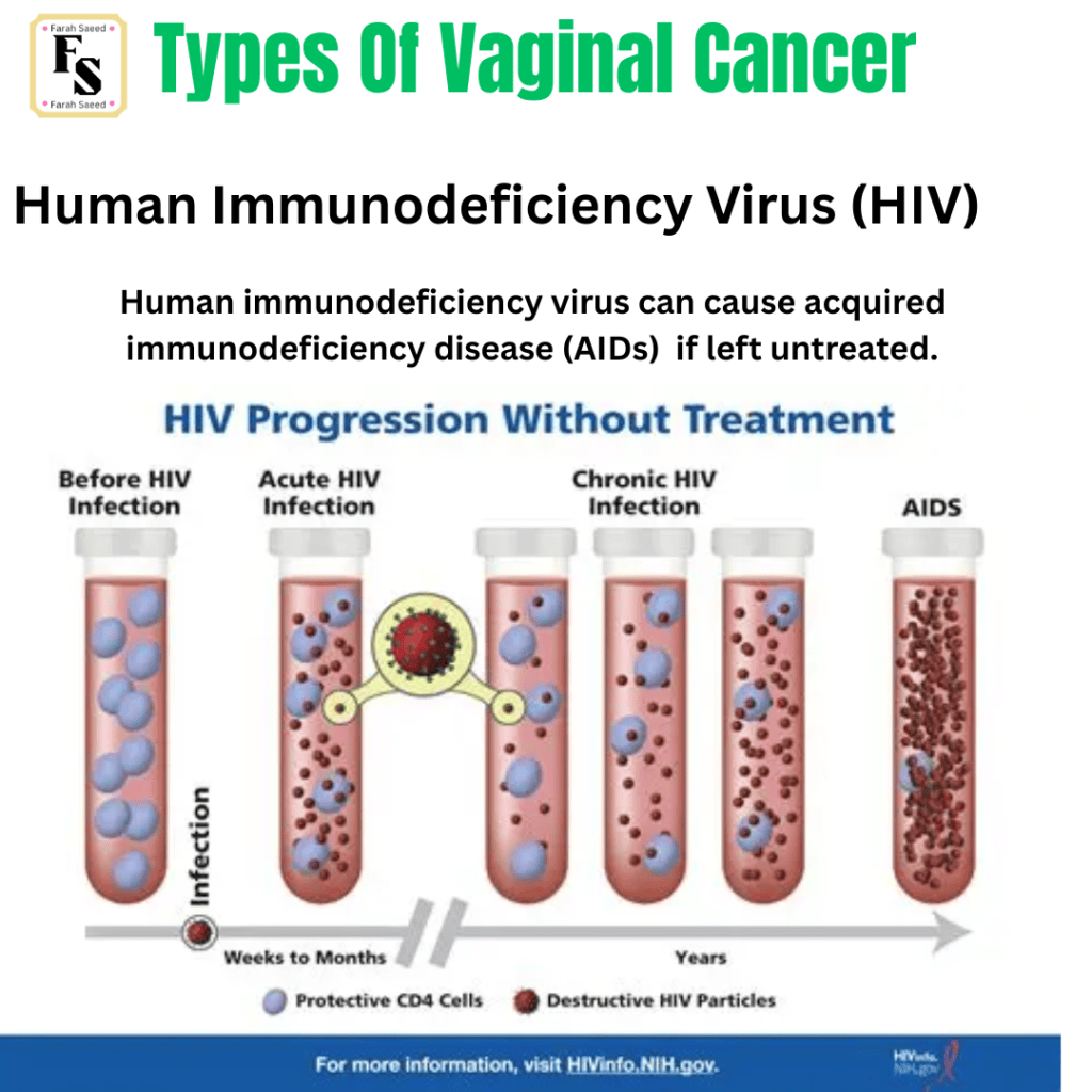
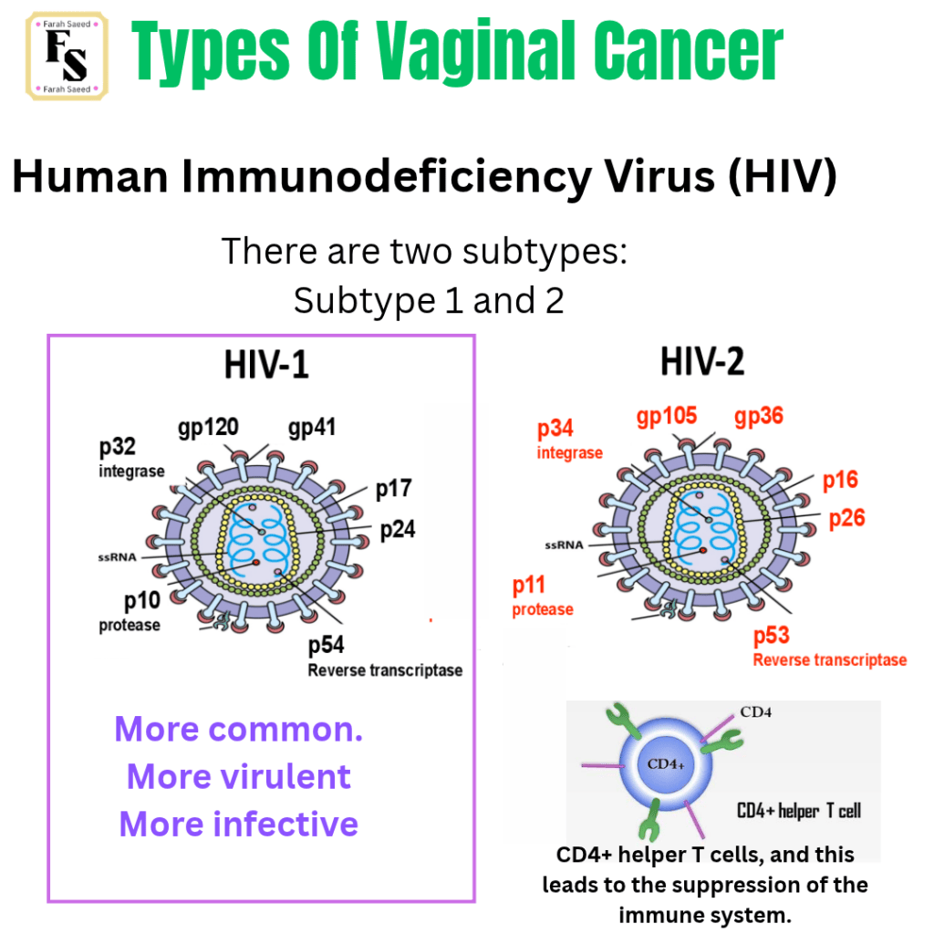
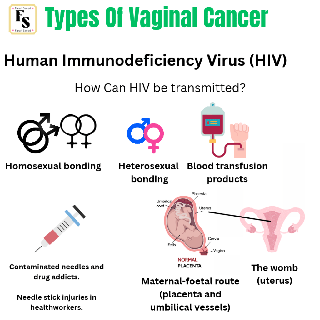
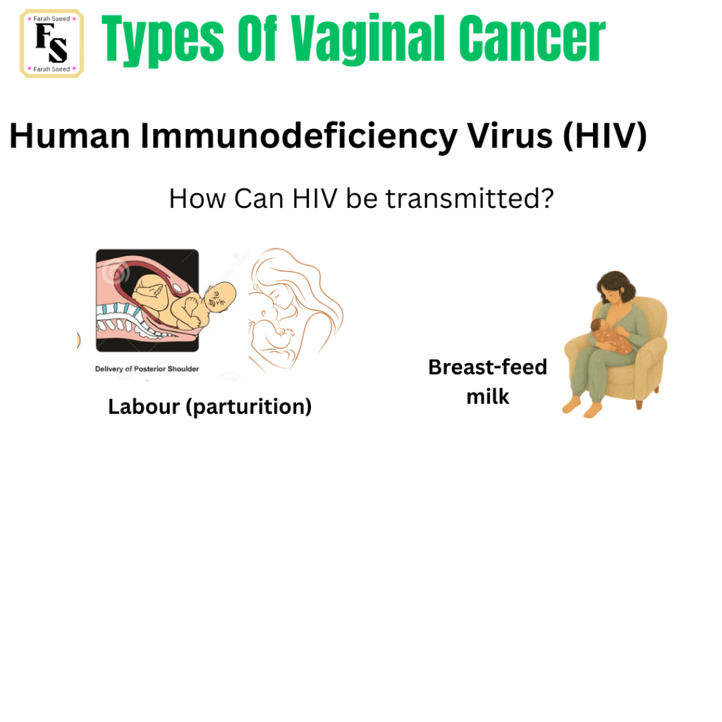
How Does HIV infection occurs?
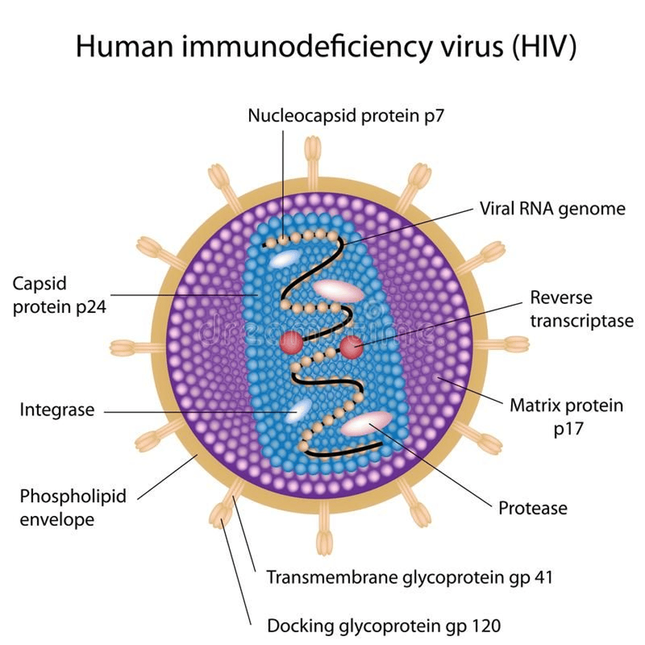
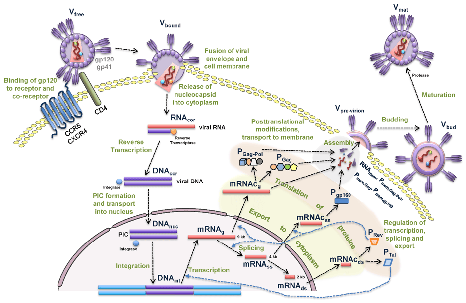
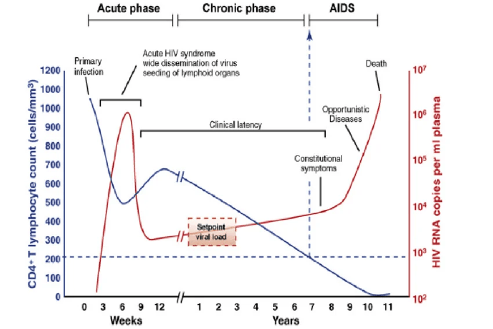
The HIV infection enters the acute phase if left untreated. The acute phase lasts between 6 and 12 weeks with flu-like symptoms, peak virus load, and low levels of CD4 T lymphocytes. There could also be mild glandular-like fever. The approximate peak level of HIV RNA copies per ml plasma is 10 to the power of 6 (viral load). The level of CD4 T lymphocytes is 500 cells per millimetre. There is an increase from week 6 to week 12, where the CD4 T lymphocytes increase from 500 to 650 cells per millimetre. The acute phase helps the virus to seed the lymphoid organs (thymus and spleen).
The level of HIV RNA copies per ml plasma steadily increases from 300 at week 9 to 500 HIV RNA copies per ml plasma at year 8. From 9 weeks to 7 years, there is a clinical latency period. The patient could be experiencing, with or without, persistent generalised abnormal lymph nodes (lymphadenopathy) which are > 1 cm in diameter at two or more extrainguinal sites (groin) for more than 3 months in the absence of causes other than HIV infection.
The chronic phase initiates after 12 weeks till 7 years. There is a descending trend from 650 CD4 + cells at week 12 till 11 years. There is a steady increase in HIV RNA copies until 7 years (setpoint viral load)
The AIDs phase then begins from year 7 to year 10. Here, there is an experience of constitutional symptoms and then opportunistic diseases in year 10, where the HIV RNA copies increase to 10 to the power of 5. This reflects viral replication and declining CD4 + T lymphocytes to <200/mm3 (time to AIDS onset varies between individuals from as little as 2 years to more than 15 years after seroconversion). The opportunistic organisms are not considered pathogenic, and susceptibility increases as the patient becomes more immunocompromised.
The HIV RNA copies reaches its midpoint between 10 to the power of 6 and 10 to the power of 7 where death is experienced.
Source: Galvis, 2014
Previous gynaecological cancers and surgical treatment
Previous gynaecological cancers can be related to the vulva, cervix, womb (endometrium), colon and rectum in the past five years. This helps to distinguish metastasis and exclude original neoplasm being in the vagina.
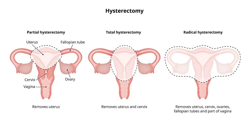
Hysterectomy is the removal of the womb that commonly occurs for non-cancerous, benign, or malignant diseases. This is especially occurs in the upper third of the vagina in patients with post-hysterectomy, but minimal vagina lesions are found for those who have not been operated.

Exposure to in utero diethylstilbestrol (DES)
It is a female hormone that is synthetically made to prevent miscarriage.
This is commonly associated with adenocarcinoma, vaginal adenosis, and endometriosis, where there is a peak incidence before aged 30 years. It was first reported in the early 1970s, when vaginal disease is highest during the first trimester (three months). It can also appear in ovarian carcinoma and cancer of the fallopian tubes/oviduct.
It is common in younger women, as adenocarcinoma is not linked with DES exposure and mainly appears after menopause (cessation or stopping of the menstrual periods).
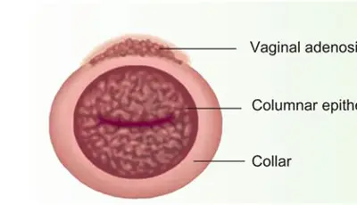
Vaginal Adenosis
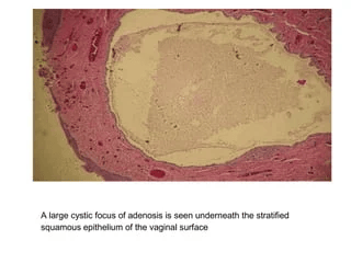
Vaginal adenosis may appear in younger women who had exposure to in utero DES and may be linked to clear cell carcinoma. Vaginal adenosis is the growth of glandular epithelium in the area rather than squamous epithelium. Vaginal adenosis rarely progresses to adenocarcinoma.
However, the role of DES in vaginal cancer is now RARE because DES is no longer prescribed.
Moreover, vaginal adenosis may be associated with hormones, drugs, trauma, inflammation, and conditions like Stevens-Johnson syndrome, otherwise known as toxic epidermal necrolysis.
What Is Stevens-Johnson syndrome?
This is a condition following a reaction to a treatment. It begins with flu-like symptoms like high temperature, sore throat, cough, and joint pain. Later, there appears to be a rash made of circular patches that are darker in the middle and lighter on the outside. The rash then progresses to the upper body, affecting the face, arms, legs, and other parts, including the genitals. This can be itchy or painful. It is common in women who have a low immune system and exposure to cancer treatment like chemotherapy. Examples of medication include antibiotics that aim to kill or decrease the growth of bacteria, painkillers, and medicines prescribed for epilepsy. Epilepsy is a neurological condition characterised by seizures with a sudden onset.
For further information, please visit: https://www.nhs.uk/conditions/stevens-johnson-syndrome/
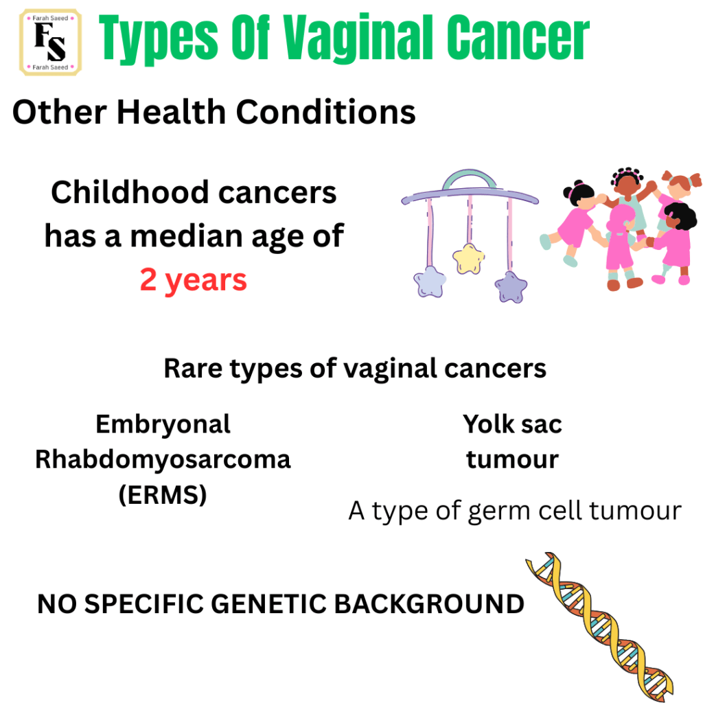
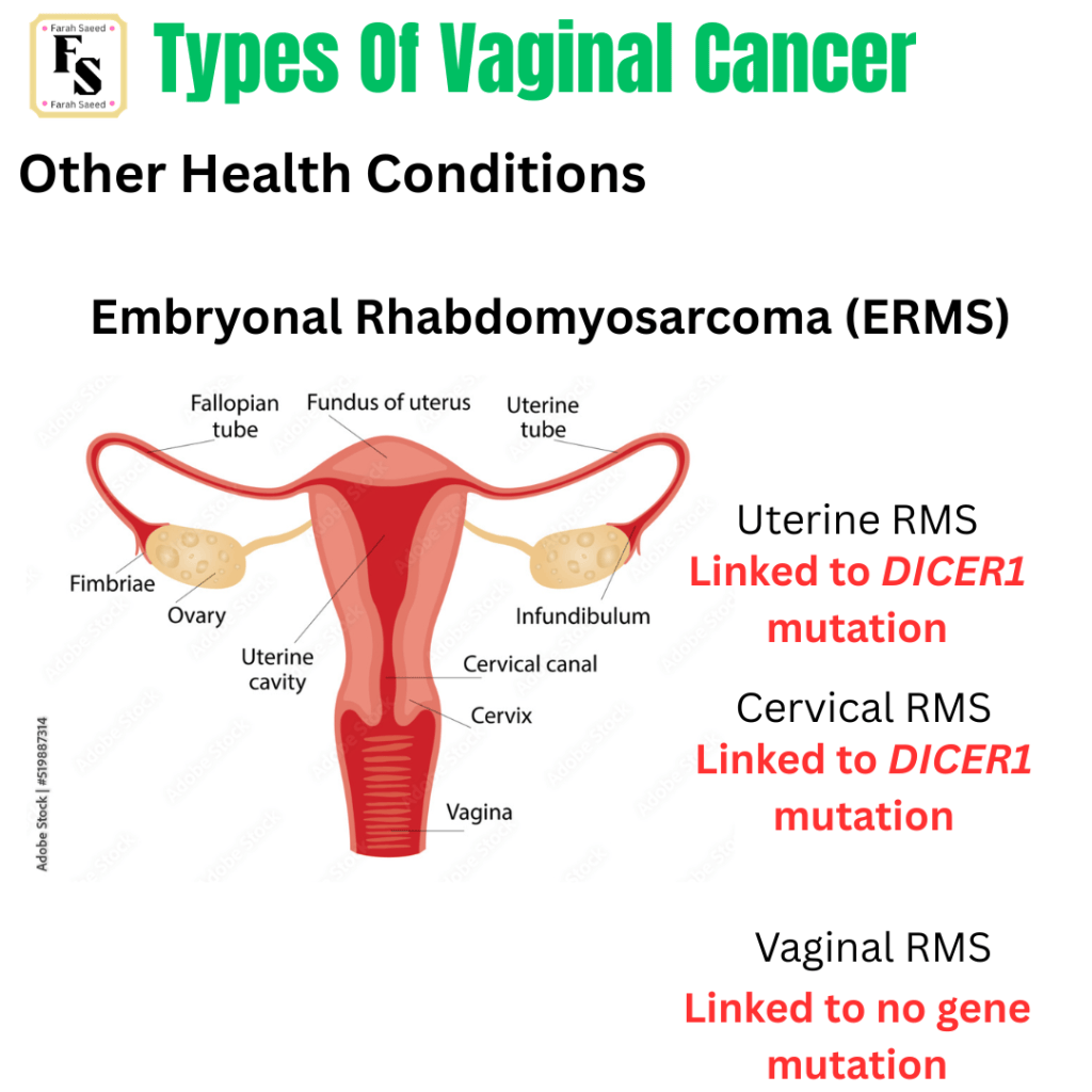
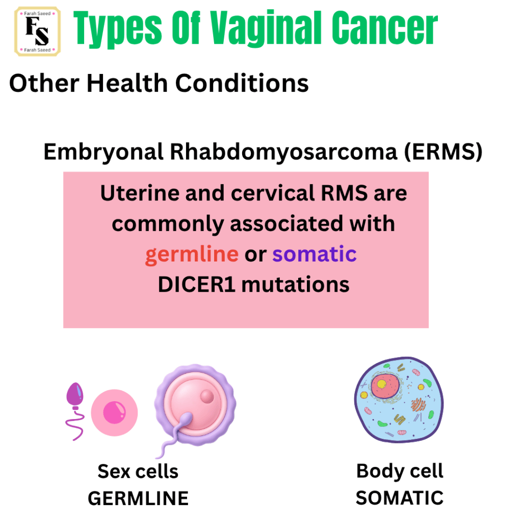
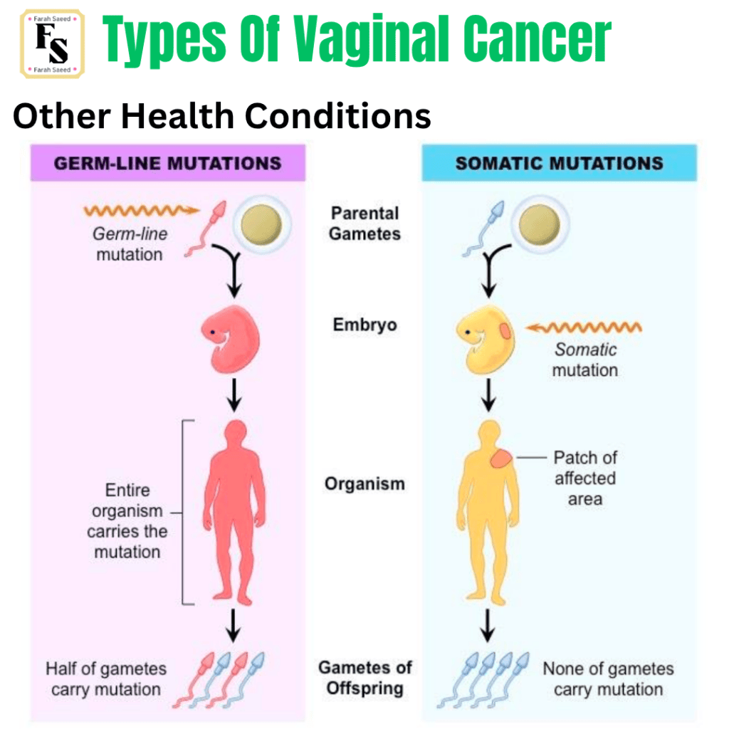
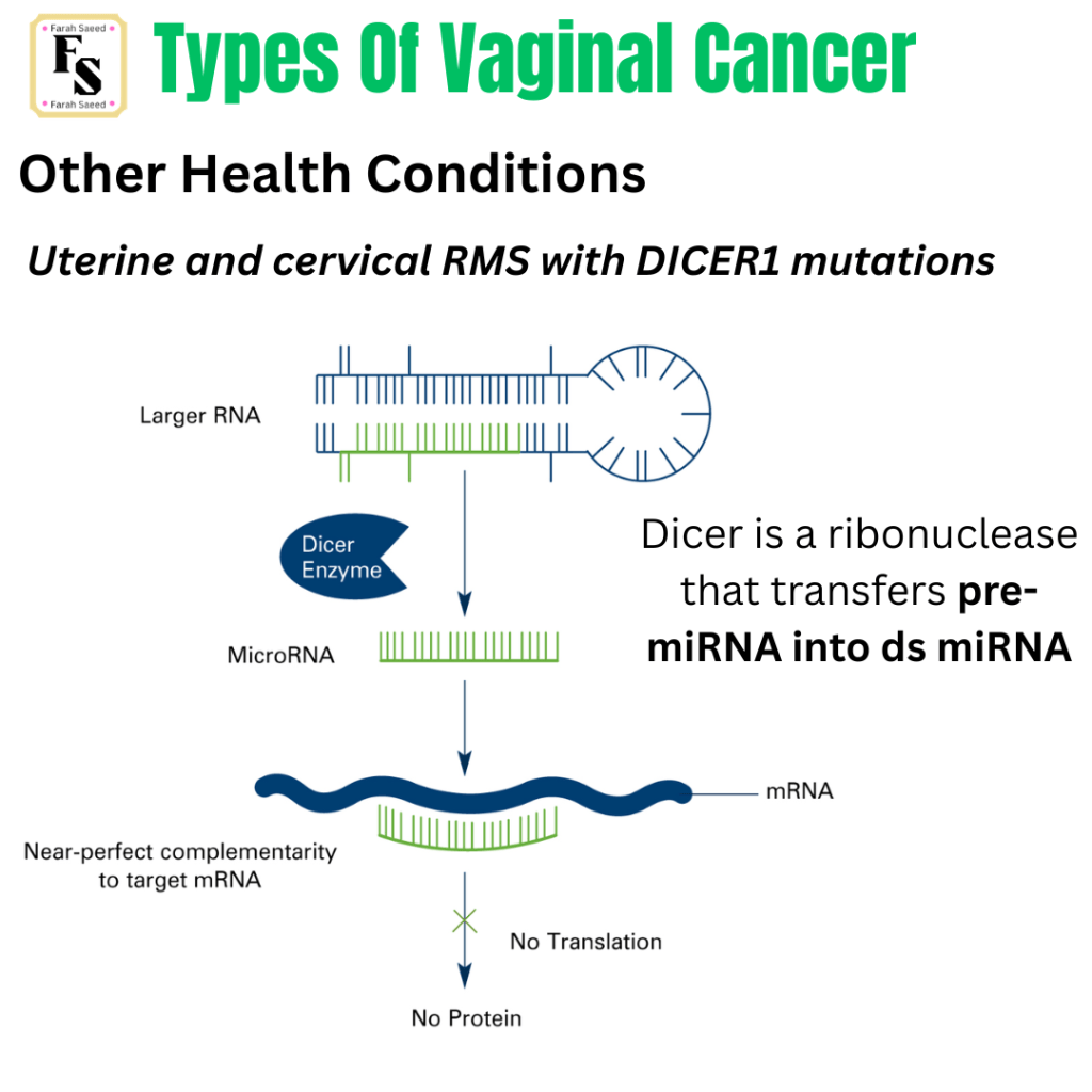
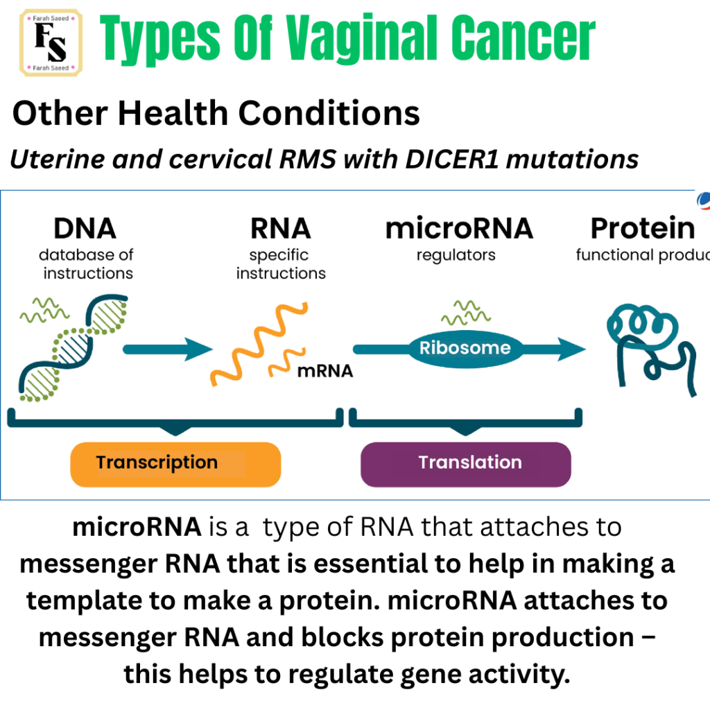
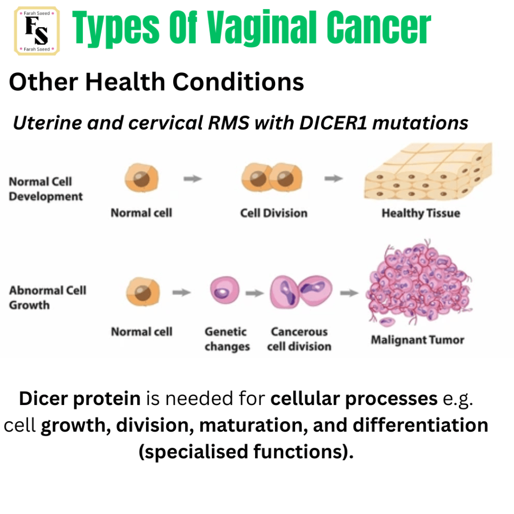
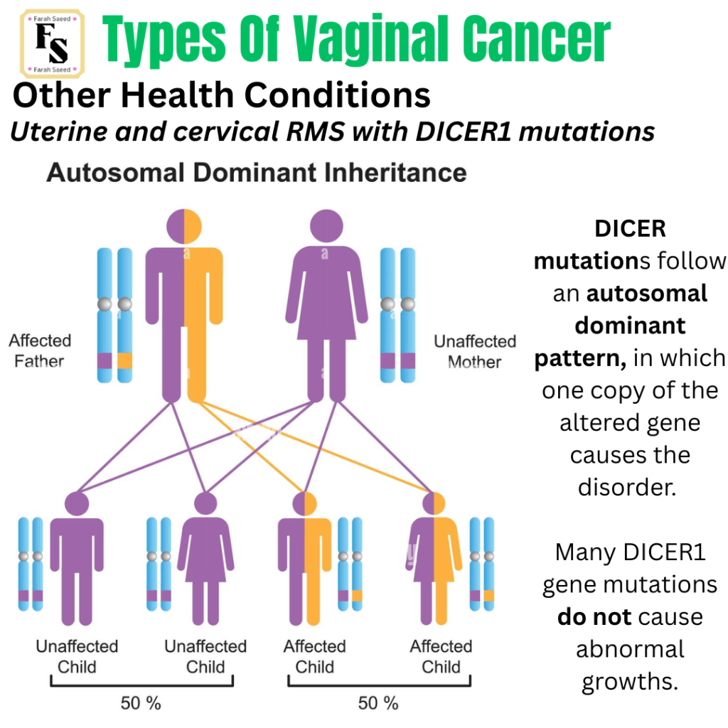
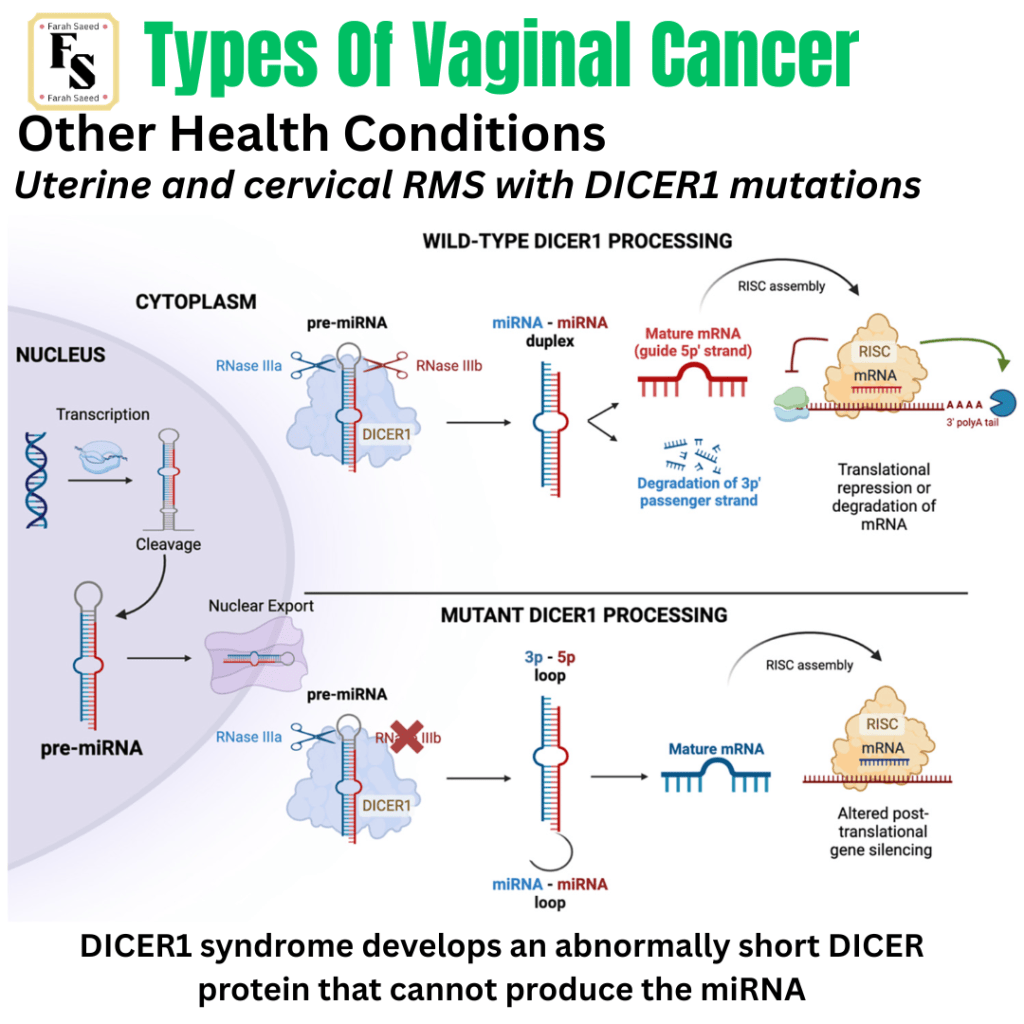
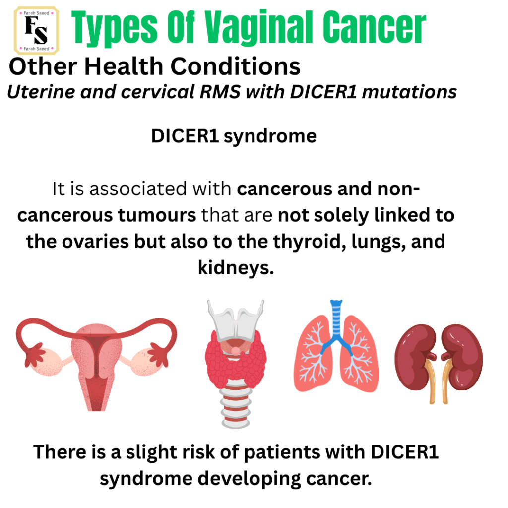
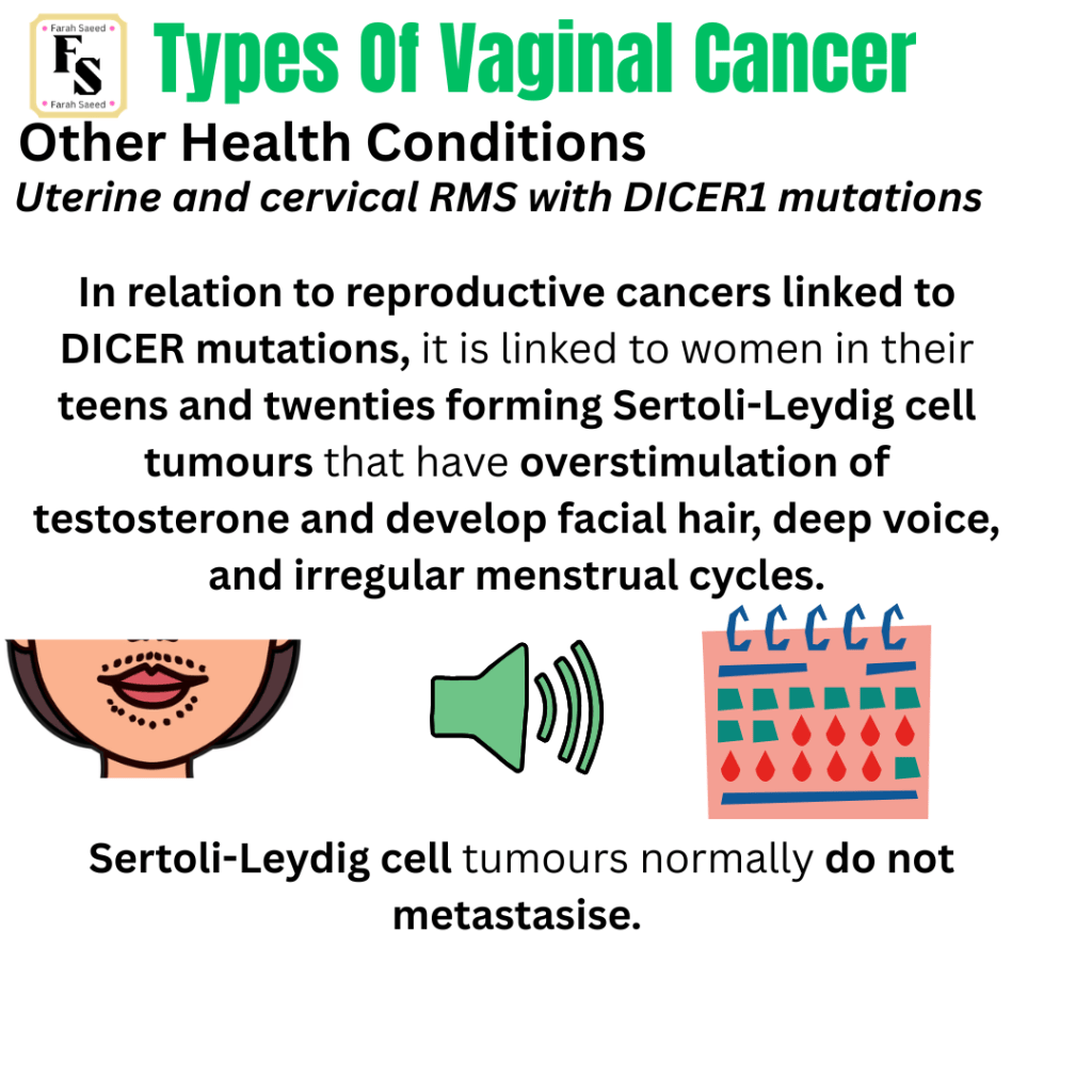
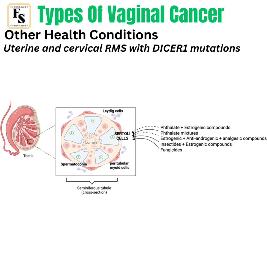
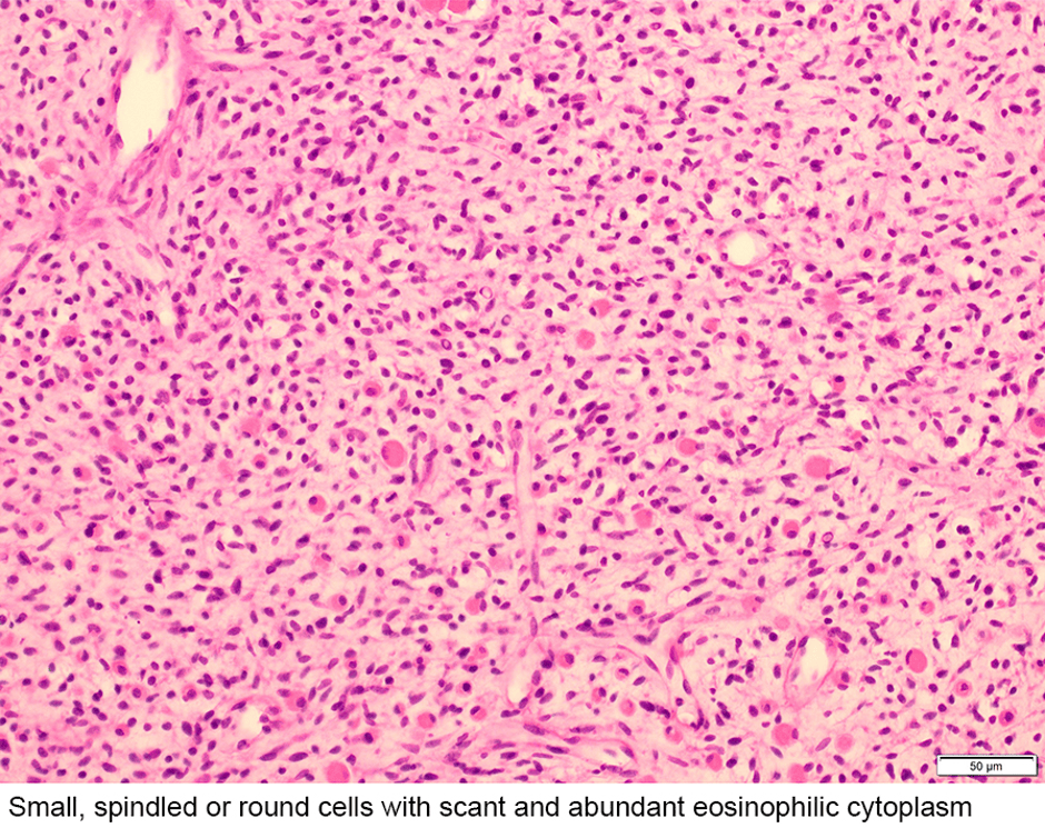
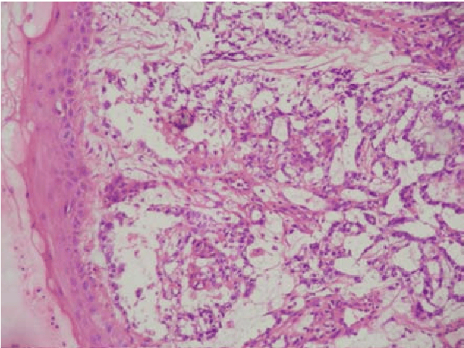
Other Health Conditions
References
Adams, T.S., Maluleke, J.C. and Cuello, M.A. (2025). Cancer of the vagina: 2025 Update. International Journal of Gynecology & Obstetrics, 171(S1), pp.48–59. doi:10.1002/ijgo.70325.
Archila, L.R., Balakrishnan, R., Boroujeni, A.M., Chiang, S., Ellenson, L., Connelly, C., Bowman, A., Vanderbilt, C., Agaram, N., Abu-Rustum, N., Hensley, M. and Park, K. (2026). 1048 HPV-Associated sarcomas of the cervix and vagina – detection of common (16, 51) and rare (30, 90, 114) HPV types, recurrent ATRX alterations and wild-type TP53/RB1 By next-generation sequencing (NGS). Laboratory Investigation, 106(3), p.105338. doi:10.1016/j.labinv.2025.105338.
Cancer Research UK (2027) Vaginal intraepithelial neoplasia (VAIN) Available at: https://www.cancerresearchuk.org/about-cancer/vaginal-cancer/vain (Accessed: 11th October 2026)
Cassidy, J., Bissett, D., Spene, R. and Payne, M. (2010) Oxford Handbook of Oncology. Oxford: Oxford University Press.
MedlinePlus (2016) DICER1 syndrome. Available at: https://medlineplus.gov/genetics/condition/dicer1-syndrome/#inheritance (Accessed: 8th October 2026)
National Cancer Institute (2025) HPV and Cancer. Available at: https://www.cancer.gov/about-cancer/causes-prevention/risk/infectious-agents/hpv-and-cancer (Accessed: 6th August 2026)
National Institute of Care and excellence Gynaecological cancers – recognition and referral: Presentation. Available at: https://cks.nice.org.uk/topics/gynaecological-cancers-recognition-referral/background-information/presentation/ Accessedl 7th August 2026.
Nout, R.A., Calaminus, G., Planchamp, F., Chargari, C., Lax, S., Martelli, H., McCluggage, W.G., Morice, P., Pakiz, M., Schmid, M.P., Stunt, J., Timmermann, B., Vokuhl, C., Orbach, D. and Fotopoulou, C. (2023). ESTRO/ESGO/SIOPe Guidelines for the management of patients with vaginal cancer. International Journal of Gynecologic Cancer, [online] 33(8). doi:10.1136/ijgc-2023-004695.
Pant, A.D., Sayami, G., Prasad, V. and Shrestha, A. (1970). A rare case of yolk sac tumor of the vagina: Cytology and histopathological findings. Nepal Journal of Obstetrics and Gynaecology, 1(2), pp.63–65. doi:10.3126/njog.v1i2.2402.
Riascos, M.C., Huynh, A., Faquin, W.C. and Nosé, V. (2024). Expanding our knowledge of DICER1 gene alterations and their role in thyroid diseases. Cancers, [online] 16(2), p.347. doi:10.3390/cancers16020347.
Torres Galvis, A.E. (2014). An RNA lariat intermediate in HIV-1 cdna synthesis. Available at: https://www.researchgate.net/publication/284360827_An_RNA_Lariat_Intermediate_in_HIV-1_cDNA_Synthesis (Accessed: 23rd Octoer 2026)


Leave a Reply